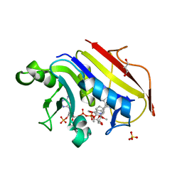
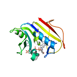
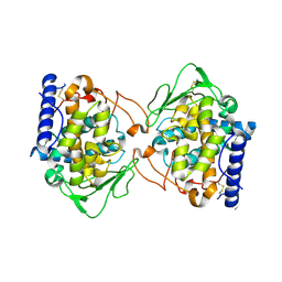
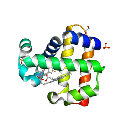
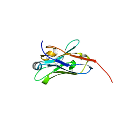
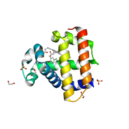
6KTQ
 
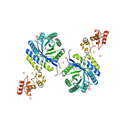 | | Crystal structure of catalytic domain of homocitrate synthase from Sulfolobus acidocaldarius (SaHCS(dRAM)) in complex with alpha-ketoglutarate/Zn2+/CoA | | 分子名称: | 2-OXOGLUTARIC ACID, COENZYME A, Homocitrate synthase, ... | | 著者 | Suzuki, T, Tomita, T, Kuzuyama, T, Nishiyama, M. | | 登録日 | 2019-08-28 | | 公開日 | 2020-09-02 | | 最終更新日 | 2023-11-22 | | 実験手法 | X-RAY DIFFRACTION (1.98 Å) | | 主引用文献 | Involvement of subdomain II in the recognition of acetyl-CoA revealed by the crystal structure of homocitrate synthase from Sulfolobus acidocaldarius.
Febs J., 288, 2021
|
|
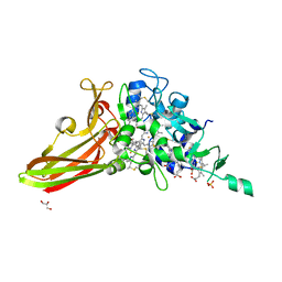
2RIU
 | | Alternative models for two crystal structures of Candida albicans 3,4-dihydroxy-2-butanone 4-phosphate synthase- alternate interpreation | | 分子名称: | 3,4-dihydroxy-2-butanone 4-phosphate synthase, RIBULOSE-5-PHOSPHATE | | 著者 | Stenkamp, R.E, Le Trong, I. | | 登録日 | 2007-10-12 | | 公開日 | 2008-01-29 | | 最終更新日 | 2024-02-21 | | 実験手法 | X-RAY DIFFRACTION (1.7 Å) | | 主引用文献 | Alternative models for two crystal structures of Candida albicans 3,4-dihydroxy-2-butanone 4-phosphate synthase.
Acta Crystallogr.,Sect.D, 64, 2008
|
|
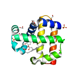
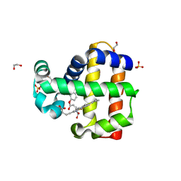
6FBP
 | | Human Methionine Adenosyltransferase II mutant (S114A) in P22121 crystal form | | 分子名称: | (DIPHOSPHONO)AMINOPHOSPHONIC ACID, 1,2-ETHANEDIOL, ACETATE ION, ... | | 著者 | Panmanee, J, Antonyuk, S.V, Hasnain, S.S. | | 登録日 | 2017-12-19 | | 公開日 | 2019-01-30 | | 最終更新日 | 2024-01-17 | | 実験手法 | X-RAY DIFFRACTION (1.65 Å) | | 主引用文献 | Control and regulation of S-Adenosylmethionine biosynthesis by the regulatory beta subunit and quinolone-based compounds.
Febs J., 286, 2019
|
|
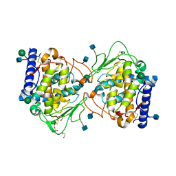
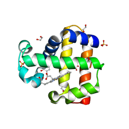
6FBN
 | | Human Methionine Adenosyltransferase II mutant (Q113A) | | 分子名称: | (DIPHOSPHONO)AMINOPHOSPHONIC ACID, 1,2-ETHANEDIOL, DI(HYDROXYETHYL)ETHER, ... | | 著者 | Panmanee, J, Antonyuk, S.V, Hasnain, S.S. | | 登録日 | 2017-12-19 | | 公開日 | 2019-01-30 | | 最終更新日 | 2024-01-17 | | 実験手法 | X-RAY DIFFRACTION (2.7 Å) | | 主引用文献 | Control and regulation of S-Adenosylmethionine biosynthesis by the regulatory beta subunit and quinolone-based compounds.
Febs J., 286, 2019
|
|
2W3A
 | |
8IGF
 | | Crystal Structure of Human Carbonic Anhydrase II In-complex with 4-Acetylphenylboronic acid at 2.6 A Resolution | | 分子名称: | (4-ethanoylphenyl)boronic acid, Carbonic anhydrase 2, GLYCEROL, ... | | 著者 | Rasheed, S, Huda, N, Fisher, S.Z, Falke, S, Gul, S, Ahmad, M.S, Choudhary, M.I. | | 登録日 | 2023-02-20 | | 公開日 | 2024-02-28 | | 最終更新日 | 2024-05-15 | | 実験手法 | X-RAY DIFFRACTION (2.6 Å) | | 主引用文献 | Identification, crystallization, and first X-ray structure analyses of phenyl boronic acid-based inhibitors of human carbonic anhydrase-II.
Int.J.Biol.Macromol., 267, 2024
|
|
6FBO
 | | Human Methionine Adenosyltransferase II mutant (S114A) in I222 crystal form | | 分子名称: | (DIPHOSPHONO)AMINOPHOSPHONIC ACID, 1,2-ETHANEDIOL, ADENOSINE, ... | | 著者 | Panmanee, J, Antonyuk, S.V, Hasnain, S.S. | | 登録日 | 2017-12-19 | | 公開日 | 2019-01-30 | | 最終更新日 | 2024-01-17 | | 実験手法 | X-RAY DIFFRACTION (1.8 Å) | | 主引用文献 | Control and regulation of S-Adenosylmethionine biosynthesis by the regulatory beta subunit and quinolone-based compounds.
Febs J., 286, 2019
|
|
6FCD
 | | Human Methionine Adenosyltransferase II mutant (R264A) | | 分子名称: | 1,2-ETHANEDIOL, ADENOSINE, DI(HYDROXYETHYL)ETHER, ... | | 著者 | Panmanee, J, Antonyuk, S.V, Hasnain, S.S. | | 登録日 | 2017-12-20 | | 公開日 | 2019-01-30 | | 最終更新日 | 2024-01-17 | | 実験手法 | X-RAY DIFFRACTION (1.7 Å) | | 主引用文献 | Control and regulation of S-Adenosylmethionine biosynthesis by the regulatory beta subunit and quinolone-based compounds.
Febs J., 286, 2019
|
|
4Y3E
 | | Endothiapepsin in complex with fragment 5 | | 分子名称: | 1H-isoindol-3-amine, ACETATE ION, DIMETHYL SULFOXIDE, ... | | 著者 | Radeva, N, Uehlein, M, Weiss, M.S, Heine, A, Klebe, G. | | 登録日 | 2015-02-10 | | 公開日 | 2016-02-17 | | 最終更新日 | 2024-11-06 | | 実験手法 | X-RAY DIFFRACTION (1.25 Å) | | 主引用文献 | Crystallographic Fragment Screening of an Entire Library
To Be Published
|
|
2W3B
 | | HUMAN DIHYDROFOLATE REDUCTASE COMPLEXED WITH NADPH AND A LIPOPHILIC ANTIFOLATE SELECTIVE FOR MYCOBACTERIUM AVIUM DHFR, 6-((2,5- DIETHOXYPHENYL)AMINOMETHYL)-2,4-DIAMINO-5-METHYLPYRIDO(2,3-D) PYRIMIDINE (SRI-8686) | | 分子名称: | 6-{[(2,5-DIETHOXYPHENYL)AMINO]METHYL}-5-METHYLPYRIDO[2,3-D]PYRIMIDINE-2,4-DIAMINE, DIHYDROFOLATE REDUCTASE, FOLIC ACID, ... | | 著者 | Leung, A.K.W, Reynolds, R.C, Borhani, D.W. | | 登録日 | 2008-11-11 | | 公開日 | 2009-11-17 | | 最終更新日 | 2024-05-01 | | 実験手法 | X-RAY DIFFRACTION (1.27 Å) | | 主引用文献 | Structural Basis for Selective Inhibition of Mycobacterium Avium Dihydrofolate Reductase by a Lipophilic Antifolate
To be Published
|
|
5OR4
 | | Crystal structure of Aspergillus oryzae catechol oxidase in deoxy-form | | 分子名称: | 2-acetamido-2-deoxy-beta-D-glucopyranose, 2-acetamido-2-deoxy-beta-D-glucopyranose-(1-4)-2-acetamido-2-deoxy-beta-D-glucopyranose, COPPER (II) ION, ... | | 著者 | Hakulinen, N, Penttinen, L, Rutanen, C, Rouvinen, J. | | 登録日 | 2017-08-15 | | 公開日 | 2018-05-09 | | 最終更新日 | 2024-10-09 | | 実験手法 | X-RAY DIFFRACTION (2.445 Å) | | 主引用文献 | A new crystal form of Aspergillus oryzae catechol oxidase and evaluation of copper site structures in coupled binuclear copper enzymes.
PLoS ONE, 13, 2018
|
|
8EE2
 | |
6G6R
 | | Human Methionine Adenosyltransferase II with SAMe and PPNP | | 分子名称: | (DIPHOSPHONO)AMINOPHOSPHONIC ACID, 1,2-ETHANEDIOL, MAGNESIUM ION, ... | | 著者 | Panmanee, J, Antonyuk, S.V, Hasnain, S.S. | | 登録日 | 2018-04-02 | | 公開日 | 2019-04-10 | | 最終更新日 | 2024-01-17 | | 実験手法 | X-RAY DIFFRACTION (1.35 Å) | | 主引用文献 | Control and regulation of S-Adenosylmethionine biosynthesis by the regulatory beta subunit and quinolone-based compounds.
Febs J., 286, 2019
|
|
6O4X
 | | Binary complex of native hAChE with 9-aminoacridine | | 分子名称: | 9-AMINOACRIDINE, Acetylcholinesterase, GLYCEROL, ... | | 著者 | Gerlits, O, Kovalevsky, A, Radic, Z. | | 登録日 | 2019-03-01 | | 公開日 | 2019-06-19 | | 最終更新日 | 2024-11-06 | | 実験手法 | X-RAY DIFFRACTION (2.3 Å) | | 主引用文献 | A new crystal form of human acetylcholinesterase for exploratory room-temperature crystallography studies.
Chem.Biol.Interact., 309, 2019
|
|
6ONG
 | | Dehaloperoxidate B in complex with substrate 4-F-cresol | | 分子名称: | 1,2-ETHANEDIOL, 4-fluoro-2-methylphenol, Dehaloperoxidase B, ... | | 著者 | Ghiladi, R.A, de Serrano, V.S, Malewschik, T, McGuire, A. | | 登録日 | 2019-04-22 | | 公開日 | 2019-09-11 | | 最終更新日 | 2024-12-25 | | 実験手法 | X-RAY DIFFRACTION (1.42 Å) | | 主引用文献 | The multifunctional globin dehaloperoxidase strikes again: Simultaneous peroxidase and peroxygenase mechanisms in the oxidation of EPA pollutants.
Arch.Biochem.Biophys., 673, 2019
|
|
8DAF
 | | Human SF-1 LBD bound to synthetic agonist 6N-10CA and bacterial phospholipid | | 分子名称: | 10-[(3aR,6S,6aR)-3-phenyl-3a-(1-phenylethenyl)-6-(sulfamoylamino)-1,3a,4,5,6,6a-hexahydropentalen-2-yl]decanoic acid (non-preferred name), DI-PALMITOYL-3-SN-PHOSPHATIDYLETHANOLAMINE, Nuclear receptor coactivator 2, ... | | 著者 | D'Agostino, E.H, Cato, M.L, Ortlund, E.A. | | 登録日 | 2022-06-13 | | 公開日 | 2023-06-28 | | 最終更新日 | 2023-10-25 | | 実験手法 | X-RAY DIFFRACTION (2.59 Å) | | 主引用文献 | Comparison of activity, structure, and dynamics of SF-1 and LRH-1 complexed with small molecule modulators.
J.Biol.Chem., 299, 2023
|
|
6ONX
 | | Dehaloperoxidase B in complex with substrate 4-Br-cresol | | 分子名称: | 1,2-ETHANEDIOL, 4-bromo-2-methylphenol, DI(HYDROXYETHYL)ETHER, ... | | 著者 | Ghiladi, R.A, de Serrano, V.S, Malewschik, T. | | 登録日 | 2019-04-22 | | 公開日 | 2019-09-25 | | 最終更新日 | 2023-10-11 | | 実験手法 | X-RAY DIFFRACTION (1.7 Å) | | 主引用文献 | The multifunctional globin dehaloperoxidase strikes again: Simultaneous peroxidase and peroxygenase mechanisms in the oxidation of EPA pollutants.
Arch.Biochem.Biophys., 673, 2019
|
|
6GOA
 | |
8E2N
 | |
5ZE8
 | | Crystal structure of a penta-heme cytochrome c552 from Thermochromatium tepidum | | 分子名称: | GLYCEROL, HEME C, SULFATE ION, ... | | 著者 | Yu, L.-J, Chen, J.-H, Shen, J.-R. | | 登録日 | 2018-02-27 | | 公開日 | 2018-04-25 | | 最終更新日 | 2024-10-16 | | 実験手法 | X-RAY DIFFRACTION (1.6 Å) | | 主引用文献 | Properties and structure of a low-potential, penta-heme cytochrome c552from a thermophilic purple sulfur photosynthetic bacterium Thermochromatium tepidum.
Photosyn. Res., 139, 2019
|
|
6ONZ
 | | Dehaloperoxidase B in complex with substtrate 4-nitro-cresol | | 分子名称: | 1,2-ETHANEDIOL, 2-methyl-4-nitrophenol, Dehaloperoxidase B, ... | | 著者 | Ghiladi, R.A, de Serrano, V.S, Malewschik, T. | | 登録日 | 2019-04-22 | | 公開日 | 2019-09-11 | | 最終更新日 | 2024-03-13 | | 実験手法 | X-RAY DIFFRACTION (1.8 Å) | | 主引用文献 | The multifunctional globin dehaloperoxidase strikes again: Simultaneous peroxidase and peroxygenase mechanisms in the oxidation of EPA pollutants.
Arch.Biochem.Biophys., 673, 2019
|
|
5OR3
 | | Crystal structure of Aspergillus oryzae catechol oxidase in met/deoxy-form | | 分子名称: | 1,2-ETHANEDIOL, 2-acetamido-2-deoxy-beta-D-glucopyranose, 2-acetamido-2-deoxy-beta-D-glucopyranose-(1-4)-2-acetamido-2-deoxy-beta-D-glucopyranose, ... | | 著者 | Hakulinen, N, Penttinen, L, Rutanen, C, Rouvinen, J. | | 登録日 | 2017-08-15 | | 公開日 | 2018-05-09 | | 最終更新日 | 2024-11-20 | | 実験手法 | X-RAY DIFFRACTION (1.795 Å) | | 主引用文献 | A new crystal form of Aspergillus oryzae catechol oxidase and evaluation of copper site structures in coupled binuclear copper enzymes.
PLoS ONE, 13, 2018
|
|
6ONK
 | | Dehaloperoxidase B in complex with substrate 4-Cl-cresol | | 分子名称: | 1,2-ETHANEDIOL, 4-chloro-2-methylphenol, Dehaloperoxidase B, ... | | 著者 | Ghiladi, R.A, de Serrano, V.S, Malewschik, T. | | 登録日 | 2019-04-22 | | 公開日 | 2019-09-11 | | 最終更新日 | 2024-03-13 | | 実験手法 | X-RAY DIFFRACTION (1.5 Å) | | 主引用文献 | The multifunctional globin dehaloperoxidase strikes again: Simultaneous peroxidase and peroxygenase mechanisms in the oxidation of EPA pollutants.
Arch.Biochem.Biophys., 673, 2019
|
|
6ONR
 | | Dehaloperoxidase B in complex with substrate 4-methyl-cresol | | 分子名称: | 1,2-ETHANEDIOL, 2,4-dimethylphenol, DI(HYDROXYETHYL)ETHER, ... | | 著者 | Ghiladi, R.A, de Serrano, V.S, Malewschik, T. | | 登録日 | 2019-04-22 | | 公開日 | 2019-09-25 | | 最終更新日 | 2023-10-11 | | 実験手法 | X-RAY DIFFRACTION (1.35 Å) | | 主引用文献 | The multifunctional globin dehaloperoxidase strikes again: Simultaneous peroxidase and peroxygenase mechanisms in the oxidation of EPA pollutants.
Arch.Biochem.Biophys., 673, 2019
|
|
6OO1
 | | Dehaloperoxidase B in complex with substrate o-cresol | | 分子名称: | 1,2-ETHANEDIOL, Dehaloperoxidase B, PROTOPORPHYRIN IX CONTAINING FE, ... | | 著者 | Ghiladi, R.A, de Serrano, V.S, Malewschik, T. | | 登録日 | 2019-04-22 | | 公開日 | 2020-07-22 | | 最終更新日 | 2023-10-11 | | 実験手法 | X-RAY DIFFRACTION (1.602 Å) | | 主引用文献 | The multifunctional globin dehaloperoxidase strikes again: Simultaneous peroxidase and peroxygenase mechanisms in the oxidation of EPA pollutants.
Arch.Biochem.Biophys., 673, 2019
|
|


