7P7S
 
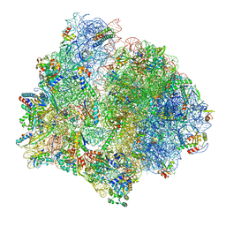 | | PoxtA-EQ2 antibiotic resistance ABCF bound to E. faecalis 70S ribosome, state II | | 分子名称: | 1,4-DIAMINOBUTANE, 16S rRNA, 23S rRNA, ... | | 著者 | Crowe-McAuliffe, C, Wilson, D.N. | | 登録日 | 2021-07-20 | | 公開日 | 2022-02-23 | | 最終更新日 | 2022-04-20 | | 実験手法 | ELECTRON MICROSCOPY (3 Å) | | 主引用文献 | Structural basis for PoxtA-mediated resistance to phenicol and oxazolidinone antibiotics.
Nat Commun, 13, 2022
|
|
7P7U
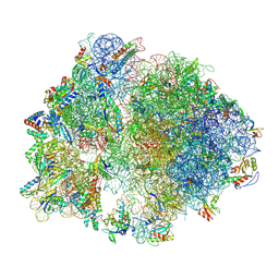 | | E. faecalis 70S ribosome with P-tRNA, state IV | | 分子名称: | 1,4-DIAMINOBUTANE, 16S rRNA, 23S rRNA, ... | | 著者 | Crowe-McAuliffe, C, Wilson, D.N. | | 登録日 | 2021-07-20 | | 公開日 | 2022-02-23 | | 最終更新日 | 2022-04-20 | | 実験手法 | ELECTRON MICROSCOPY (3.1 Å) | | 主引用文献 | Structural basis for PoxtA-mediated resistance to phenicol and oxazolidinone antibiotics.
Nat Commun, 13, 2022
|
|
7P7Q
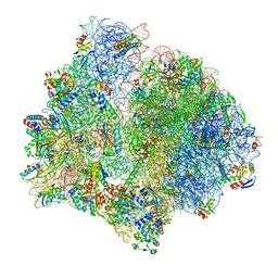 | | E. faecalis 70S ribosome bound by PoxtA-EQ2, high-resolution combined volume | | 分子名称: | 1,4-DIAMINOBUTANE, 16S rRNA, 23S rRNA, ... | | 著者 | Crowe-McAuliffe, C, Wilson, D.N. | | 登録日 | 2021-07-20 | | 公開日 | 2022-02-23 | | 最終更新日 | 2024-04-24 | | 実験手法 | ELECTRON MICROSCOPY (2.4 Å) | | 主引用文献 | Structural basis for PoxtA-mediated resistance to phenicol and oxazolidinone antibiotics.
Nat Commun, 13, 2022
|
|
7P7R
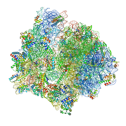 | | PoxtA-EQ2 antibiotic resistance ABCF bound to E. faecalis 70S ribosome, state I | | 分子名称: | 1,4-DIAMINOBUTANE, 16S rRNA, 23S rRNA, ... | | 著者 | Crowe-McAuliffe, C, Wilson, D.N. | | 登録日 | 2021-07-20 | | 公開日 | 2022-03-23 | | 最終更新日 | 2022-04-20 | | 実験手法 | ELECTRON MICROSCOPY (2.9 Å) | | 主引用文献 | Structural basis for PoxtA-mediated resistance to phenicol and oxazolidinone antibiotics.
Nat Commun, 13, 2022
|
|
7Q37
 | | Crystal structure of proton pump MAR rhodopsin pressurized with krypton | | 分子名称: | Bacteriorhodopsin, EICOSANE, HEXANE, ... | | 著者 | Melnikov, I, Rulev, M, Astashkin, R, Kovalev, K, Carpentier, P, Gordeliy, V, Popov, A. | | 登録日 | 2021-10-27 | | 公開日 | 2022-04-27 | | 最終更新日 | 2024-01-31 | | 実験手法 | X-RAY DIFFRACTION (2.25 Å) | | 主引用文献 | High-pressure crystallography shows noble gas intervention into protein-lipid interaction and suggests a model for anaesthetic action.
Commun Biol, 5, 2022
|
|
5O0S
 | | Crystal structure of txGH116 (beta-glucosidase from Thermoanaerobacterium xylolyticum) in complex with unreacted beta Cyclophellitol Cyclosulfate probe ME711 | | 分子名称: | (3~{a}~{S},4~{R},5~{S},6~{R},7~{R},7~{a}~{R})-7-(hydroxymethyl)-2,2-bis(oxidanylidene)-3~{a},4,5,6,7,7~{a}-hexahydrobenzo[d][1,3,2]dioxathiole-4,5,6-triol, 1,2-ETHANEDIOL, CALCIUM ION, ... | | 著者 | Wu, L, Offen, W.A, Breen, I.Z, Davies, G.J. | | 登録日 | 2017-05-16 | | 公開日 | 2017-08-09 | | 最終更新日 | 2024-01-17 | | 実験手法 | X-RAY DIFFRACTION (1.16 Å) | | 主引用文献 | 1,6-Cyclophellitol Cyclosulfates: A New Class of Irreversible Glycosidase Inhibitor.
ACS Cent Sci, 3, 2017
|
|
5OLE
 | |
5O4W
 | | Protein structure determination by electron diffraction using a single three-dimensional nanocrystal | | 分子名称: | Lysozyme C | | 著者 | Clabbers, M.T.B, van Genderen, E, Wan, W, Wiegers, E.L, Gruene, T, Abrahams, J.P. | | 登録日 | 2017-05-31 | | 公開日 | 2017-08-23 | | 最終更新日 | 2024-01-17 | | 実験手法 | ELECTRON CRYSTALLOGRAPHY (2.11 Å) | | 主引用文献 | Protein structure determination by electron diffraction using a single three-dimensional nanocrystal.
Acta Crystallogr D Struct Biol, 73, 2017
|
|
5O4X
 | | Protein structure determination by electron diffraction using a single three-dimensional nanocrystal | | 分子名称: | Lysozyme C | | 著者 | Clabbers, M.T.B, van Genderen, E, Wan, W, Wiegers, E.L, Gruene, T, Abrahams, J.P. | | 登録日 | 2017-05-31 | | 公開日 | 2017-08-23 | | 最終更新日 | 2024-01-17 | | 実験手法 | ELECTRON CRYSTALLOGRAPHY (2.11 Å) | | 主引用文献 | Protein structure determination by electron diffraction using a single three-dimensional nanocrystal.
Acta Crystallogr D Struct Biol, 73, 2017
|
|
5NPF
 | | Crystal structure of txGH116 (beta-glucosidase from Thermoanaerobacterium xylolyticum) in complex with beta Cyclophellitol Cyclosulfate probe ME594 | | 分子名称: | 1,2-ETHANEDIOL, CALCIUM ION, Glucosylceramidase, ... | | 著者 | Wu, L, Offen, W.A, Breen, I.Z, Davies, G.J. | | 登録日 | 2017-04-16 | | 公開日 | 2017-08-09 | | 最終更新日 | 2024-01-17 | | 実験手法 | X-RAY DIFFRACTION (1.38 Å) | | 主引用文献 | 1,6-Cyclophellitol Cyclosulfates: A New Class of Irreversible Glycosidase Inhibitor.
ACS Cent Sci, 3, 2017
|
|
7O6N
 | |
7OCX
 | |
7OCZ
 | |
7O6L
 | |
7O7K
 | | Crystal structure of the human DYRK1A kinase domain bound to abemaciclib | | 分子名称: | 1,2-ETHANEDIOL, CITRATE ANION, DI(HYDROXYETHYL)ETHER, ... | | 著者 | Kaltheuner, I.H, Anand, K, Geyer, M. | | 登録日 | 2021-04-13 | | 公開日 | 2021-11-24 | | 最終更新日 | 2024-01-31 | | 実験手法 | X-RAY DIFFRACTION (1.82 Å) | | 主引用文献 | Abemaciclib is a potent inhibitor of DYRK1A and HIP kinases involved in transcriptional regulation.
Nat Commun, 12, 2021
|
|
4H4A
 | |
8EUA
 | | Structure of SARS-CoV2 PLpro bound to a covalent inhibitor | | 分子名称: | Papain-like protease nsp3, SULFATE ION, ZINC ION, ... | | 著者 | Mathews, I.I, Pokhrel, S, Wakatsuki, S. | | 登録日 | 2022-10-18 | | 公開日 | 2023-04-05 | | 最終更新日 | 2023-10-25 | | 実験手法 | X-RAY DIFFRACTION (3.1 Å) | | 主引用文献 | Potent and selective covalent inhibition of the papain-like protease from SARS-CoV-2.
Nat Commun, 14, 2023
|
|
5XYW
 | |
5FIQ
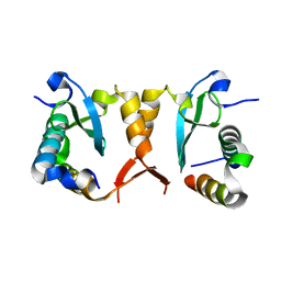
 | | Exonuclease domain-containing 1 (Exd1) in the native conformation | | 分子名称: | EXD1 | | 著者 | Yang, Z, Chen, K.M, Pandey, R.R, Homolka, D, Reuter, M, Rodino Janeiro, B.K, Sachidanandam, R, Fauvarque, M.O, McCarthy, A.A, Pillai, R.S. | | 登録日 | 2015-10-01 | | 公開日 | 2015-12-23 | | 最終更新日 | 2024-01-10 | | 実験手法 | X-RAY DIFFRACTION (2.4 Å) | | 主引用文献 | Piwi Slicing and Exd1 Drive Biogenesis of Nuclear Pirnas from Cytosolic Targets of the Mouse Pirna Pathway
Mol.Cell, 61, 2016
|
|
5XYV
 | |
5FIS
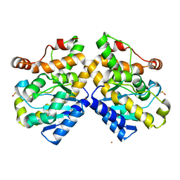 | | Exonuclease domain-containing 1 (Exd1) in the Gd bound conformation | | 分子名称: | EXD1, GADOLINIUM ATOM | | 著者 | Yang, Z, Chen, K.M, Pandey, R.R, Homolka, D, Reuter, M, Rodino Janeiro, B.K, Sachidanandam, R, Fauvarque, M.O, McCarthy, A.A, Pillai, R.S. | | 登録日 | 2015-10-02 | | 公開日 | 2015-12-23 | | 最終更新日 | 2016-01-20 | | 実験手法 | X-RAY DIFFRACTION (1.6 Å) | | 主引用文献 | Piwi Slicing and Exd1 Drive Biogenesis of Nuclear Pirnas from Cytosolic Targets of the Mouse Pirna Pathway
Mol.Cell, 61, 2016
|
|
8DL9
 | |
8DLB
 | | Room temperature X-ray structure of SARS-CoV-2 main protease in complex with compound Z2799209083 | | 分子名称: | 1-[(5S)-5-(3,4-dimethoxyphenyl)-3-phenyl-4,5-dihydro-1H-pyrazol-1-yl]ethan-1-one, 3C-like proteinase | | 著者 | Kovalevsky, A.Y, Coates, L, Kneller, D.W. | | 登録日 | 2022-07-07 | | 公開日 | 2023-05-17 | | 最終更新日 | 2023-10-25 | | 実験手法 | X-RAY DIFFRACTION (1.9 Å) | | 主引用文献 | AI-Accelerated Design of Targeted Covalent Inhibitors for SARS-CoV-2.
J.Chem.Inf.Model., 63, 2023
|
|
8DMD
 | |
6AKL
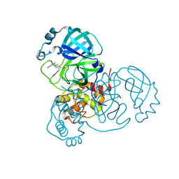
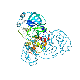
 | | Crystal structure of Striatin3 in complex with SIKE1 Coiled-coil domain | | 分子名称: | Striatin-3, Suppressor of IKBKE 1 | | 著者 | Zhou, L, Chen, M, Zhou, Z.C. | | 登録日 | 2018-09-02 | | 公開日 | 2019-01-16 | | 最終更新日 | 2023-11-22 | | 実験手法 | X-RAY DIFFRACTION (1.75 Å) | | 主引用文献 | Architecture, substructures, and dynamic assembly of STRIPAK complexes in Hippo signaling.
Cell Discov, 5, 2019
|
|


