3WGX
 
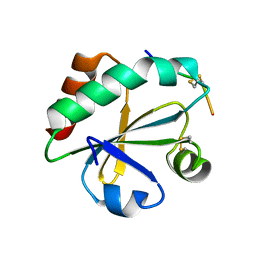 | | Crystal structure of ERp46 Trx2 in a complex with Prx4 C-term | | 分子名称: | GLYCEROL, Peroxiredoxin-4, Thioredoxin domain-containing protein 5 | | 著者 | Inaba, K, Suzuki, M, Kojima, R. | | 登録日 | 2013-08-13 | | 公開日 | 2014-06-25 | | 実験手法 | X-RAY DIFFRACTION (0.92 Å) | | 主引用文献 | Radically different thioredoxin domain arrangement of ERp46, an efficient disulfide bond introducer of the mammalian PDI family
Structure, 22, 2014
|
|
2FVY
 
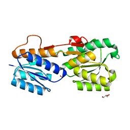 | | High Resolution Glucose Bound Crystal Structure of GGBP | | 分子名称: | ACETATE ION, CALCIUM ION, CARBON DIOXIDE, ... | | 著者 | Borrok, M.J, Kiessling, L.L, Forest, K.T. | | 登録日 | 2006-01-31 | | 公開日 | 2007-02-06 | | 最終更新日 | 2024-04-03 | | 実験手法 | X-RAY DIFFRACTION (0.92 Å) | | 主引用文献 | Conformational changes of glucose/galactose-binding protein illuminated by open, unliganded, and ultra-high-resolution ligand-bound structures.
Protein Sci., 16, 2007
|
|
2G6F
 
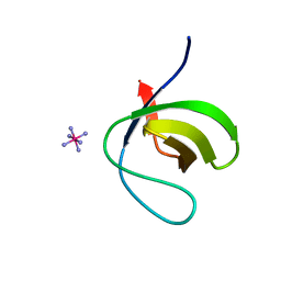 | |
6TJ8
 
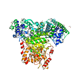 | | Escherichia coli transketolase in complex with cofactor analog 2'-methoxythiamine diphosphate | | 分子名称: | 1,2-ETHANEDIOL, 2-[3-[(4-azanyl-2-methoxy-pyrimidin-5-yl)methyl]-4-methyl-1,3-thiazol-5-yl]ethyl phosphono hydrogen phosphate, CALCIUM ION, ... | | 著者 | Rabe von Pappenheim, F, Tittmann, K. | | 登録日 | 2019-11-25 | | 公開日 | 2020-07-08 | | 最終更新日 | 2024-01-24 | | 実験手法 | X-RAY DIFFRACTION (0.921 Å) | | 主引用文献 | Structural basis for antibiotic action of the B 1 antivitamin 2'-methoxy-thiamine.
Nat.Chem.Biol., 16, 2020
|
|
6TE2
 
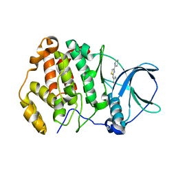 | | Crystal structure of human protein kinase CK2alpha' (CSNK2A2 gene product) in complex with the 2-aminothiazole-type inhibitor 17 | | 分子名称: | 3-[(4-pyridin-2-yl-1,3-thiazol-2-yl)amino]benzoic acid, Casein kinase II subunit alpha' | | 著者 | Niefind, K, Lindenblatt, D, Jose, J, Applegate, V.M, Nickelsen, A. | | 登録日 | 2019-11-11 | | 公開日 | 2020-07-08 | | 最終更新日 | 2024-05-15 | | 実験手法 | X-RAY DIFFRACTION (0.922 Å) | | 主引用文献 | Structural and Mechanistic Basis of the Inhibitory Potency of Selected 2-Aminothiazole Compounds on Protein Kinase CK2.
J.Med.Chem., 63, 2020
|
|
6UMY
 
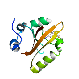 | |
7MBO
 
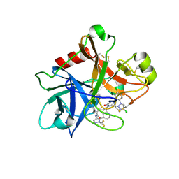 | | FACTOR XIA (PICHIA PASTORIS; C500S [C122S]) IN COMPLEX WITH THE INHIBITOR Milvexian (BMS-986177), IUPAC NAME:(6R,10S)-10-{4-[5-chloro-2-(4-chloro-1H-1,2,3-triazol-1-yl)phenyl]-6- oxopyrimidin-1(6H)-yl}-1-(difluoromethyl)-6-methyl-1,4,7,8,9,10-hexahydro-15,11- (metheno)pyrazolo[4,3-b][1,7]diazacyclotetradecin-5(6H)-one | | 分子名称: | 2-acetamido-2-deoxy-beta-D-glucopyranose, Coagulation factor XIa light chain, Milvexian | | 著者 | Sheriff, S. | | 登録日 | 2021-04-01 | | 公開日 | 2021-09-15 | | 最終更新日 | 2023-10-18 | | 実験手法 | X-RAY DIFFRACTION (0.924 Å) | | 主引用文献 | Discovery of Milvexian, a High-Affinity, Orally Bioavailable Inhibitor of Factor XIa in Clinical Studies for Antithrombotic Therapy.
J.Med.Chem., 65, 2022
|
|
3LZT
 
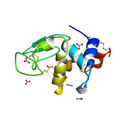 | | REFINEMENT OF TRICLINIC LYSOZYME AT ATOMIC RESOLUTION | | 分子名称: | ACETATE ION, LYSOZYME, NITRATE ION | | 著者 | Walsh, M.A, Schneider, T, Sieker, L.C, Dauter, Z, Lamzin, V, Wilson, K.S. | | 登録日 | 1997-03-23 | | 公開日 | 1998-03-25 | | 最終更新日 | 2023-08-09 | | 実験手法 | X-RAY DIFFRACTION (0.925 Å) | | 主引用文献 | Refinement of triclinic hen egg-white lysozyme at atomic resolution.
Acta Crystallogr.,Sect.D, 54, 1998
|
|
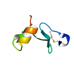
6ZSY
 
 | |
6ROB
 
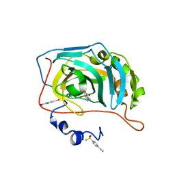 | | Human Carbonic Anhydrase II in complex with 4-cyanobenzenesulfonamide | | 分子名称: | (4-CARBOXYPHENYL)(CHLORO)MERCURY, 4-cyanobenzenesulfonamide, Carbonic anhydrase 2, ... | | 著者 | Gloeckner, S, Heine, A, Klebe, G. | | 登録日 | 2019-05-10 | | 公開日 | 2020-04-15 | | 最終更新日 | 2024-01-24 | | 実験手法 | X-RAY DIFFRACTION (0.929 Å) | | 主引用文献 | The Influence of Varying Fluorination Patterns on the Thermodynamics and Kinetics of Benzenesulfonamide Binding to Human Carbonic Anhydrase II.
Biomolecules, 10, 2020
|
|
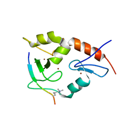
3QL9
 
 | |
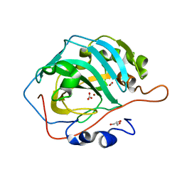
6KM0
 
 | |
3M5Q
 
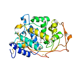 | | 0.93 A Structure of Manganese-Bound Manganese Peroxidase | | 分子名称: | 2-acetamido-2-deoxy-beta-D-glucopyranose-(1-4)-2-acetamido-2-deoxy-beta-D-glucopyranose, CALCIUM ION, GLYCEROL, ... | | 著者 | Sundaramoorthy, M, Gold, M.H, Poulos, T.L. | | 登録日 | 2010-03-12 | | 公開日 | 2010-04-14 | | 最終更新日 | 2023-09-06 | | 実験手法 | X-RAY DIFFRACTION (0.93 Å) | | 主引用文献 | Ultrahigh (0.93A) resolution structure of manganese peroxidase from Phanerochaete chrysosporium: implications for the catalytic mechanism.
J.Inorg.Biochem., 104, 2010
|
|
5X9M
 
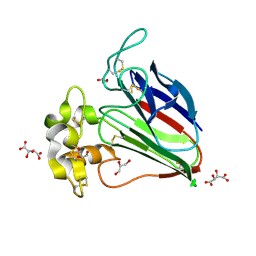 | | Structure of hyper-sweet thaumatin (D21N) | | 分子名称: | GLYCEROL, L(+)-TARTARIC ACID, Thaumatin I | | 著者 | Masuda, T, Okubo, K, Sugahara, M, Suzuki, M, Mikami, B. | | 登録日 | 2017-03-08 | | 公開日 | 2018-03-14 | | 最終更新日 | 2023-11-22 | | 実験手法 | X-RAY DIFFRACTION (0.93 Å) | | 主引用文献 | Subatomic structure of hyper-sweet thaumatin D21N mutant reveals the importance of flexible conformations for enhanced sweetness.
Biochimie, 157, 2019
|
|
8PB6
 
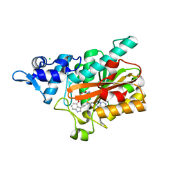 | | PsiM in complex with SAH and baeocystin | | 分子名称: | Baeocystin, CHLORIDE ION, Psilocybin synthase, ... | | 著者 | Werten, S, Hudspeth, J, Rupp, B. | | 登録日 | 2023-06-08 | | 公開日 | 2024-04-03 | | 最終更新日 | 2024-04-10 | | 実験手法 | X-RAY DIFFRACTION (0.93 Å) | | 主引用文献 | Methyl transfer in psilocybin biosynthesis.
Nat Commun, 15, 2024
|
|
5MNH
 
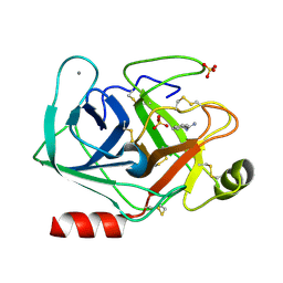 | | Cationic trypsin in complex with benzamidine (deuterated sample at 295 K) | | 分子名称: | BENZAMIDINE, CALCIUM ION, Cationic trypsin, ... | | 著者 | Schiebel, J, Heine, A, Klebe, G. | | 登録日 | 2016-12-13 | | 公開日 | 2018-01-17 | | 最終更新日 | 2024-01-17 | | 実験手法 | X-RAY DIFFRACTION (0.93 Å) | | 主引用文献 | Intriguing role of water in protein-ligand binding studied by neutron crystallography on trypsin complexes.
Nat Commun, 9, 2018
|
|
5MOQ
 
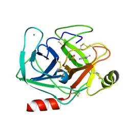 | | Joint X-ray/neutron structure of cationic trypsin in complex with benzamidine | | 分子名称: | BENZAMIDINE, CALCIUM ION, Cationic trypsin, ... | | 著者 | Schiebel, J, Schrader, T.E, Ostermann, A, Heine, A, Klebe, G. | | 登録日 | 2016-12-14 | | 公開日 | 2018-02-28 | | 最終更新日 | 2024-05-01 | | 実験手法 | NEUTRON DIFFRACTION (0.93 Å), X-RAY DIFFRACTION | | 主引用文献 | Intriguing role of water in protein-ligand binding studied by neutron crystallography on trypsin complexes.
Nat Commun, 9, 2018
|
|
7VDN
 
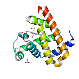 | | High resolution crystal structure of Sperm Whale Myoglobin in the carbonmonoxy form | | 分子名称: | CARBON MONOXIDE, Myoglobin, PROTOPORPHYRIN IX CONTAINING FE, ... | | 著者 | Shibayama, N, Sato-Tomita, A, Ishimoto, N, Park, S.Y. | | 登録日 | 2021-09-07 | | 公開日 | 2022-09-14 | | 最終更新日 | 2023-11-29 | | 実験手法 | X-RAY DIFFRACTION (0.93 Å) | | 主引用文献 | X-ray fluorescence holography of biological metal sites: Application to myoglobin.
Biochem.Biophys.Res.Commun., 635, 2022
|
|
7KQW
 
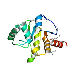 | | Crystal structure of SARS-CoV-2 NSP3 macrodomain (C2 crystal form, methylated) | | 分子名称: | Non-structural protein 3 | | 著者 | Correy, G.J, Young, I.D, Thompson, M.C, Fraser, J.S. | | 登録日 | 2020-11-17 | | 公開日 | 2020-12-09 | | 最終更新日 | 2023-11-15 | | 実験手法 | X-RAY DIFFRACTION (0.93 Å) | | 主引用文献 | Fragment binding to the Nsp3 macrodomain of SARS-CoV-2 identified through crystallographic screening and computational docking.
Sci Adv, 7, 2021
|
|
1OK0
 
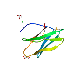 | | Crystal Structure of Tendamistat | | 分子名称: | ALPHA-AMYLASE INHIBITOR HOE-467A, CHLORIDE ION, GLYCEROL | | 著者 | Koenig, V, Vertesy, L, Schneider, T.R. | | 登録日 | 2003-07-16 | | 公開日 | 2004-01-15 | | 最終更新日 | 2023-12-13 | | 実験手法 | X-RAY DIFFRACTION (0.93 Å) | | 主引用文献 | Crystal Structure of the Alpha-Amylase Inhibitor Tendamistat at 0.93 A
Acta Crystallogr.,Sect.D, 59, 2003
|
|
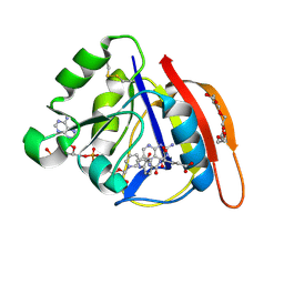
8UVZ
 
 | |
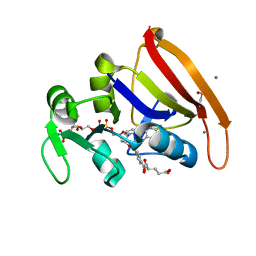
8UW0
 
 | |
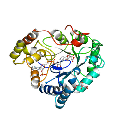
8A4N
 
 | |
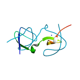
5QU8
 
 | |
3FX5
 
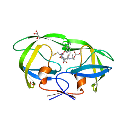 | | Structure of HIV-1 Protease in Complex with Potent Inhibitor KNI-272 Determined by High Resolution X-ray Crystallography | | 分子名称: | (4R)-N-tert-butyl-3-[(2S,3S)-2-hydroxy-3-({N-[(isoquinolin-5-yloxy)acetyl]-S-methyl-L-cysteinyl}amino)-4-phenylbutanoyl]-1,3-thiazolidine-4-carboxamide, GLYCEROL, protease | | 著者 | Adachi, M, Ohhara, T, Tamada, T, Okazaki, N, Kuroki, R. | | 登録日 | 2009-01-20 | | 公開日 | 2009-03-24 | | 最終更新日 | 2023-11-01 | | 実験手法 | X-RAY DIFFRACTION (0.93 Å) | | 主引用文献 | Structure of HIV-1 protease in complex with potent inhibitor KNI-272 determined by high-resolution X-ray and neutron crystallography.
Proc.Natl.Acad.Sci.USA, 2009
|
|
