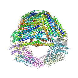
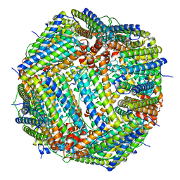
6WRW
 
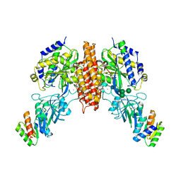 | | Crystal structure of computationally designed protein 2DS25.5 in complex with the human Transferrin receptor ectodomain | | 分子名称: | 2-acetamido-2-deoxy-beta-D-glucopyranose-(1-4)-2-acetamido-2-deoxy-beta-D-glucopyranose, CALCIUM ION, Computationally designed protein 2DS25.5, ... | | 著者 | Abraham, J, Coscia, A, Olal, D, Sahtoe, D.D, Baker, D, Clark, L. | | 登録日 | 2020-04-30 | | 公開日 | 2021-04-28 | | 最終更新日 | 2023-10-18 | | 実験手法 | X-RAY DIFFRACTION (2.84 Å) | | 主引用文献 | Transferrin receptor targeting by de novo sheet extension.
Proc.Natl.Acad.Sci.USA, 118, 2021
|
|
1TFA
 | | OVOTRANSFERRIN, N-TERMINAL LOBE, APO FORM | | 分子名称: | PROTEIN (OVOTRANSFERRIN), SULFATE ION | | 著者 | Mizutani, K, Yamashita, H, Mikami, B, Hirose, M. | | 登録日 | 1999-01-07 | | 公開日 | 1999-01-13 | | 最終更新日 | 2018-04-04 | | 実験手法 | X-RAY DIFFRACTION (1.9 Å) | | 主引用文献 | Alternative structural state of transferrin. The crystallographic analysis of iron-loaded but domain-opened ovotransferrin N-lobe.
J.Biol.Chem., 274, 1999
|
|
1NFT
 | | OVOTRANSFERRIN, N-TERMINAL LOBE, IRON LOADED OPEN FORM | | 分子名称: | FE (III) ION, NITRILOTRIACETIC ACID, PROTEIN (OVOTRANSFERRIN), ... | | 著者 | Mizutani, K, Yamashita, H, Kurokawa, H, Mikami, B, Hirose, M. | | 登録日 | 1999-01-07 | | 公開日 | 1999-01-13 | | 最終更新日 | 2023-11-15 | | 実験手法 | X-RAY DIFFRACTION (2.1 Å) | | 主引用文献 | Alternative structural state of transferrin. The crystallographic analysis of iron-loaded but domain-opened ovotransferrin N-lobe.
J.Biol.Chem., 274, 1999
|
|
3E1P
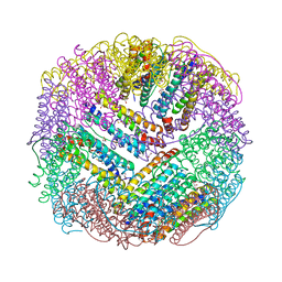
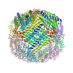 | | Crystal structure of E. coli Bacterioferritin (BFR) in which the Ferroxidase centre is inhibited with ZN(II) and high occupancy iron is bound within the cavity. | | 分子名称: | BACTERIOFERRITIN, FE (II) ION, PROTOPORPHYRIN IX CONTAINING FE, ... | | 著者 | Crow, A, Lawson, T, Lewin, A, Moore, G.R, Le Brun, N. | | 登録日 | 2008-08-04 | | 公開日 | 2009-05-05 | | 最終更新日 | 2023-08-30 | | 実験手法 | X-RAY DIFFRACTION (2.4 Å) | | 主引用文献 | Structural basis for iron mineralization by bacterioferritin
J.Am.Chem.Soc., 131, 2009
|
|
3E1L
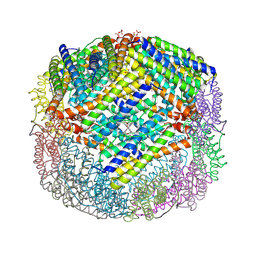
 | | Crystal structure of E. coli Bacterioferritin (BFR) soaked in phosphate with an alternative conformation of the unoccupied Ferroxidase centre (APO-BFR II). | | 分子名称: | BACTERIOFERRITIN, PROTOPORPHYRIN IX CONTAINING FE, SULFATE ION | | 著者 | Crow, A, Lawson, T, Lewin, A, Moore, G.R, Le Brun, N. | | 登録日 | 2008-08-04 | | 公開日 | 2009-05-05 | | 最終更新日 | 2023-08-30 | | 実験手法 | X-RAY DIFFRACTION (2.5 Å) | | 主引用文献 | Structural basis for iron mineralization by bacterioferritin
J.Am.Chem.Soc., 131, 2009
|
|
3E1N
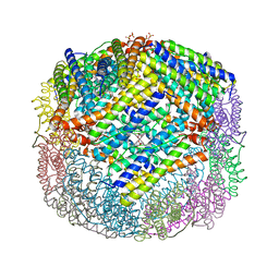
 | | Crystal structure of E. coli Bacterioferritin (BFR) after a 65 minute (aerobic) exposure to FE(II) revealing a possible MU-OXO bridge/MU-Hydroxy bridged DIIRON intermediate at the ferroxidase centre. (FE(III)-O-FE(III)-BFR). | | 分子名称: | BACTERIOFERRITIN, FE (II) ION, PROTOPORPHYRIN IX CONTAINING FE, ... | | 著者 | Crow, A, Lawson, T, Lewin, A, Moore, G.R, Le Brun, N. | | 登録日 | 2008-08-04 | | 公開日 | 2009-05-05 | | 最終更新日 | 2023-08-30 | | 実験手法 | X-RAY DIFFRACTION (2.8 Å) | | 主引用文献 | Structural basis for iron mineralization by bacterioferritin
J.Am.Chem.Soc., 131, 2009
|
|
3E1O
 | | Crystal structure of E. coli Bacterioferritin (BFR) with two ZN(II) ION sites at the Ferroxidase centre (ZN-BFR). | | 分子名称: | BACTERIOFERRITIN, PROTOPORPHYRIN IX CONTAINING FE, SULFATE ION, ... | | 著者 | Crow, A, Lawson, T, Lewin, A, Moore, G.R, Le Brun, N. | | 登録日 | 2008-08-04 | | 公開日 | 2009-05-05 | | 最終更新日 | 2023-08-30 | | 実験手法 | X-RAY DIFFRACTION (2.95 Å) | | 主引用文献 | Structural basis for iron mineralization by bacterioferritin
J.Am.Chem.Soc., 131, 2009
|
|
3E1M
 | | Crystal structure of E. coli Bacterioferritin (BFR) obtained after soaking APO-BFR crystals for 2.5 minutes in FE2+ (2.5M FE(II)-BFR) | | 分子名称: | BACTERIOFERRITIN, FE (II) ION, PROTOPORPHYRIN IX CONTAINING FE, ... | | 著者 | Crow, A, Lawson, T, Lewin, A, Moore, G.R, Le Brun, N. | | 登録日 | 2008-08-04 | | 公開日 | 2009-05-05 | | 最終更新日 | 2024-02-21 | | 実験手法 | X-RAY DIFFRACTION (2.7 Å) | | 主引用文献 | Structural basis for iron mineralization by bacterioferritin
J.Am.Chem.Soc., 131, 2009
|
|
3E1J
 | | Crystal structure of E. coli Bacterioferritin (BFR) with an unoccupied ferroxidase centre (APO-BFR). | | 分子名称: | Bacterioferritin, PROTOPORPHYRIN IX CONTAINING FE, SULFATE ION | | 著者 | Crow, A, Lawson, T, Lewin, A, Moore, G.R, Le Brun, N. | | 登録日 | 2008-08-04 | | 公開日 | 2009-05-05 | | 最終更新日 | 2023-08-30 | | 実験手法 | X-RAY DIFFRACTION (2.7 Å) | | 主引用文献 | Structural basis for iron mineralization by bacterioferritin
J.Am.Chem.Soc., 131, 2009
|
|
4U3G
 | | Crystal structure of Escherichia coli bacterioferritin mutant D132F | | 分子名称: | Bacterioferritin, SULFATE ION | | 著者 | Wong, S.G, Grigg, J.C, Le Brun, N.E, Moore, G.R, Murphy, M.E.P, Mauk, A.G. | | 登録日 | 2014-07-21 | | 公開日 | 2014-12-24 | | 最終更新日 | 2023-09-27 | | 実験手法 | X-RAY DIFFRACTION (2 Å) | | 主引用文献 | The B-type Channel Is a Major Route for Iron Entry into the Ferroxidase Center and Central Cavity of Bacterioferritin.
J.Biol.Chem., 290, 2015
|
|
4AM2
 | | Bacterioferritin from Blastochloris viridis | | 分子名称: | BACTERIOFERRITIN, FE (III) ION, PROTOPORPHYRIN IX CONTAINING FE | | 著者 | Wahlgren, W.Y, Omran, H, von Stetten, D, Royant, A, van der Post, S, Katona, G. | | 登録日 | 2012-03-07 | | 公開日 | 2012-10-31 | | 最終更新日 | 2023-12-20 | | 実験手法 | X-RAY DIFFRACTION (1.8 Å) | | 主引用文献 | Structural Characterization of Bacterioferritin from Blastochloris Viridis.
Plos One, 7, 2012
|
|
4AM4
 | | Bacterioferritin from Blastochloris viridis | | 分子名称: | BACTERIOFERRITIN, FE (III) ION, PROTOPORPHYRIN IX CONTAINING FE | | 著者 | Wahlgren, W.Y, Omran, H, von Stetten, D, Royant, A, van der Post, S, Katona, G. | | 登録日 | 2012-03-07 | | 公開日 | 2012-10-31 | | 最終更新日 | 2023-12-20 | | 実験手法 | X-RAY DIFFRACTION (1.68 Å) | | 主引用文献 | Structural Characterization of Bacterioferritin from Blastochloris Viridis.
Plos One, 7, 2012
|
|
4AM5
 | | Bacterioferritin from Blastochloris viridis | | 分子名称: | BACTERIOFERRITIN, FE (III) ION, PROTOPORPHYRIN IX CONTAINING FE | | 著者 | Wahlgren, W.Y, Omran, H, von Stetten, D, Royant, A, van der Post, S, Katona, G. | | 登録日 | 2012-03-07 | | 公開日 | 2012-10-31 | | 最終更新日 | 2023-12-20 | | 実験手法 | X-RAY DIFFRACTION (1.58 Å) | | 主引用文献 | Structural Characterization of Bacterioferritin from Blastochloris Viridis.
Plos One, 7, 2012
|
|
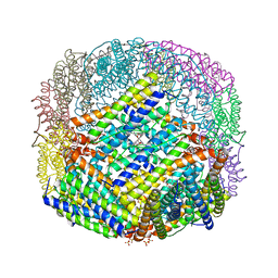
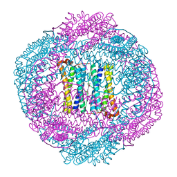
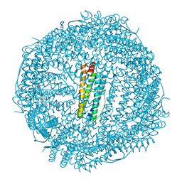
6H5I
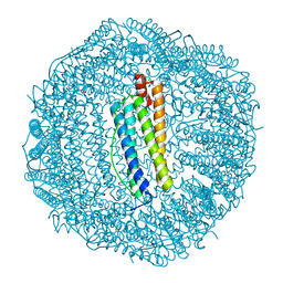
 | | Single Particle Cryo-EM map of human Transferrin receptor 1 - H-Ferritin complex. | | 分子名称: | Ferritin heavy chain, Transferrin receptor protein 1 | | 著者 | Testi, C, Montemiglio, L.C, Vallone, B, Des Georges, A, Boffi, A, Mancia, F, Baiocco, P, Savino, C. | | 登録日 | 2018-07-24 | | 公開日 | 2019-03-27 | | 最終更新日 | 2019-12-18 | | 実験手法 | ELECTRON MICROSCOPY (3.9 Å) | | 主引用文献 | Cryo-EM structure of the human ferritin-transferrin receptor 1 complex.
Nat Commun, 10, 2019
|
|
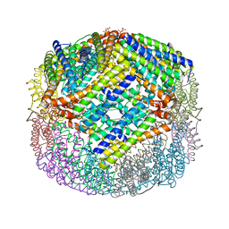
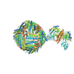
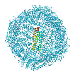
6GSR
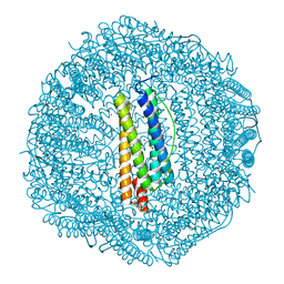
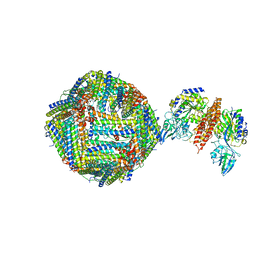 | | Single Particle Cryo-EM map of human Transferrin receptor 1 - H-Ferritin complex at 5.5 Angstrom resolution. | | 分子名称: | Ferritin heavy chain, Transferrin receptor protein 1 | | 著者 | Testi, C, Montemiglio, L.C, Vallone, B, Des Georges, A, Boffi, A, Mancia, F, Baiocco, P. | | 登録日 | 2018-06-15 | | 公開日 | 2019-03-27 | | 最終更新日 | 2019-05-08 | | 実験手法 | ELECTRON MICROSCOPY (5.5 Å) | | 主引用文献 | Cryo-EM structure of the human ferritin-transferrin receptor 1 complex.
Nat Commun, 10, 2019
|
|
5LS9
 | |
5OBA
 | |
4Y08
 | | ONE MINUTE IRON LOADED HUMAN H FERRITIN | | 分子名称: | CHLORIDE ION, FE (II) ION, Ferritin heavy chain, ... | | 著者 | Pozzi, C, Di Pisa, F, Mangani, S. | | 登録日 | 2015-02-05 | | 公開日 | 2015-09-23 | | 最終更新日 | 2024-01-10 | | 実験手法 | X-RAY DIFFRACTION (1.34 Å) | | 主引用文献 | Iron binding to human heavy-chain ferritin.
Acta Crystallogr.,Sect.D, 71, 2015
|
|
4YKH
 | | Thirty minutes iron loaded human H ferritin | | 分子名称: | BICINE, CHLORIDE ION, FE (II) ION, ... | | 著者 | Pozzi, C, Di Pisa, F, Mangani, S. | | 登録日 | 2015-03-04 | | 公開日 | 2015-09-09 | | 最終更新日 | 2024-01-10 | | 実験手法 | X-RAY DIFFRACTION (1.52 Å) | | 主引用文献 | Iron binding to human heavy-chain ferritin.
Acta Crystallogr.,Sect.D, 71, 2015
|
|
4XGS
 | |
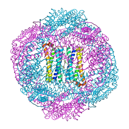
7O6E
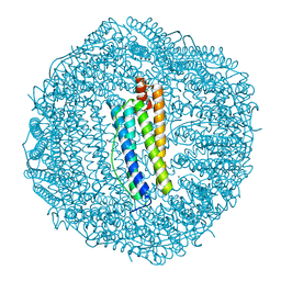
 | | 2.12 A cryo-EM structure of Mycobacterium tuberculosis Ferritin | | 分子名称: | Ferritin BfrB | | 著者 | Gijsbers, A, Zhang, Y, Gao, Y, Peters, P.J, Ravelli, R.B.G. | | 登録日 | 2021-04-10 | | 公開日 | 2021-05-19 | | 最終更新日 | 2024-07-10 | | 実験手法 | ELECTRON MICROSCOPY (2.1 Å) | | 主引用文献 | Mycobacterium tuberculosis ferritin: a suitable workhorse protein for cryo-EM development.
Acta Crystallogr D Struct Biol, 77, 2021
|
|
3KA4
 | | Frog M-ferritin with cobalt | | 分子名称: | CHLORIDE ION, COBALT (II) ION, Ferritin, ... | | 著者 | Tosha, T, Ng, H.L, Theil, E, Alber, T, Bhattasali, O. | | 登録日 | 2009-10-18 | | 公開日 | 2010-10-06 | | 最終更新日 | 2023-09-06 | | 実験手法 | X-RAY DIFFRACTION (1.4 Å) | | 主引用文献 | Moving Metal Ions through Ferritin-Protein Nanocages from Three-Fold Pores to Catalytic Sites.
J.Am.Chem.Soc., 132, 2010
|
|
3KA6
 | | Frog M-ferritin, EED mutant, with cobalt | | 分子名称: | CHLORIDE ION, COBALT (II) ION, Ferritin, ... | | 著者 | Tosha, T, Ng, H.L, Theil, E, Alber, T, Bhattasali, O. | | 登録日 | 2009-10-18 | | 公開日 | 2010-10-06 | | 最終更新日 | 2023-09-06 | | 実験手法 | X-RAY DIFFRACTION (1.4 Å) | | 主引用文献 | Moving Metal Ions through Ferritin-Protein Nanocages from Three-Fold Pores to Catalytic Sites.
J.Am.Chem.Soc., 132, 2010
|
|
3KA8
 | | Frog M-ferritin, EQH mutant, with cobalt | | 分子名称: | CHLORIDE ION, COBALT (II) ION, Ferritin, ... | | 著者 | Tosha, T, Ng, H.L, Theil, E, Alber, T, Bhattasali, O. | | 登録日 | 2009-10-19 | | 公開日 | 2010-10-06 | | 最終更新日 | 2023-09-06 | | 実験手法 | X-RAY DIFFRACTION (1.35 Å) | | 主引用文献 | Moving Metal Ions through Ferritin-Protein Nanocages from Three-Fold Pores to Catalytic Sites.
J.Am.Chem.Soc., 132, 2010
|
|
3KA3
 | | Frog M-ferritin with magnesium | | 分子名称: | CHLORIDE ION, Ferritin, middle subunit, ... | | 著者 | Tosha, T, Ng, H.L, Theil, E, Alber, T, Bhattasali, O. | | 登録日 | 2009-10-18 | | 公開日 | 2010-10-06 | | 最終更新日 | 2024-02-21 | | 実験手法 | X-RAY DIFFRACTION (1.4 Å) | | 主引用文献 | Moving Metal Ions through Ferritin-Protein Nanocages from Three-Fold Pores to Catalytic Sites.
J.Am.Chem.Soc., 132, 2010
|
|


