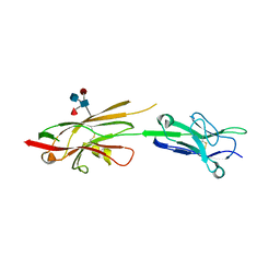
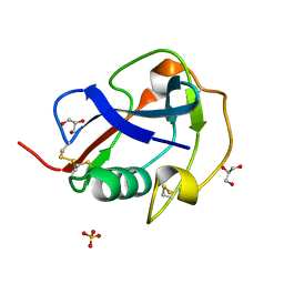
4OFD
 
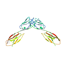 | | Crystal Structure of mouse Neph1 D1-D2 | | 分子名称: | 2-acetamido-2-deoxy-beta-D-glucopyranose-(1-4)-2-acetamido-2-deoxy-beta-D-glucopyranose, Kin of IRRE-like protein 1, beta-D-mannopyranose-(1-4)-2-acetamido-2-deoxy-beta-D-glucopyranose-(1-4)-2-acetamido-2-deoxy-beta-D-glucopyranose | | 著者 | Ozkan, E, Garcia, K.C. | | 登録日 | 2014-01-14 | | 公開日 | 2014-02-19 | | 最終更新日 | 2023-09-20 | | 実験手法 | X-RAY DIFFRACTION (3.94 Å) | | 主引用文献 | Extracellular Architecture of the SYG-1/SYG-2 Adhesion Complex Instructs Synaptogenesis.
Cell(Cambridge,Mass.), 156, 2014
|
|
6X33
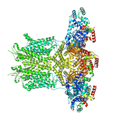
 | | Wt pig RyR1 in complex with apoCaM, EGTA condition (class 3, open) | | 分子名称: | Calmodulin-1, Peptidyl-prolyl cis-trans isomerase FKBP1B, Ryanodine Receptor, ... | | 著者 | Woll, K.W, Haji-Ghassemi, O, Van Petegem, F. | | 登録日 | 2020-05-21 | | 公開日 | 2021-01-13 | | 最終更新日 | 2024-03-06 | | 実験手法 | ELECTRON MICROSCOPY (4.2 Å) | | 主引用文献 | Pathological conformations of disease mutant Ryanodine Receptors revealed by cryo-EM.
Nat Commun, 12, 2021
|
|
4OAU
 | | Complete human RNase L in complex with biological activators. | | 分子名称: | 2-5A-dependent ribonuclease, ADENOSINE-5'-DIPHOSPHATE, MAGNESIUM ION, ... | | 著者 | Han, Y, Donovan, J, Rath, S, Whitney, G, Chitrakar, A, Korennykh, A. | | 登録日 | 2014-01-06 | | 公開日 | 2014-03-12 | | 最終更新日 | 2024-02-28 | | 実験手法 | X-RAY DIFFRACTION (2.6 Å) | | 主引用文献 | Structure of human RNase L reveals the basis for regulated RNA decay in the IFN response.
Science, 343, 2014
|
|
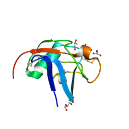
4OFI
 | | Crystal Structure of Duf (Kirre) D1 | | 分子名称: | 2-acetamido-2-deoxy-beta-D-glucopyranose, Kin of irre, isoform A, ... | | 著者 | Ozkan, E, Garcia, K.C. | | 登録日 | 2014-01-14 | | 公開日 | 2014-02-19 | | 最終更新日 | 2023-09-20 | | 実験手法 | X-RAY DIFFRACTION (2.3 Å) | | 主引用文献 | Extracellular Architecture of the SYG-1/SYG-2 Adhesion Complex Instructs Synaptogenesis.
Cell(Cambridge,Mass.), 156, 2014
|
|
4OF3
 | | Crystal Structure of SYG-1 D1-D2, Glycosylated | | 分子名称: | 2-acetamido-2-deoxy-beta-D-glucopyranose-(1-4)-[alpha-L-fucopyranose-(1-6)]2-acetamido-2-deoxy-beta-D-glucopyranose, Protein SYG-1, isoform b, ... | | 著者 | Ozkan, E, Garcia, K.C. | | 登録日 | 2014-01-14 | | 公開日 | 2014-02-19 | | 最終更新日 | 2023-09-20 | | 実験手法 | X-RAY DIFFRACTION (2.5 Å) | | 主引用文献 | Extracellular Architecture of the SYG-1/SYG-2 Adhesion Complex Instructs Synaptogenesis.
Cell(Cambridge,Mass.), 156, 2014
|
|
7YWQ
 | |
4OFP
 | | Crystal Structure of SYG-2 D3-D4 | | 分子名称: | 2-acetamido-2-deoxy-beta-D-glucopyranose, Protein SYG-2 | | 著者 | Ozkan, E, Garcia, K.C. | | 登録日 | 2014-01-15 | | 公開日 | 2014-02-19 | | 最終更新日 | 2023-09-20 | | 実験手法 | X-RAY DIFFRACTION (3 Å) | | 主引用文献 | Extracellular Architecture of the SYG-1/SYG-2 Adhesion Complex Instructs Synaptogenesis.
Cell(Cambridge,Mass.), 156, 2014
|
|
8EP1
 | |
8EOW
 | |
8EP0
 | |
5MSK
 | | Mouse PA28beta | | 分子名称: | Proteasome activator complex subunit 2 | | 著者 | Huber, E.M, Groll, M. | | 登録日 | 2017-01-05 | | 公開日 | 2017-09-13 | | 最終更新日 | 2024-01-17 | | 実験手法 | X-RAY DIFFRACTION (3.6 Å) | | 主引用文献 | The Mammalian Proteasome Activator PA28 Forms an Asymmetric alpha 4 beta 3 Complex.
Structure, 25, 2017
|
|
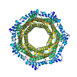
5MX5
 | | Mouse PA28alpha-beta | | 分子名称: | PHOSPHATE ION, Proteasome activator complex subunit 1, Proteasome activator complex subunit 2 | | 著者 | Huber, E.M, Groll, M. | | 登録日 | 2017-01-20 | | 公開日 | 2017-09-13 | | 最終更新日 | 2024-01-17 | | 実験手法 | X-RAY DIFFRACTION (2.9 Å) | | 主引用文献 | The Mammalian Proteasome Activator PA28 Forms an Asymmetric alpha 4 beta 3 Complex.
Structure, 25, 2017
|
|
6X36
 | | Pig R615C RyR1 in complex with CaM, EGTA (class 3, closed) | | 分子名称: | Calmodulin-1, Peptidyl-prolyl cis-trans isomerase FKBP1B, Ryanodine Receptor | | 著者 | Woll, K.W, Haji-Ghassemi, O, Van Petegem, F. | | 登録日 | 2020-05-21 | | 公開日 | 2021-01-13 | | 最終更新日 | 2024-03-06 | | 実験手法 | ELECTRON MICROSCOPY (4.7 Å) | | 主引用文献 | Pathological conformations of disease mutant Ryanodine Receptors revealed by cryo-EM.
Nat Commun, 12, 2021
|
|
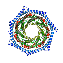
5MSJ
 | | Mouse PA28alpha | | 分子名称: | Proteasome activator complex subunit 1 | | 著者 | Huber, E.M, Groll, M. | | 登録日 | 2017-01-05 | | 公開日 | 2017-09-13 | | 最終更新日 | 2024-01-17 | | 実験手法 | X-RAY DIFFRACTION (3.5 Å) | | 主引用文献 | The Mammalian Proteasome Activator PA28 Forms an Asymmetric alpha 4 beta 3 Complex.
Structure, 25, 2017
|
|
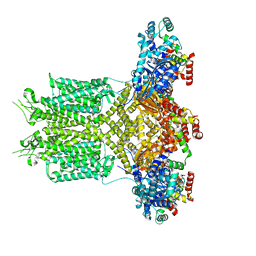
6X32
 | | Wt pig RyR1 in complex with apoCaM, EGTA condition (class 1 and 2, closed) | | 分子名称: | Calmodulin-1, Peptidyl-prolyl cis-trans isomerase FKBP1B, Ryanodine Receptor, ... | | 著者 | Woll, K.W, Haji-Ghassemi, O, Van Petegem, F. | | 登録日 | 2020-05-21 | | 公開日 | 2021-01-13 | | 最終更新日 | 2024-03-06 | | 実験手法 | ELECTRON MICROSCOPY (3.8 Å) | | 主引用文献 | Pathological conformations of disease mutant Ryanodine Receptors revealed by cryo-EM.
Nat Commun, 12, 2021
|
|
5NIN
 | |
7UJN
 | |
6TP8
 | |
1MK6
 | | SOLUTION STRUCTURE OF THE 8,9-DIHYDRO-8-(N7-GUANYL)-9-HYDROXY-AFLATOXIN B1 ADDUCT MISPAIRED WITH DEOXYADENOSINE | | 分子名称: | 5'-D(*AP*CP*AP*TP*CP*GP*AP*TP*CP*T)-3', 5'-D(*AP*GP*AP*TP*AP*GP*AP*TP*GP*T)-3', 8,9-DIHYDRO-9-HYDROXY-AFLATOXIN B1 | | 著者 | Giri, I, Johnston, D.S, Stone, M.P. | | 登録日 | 2002-08-28 | | 公開日 | 2002-10-16 | | 最終更新日 | 2024-05-01 | | 実験手法 | SOLUTION NMR | | 主引用文献 | MISPAIRING OF THE 8,9-DIHYDRO-8-(N7-GUANYL)-9-HYDROXY-AFLATOXIN B1 ADDUCT WITH DEOXYADENOSINE RESULTS IN EXTRUSION OF THE MISMATCHED DA TOWARD THE MAJOR GROOVE
Biochemistry, 41, 2002
|
|
6SAN
 | | SALSA / DMBT1 / GP340 SRCR domain 8 soaked in calcium and magnesium | | 分子名称: | CHLORIDE ION, Deleted in malignant brain tumors 1 protein, GLYCEROL, ... | | 著者 | Reichhardt, M.P, Johnson, S, Loimaranta, V, Lea, S.M. | | 登録日 | 2019-07-17 | | 公開日 | 2020-03-18 | | 最終更新日 | 2024-01-24 | | 実験手法 | X-RAY DIFFRACTION (1.36 Å) | | 主引用文献 | Structures of SALSA/DMBT1 SRCR domains reveal the conserved ligand-binding mechanism of the ancient SRCR fold.
Life Sci Alliance, 3, 2020
|
|
4V46
 | | Crystal structure of the BAFF-BAFF-R complex | | 分子名称: | MAGNESIUM ION, Tumor necrosis factor ligand superfamily member 13B, Tumor necrosis factor receptor superfamily member 13C | | 著者 | Kim, H.M, Yu, K.S, Lee, M.E, Shin, D.R, Kim, Y.S, Paik, S.G, Yoo, O.J, Lee, H, Lee, J.-O. | | 登録日 | 2003-03-23 | | 公開日 | 2014-07-09 | | 最終更新日 | 2014-12-10 | | 実験手法 | X-RAY DIFFRACTION (3.3 Å) | | 主引用文献 | Crystal structure of the BAFF-BAFF-R complex and its implications for receptor activation
NAT.STRUCT.BIOL., 10, 2003
|
|
6SA5
 | | SALSA / DMBT1 / GP340 SRCR domain 8 | | 分子名称: | Deleted in malignant brain tumors 1 protein, GLYCEROL, MAGNESIUM ION, ... | | 著者 | Reichhardt, M.P, Johnson, S, Loimaranta, V, Lea, S.M. | | 登録日 | 2019-07-16 | | 公開日 | 2020-03-18 | | 最終更新日 | 2024-01-24 | | 実験手法 | X-RAY DIFFRACTION (1.29 Å) | | 主引用文献 | Structures of SALSA/DMBT1 SRCR domains reveal the conserved ligand-binding mechanism of the ancient SRCR fold.
Life Sci Alliance, 3, 2020
|
|
6SA4
 | | SALSA / DMBT1 / GP340 SRCR domain 1 | | 分子名称: | CHLORIDE ION, Deleted in malignant brain tumors 1 protein, GLYCEROL, ... | | 著者 | Reichhardt, M.P, Lea, S.M, Johnson, S. | | 登録日 | 2019-07-16 | | 公開日 | 2020-03-18 | | 最終更新日 | 2024-01-24 | | 実験手法 | X-RAY DIFFRACTION (1.77 Å) | | 主引用文献 | Structures of SALSA/DMBT1 SRCR domains reveal the conserved ligand-binding mechanism of the ancient SRCR fold.
Life Sci Alliance, 3, 2020
|
|
6S8C
 | |
1D0G
 | | CRYSTAL STRUCTURE OF DEATH RECEPTOR 5 (DR5) BOUND TO APO2L/TRAIL | | 分子名称: | APOPTOSIS-2 LIGAND, CHLORIDE ION, DEATH RECEPTOR-5, ... | | 著者 | Hymowitz, S.G, Christinger, H.W, Fuh, G, O'Connell, M.P, Kelley, R.F, Ashkenazi, A, de Vos, A.M. | | 登録日 | 1999-09-09 | | 公開日 | 1999-10-22 | | 最終更新日 | 2018-01-31 | | 実験手法 | X-RAY DIFFRACTION (2.4 Å) | | 主引用文献 | Triggering cell death: the crystal structure of Apo2L/TRAIL in a complex with death receptor 5.
Mol.Cell, 4, 1999
|
|


