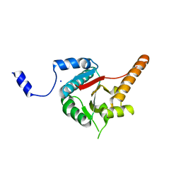
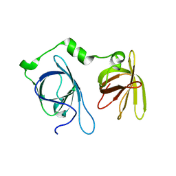
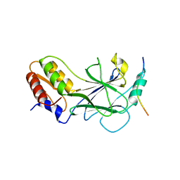
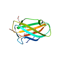
6ERH
 
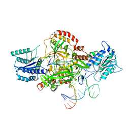 | | Complex of XLF and heterodimer Ku bound to DNA | | 分子名称: | DNA (21-MER), DNA (34-MER), Non-homologous end-joining factor 1, ... | | 著者 | Nemoz, C, Legrand, P, Ropars, V, Charbonnier, J.B. | | 登録日 | 2017-10-18 | | 公開日 | 2018-10-17 | | 最終更新日 | 2024-01-17 | | 実験手法 | X-RAY DIFFRACTION (2.8 Å) | | 主引用文献 | XLF and APLF bind Ku80 at two remote sites to ensure DNA repair by non-homologous end joining.
Nat. Struct. Mol. Biol., 25, 2018
|
|
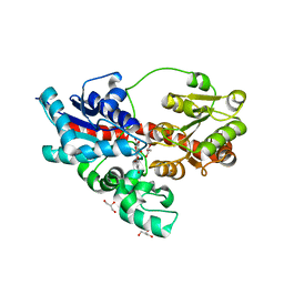
5W5H
 | |
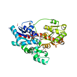
5W5I
 | |
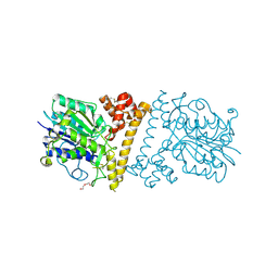
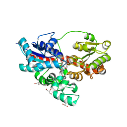
4J0U
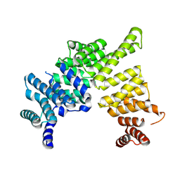 | | Crystal structure of IFIT5/ISG58 | | 分子名称: | Interferon-induced protein with tetratricopeptide repeats 5 | | 著者 | Liu, Y, Liang, H, Feng, F, Yuan, L, Wang, Y.E, Crowley, C, Lv, Z, Li, J, Zeng, S, Cheng, G. | | 登録日 | 2013-01-31 | | 公開日 | 2013-02-13 | | 実験手法 | X-RAY DIFFRACTION (1.969 Å) | | 主引用文献 | Crystal Structure of IFIT5
To be Published
|
|
3B6E
 | | Crystal structure of human DECH-box RNA Helicase MDA5 (Melanoma differentiation-associated protein 5), DECH-domain | | 分子名称: | Interferon-induced helicase C domain-containing protein 1, SODIUM ION | | 著者 | Karlberg, T, Welin, M, Arrowsmith, C.H, Berglund, H, Busam, R.D, Collins, R, Dahlgren, L.G, Edwards, A.M, Flodin, S, Flores, A, Graslund, S, Hammarstrom, M, Johansson, I, Kallas, A, Kotenyova, T, Lehtio, L, Moche, M, Nilsson, M.E, Nordlund, P, Nyman, T, Persson, C, Sagemark, J, Svensson, L, Thorsell, A.G, Tresaugues, L, Van Den Berg, S, Weigelt, J, Holmberg-Schiavone, L, Structural Genomics Consortium (SGC) | | 登録日 | 2007-10-29 | | 公開日 | 2007-11-13 | | 最終更新日 | 2024-02-21 | | 実験手法 | X-RAY DIFFRACTION (1.6 Å) | | 主引用文献 | Human DECH-box RNA Helicase MDA5 (Melanoma differentiation-associated protein 5), DECH-domain.
To be Published
|
|
4GL2
 | | Structural Basis for dsRNA duplex backbone recognition by MDA5 | | 分子名称: | Interferon-induced helicase C domain-containing protein 1, PHOSPHOAMINOPHOSPHONIC ACID-ADENYLATE ESTER, RNA (5'-R(*AP*UP*CP*CP*GP*CP*GP*GP*CP*CP*CP*U)-3'), ... | | 著者 | Wu, B, Hur, S. | | 登録日 | 2012-08-13 | | 公開日 | 2013-01-09 | | 最終更新日 | 2013-02-06 | | 実験手法 | X-RAY DIFFRACTION (3.557 Å) | | 主引用文献 | Structural Basis for dsRNA Recognition, Filament Formation, and Antiviral Signal Activation by MDA5.
Cell(Cambridge,Mass.), 152, 2013
|
|
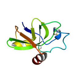
2N00
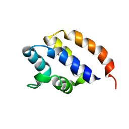 | | NMR Solution structure of AIM2 PYD from Mus musculus | | 分子名称: | Interferon-inducible protein AIM2 | | 著者 | Hou, X, Niu, X. | | 登録日 | 2015-03-01 | | 公開日 | 2015-05-27 | | 最終更新日 | 2024-05-15 | | 実験手法 | SOLUTION NMR | | 主引用文献 | The NMR solution structure of AIM2 PYD domain from Mus musculus reveals a distinct alpha 2-alpha 3 helix conformation from its human homologues
Biochem.Biophys.Res.Commun., 461, 2015
|
|
7S55
 | |
4JBJ
 | | Structural mimicry for functional antagonism | | 分子名称: | Interferon-activable protein 202 | | 著者 | Ru, H, Ni, X, Ma, F, Zhao, L, Ding, W, Hung, L.-W, Shaw, N, Cheng, G, Liu, Z.-J. | | 登録日 | 2013-02-19 | | 公開日 | 2013-06-26 | | 最終更新日 | 2023-11-08 | | 実験手法 | X-RAY DIFFRACTION (2.692 Å) | | 主引用文献 | Structural basis for termination of AIM2-mediated signaling by p202
Cell Res., 23, 2013
|
|
4Q2P
 | |
3GA3
 | |
4WHM
 | | Crystal structure of UDP-glucose: anthocyanidin 3-O-glucosyltransferase in complex with UDP | | 分子名称: | ACETATE ION, GLYCEROL, UDP-glucose:anthocyanidin 3-O-glucosyltransferase, ... | | 著者 | Hiromoto, T, Honjo, E, Tamada, T, Kuroki, R. | | 登録日 | 2014-09-23 | | 公開日 | 2015-01-21 | | 最終更新日 | 2023-11-08 | | 実験手法 | X-RAY DIFFRACTION (1.851 Å) | | 主引用文献 | Structural basis for acceptor-substrate recognition of UDP-glucose: anthocyanidin 3-O-glucosyltransferase from Clitoria ternatea
Protein Sci., 24, 2015
|
|
5JER
 | | Structure of Rotavirus NSP1 bound to IRF-3 | | 分子名称: | Interferon regulatory factor 3, Rotavirus NSP1 peptide | | 著者 | Zhao, B, Li, P. | | 登録日 | 2016-04-18 | | 公開日 | 2016-06-15 | | 最終更新日 | 2024-03-06 | | 実験手法 | X-RAY DIFFRACTION (2.913 Å) | | 主引用文献 | Structural basis for concerted recruitment and activation of IRF-3 by innate immune adaptor proteins.
Proc.Natl.Acad.Sci.USA, 113, 2016
|
|
3TS9
 | |
6L4Y
 | |
6L4X
 | |
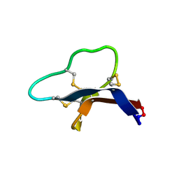
1WRF
 | | Refined solution structure of Der f 2, The Major Mite Allergen from Dermatophagoides farinae | | 分子名称: | Mite group 2 allergen Der f 2 | | 著者 | Ichikawa, S, Takai, T, Inoue, T, Yuuki, T, Okumura, Y, Ogura, K, Inagaki, F, Hatanaka, H. | | 登録日 | 2004-10-15 | | 公開日 | 2005-04-19 | | 最終更新日 | 2022-03-02 | | 実験手法 | SOLUTION NMR | | 主引用文献 | NMR Study on the Major Mite Allergen Der f 2: Its Refined Tertiary Structure, Epitopes for Monoclonal Antibodies and Characteristics Shared by ML Protein Group Members
J.Biochem.(Tokyo), 137, 2005
|
|
1XWV
 | | Structure of the house dust mite allergen Der f 2: Implications for function and molecular basis of IgE cross-reactivity | | 分子名称: | 3,6,9,12,15,18,21,24,27,30,33,36,39-TRIDECAOXAHENTETRACONTANE-1,41-DIOL, 3,6,9,12,15,18,21,24,27-NONAOXANONACOSANE-1,29-DIOL, Der f II | | 著者 | Johannessen, B.R, Skov, L.K, Kastrup, J.S, Kristensen, O, Bolwig, C, Larsen, J.N, Spangfort, M, Lund, K, Gajhede, M. | | 登録日 | 2004-11-02 | | 公開日 | 2004-12-14 | | 最終更新日 | 2023-10-25 | | 実験手法 | X-RAY DIFFRACTION (1.83 Å) | | 主引用文献 | Structure of the house dust mite allergen Der f 2: implications for function and molecular basis of IgE cross-reactivity.
Febs Lett., 579, 2005
|
|
6LKO
 | |
4OW4
 | |
1N0S
 | | ENGINEERED LIPOCALIN FLUA IN COMPLEX WITH FLUORESCEIN | | 分子名称: | 2-(6-HYDROXY-3-OXO-3H-XANTHEN-9-YL)-BENZOIC ACID, Bilin-binding protein, SULFATE ION | | 著者 | Korndoerfer, I.P, Skerra, A. | | 登録日 | 2002-10-15 | | 公開日 | 2003-08-05 | | 最終更新日 | 2017-10-11 | | 実験手法 | X-RAY DIFFRACTION (2 Å) | | 主引用文献 | Crystallographic analysis of an "anticalin" with tailored specificity for fluorescein reveals high structural plasticity of the lipocalin loop region.
Proteins, 53, 2003
|
|
4REN
 | | Crystal structure of UDP-glucose: anthocyanidin 3-O-glucosyltransferase in complex with petunidin | | 分子名称: | 2-(3,4-dihydroxy-5-methoxyphenyl)-3,5,7-trihydroxychromenium, GLYCEROL, UDP-glucose:anthocyanidin 3-O-glucosyltransferase | | 著者 | Hiromoto, T, Honjo, E, Tamada, T, Kuroki, R. | | 登録日 | 2014-09-23 | | 公開日 | 2015-01-21 | | 最終更新日 | 2023-11-08 | | 実験手法 | X-RAY DIFFRACTION (2.704 Å) | | 主引用文献 | Structural basis for acceptor-substrate recognition of UDP-glucose: anthocyanidin 3-O-glucosyltransferase from Clitoria ternatea
Protein Sci., 24, 2015
|
|
4REM
 | | Crystal structure of UDP-glucose: anthocyanidin 3-O-glucosyltransferase in complex with delphinidin | | 分子名称: | 3,5,7-trihydroxy-2-(3,4,5-trihydroxyphenyl)chromenium, GLYCEROL, UDP-glucose:anthocyanidin 3-O-glucosyltransferase | | 著者 | Hiromoto, T, Honjo, E, Tamada, T, Kuroki, R. | | 登録日 | 2014-09-23 | | 公開日 | 2015-01-21 | | 最終更新日 | 2023-11-08 | | 実験手法 | X-RAY DIFFRACTION (2.55 Å) | | 主引用文献 | Structural basis for acceptor-substrate recognition of UDP-glucose: anthocyanidin 3-O-glucosyltransferase from Clitoria ternatea
Protein Sci., 24, 2015
|
|
4REL
 | | Crystal structure of UDP-glucose: anthocyanidin 3-O-glucosyltransferase in complex with kaempferol | | 分子名称: | 3,5,7-TRIHYDROXY-2-(4-HYDROXYPHENYL)-4H-CHROMEN-4-ONE, ACETATE ION, GLYCEROL, ... | | 著者 | Hiromoto, T, Honjo, E, Tamada, T, Kuroki, R. | | 登録日 | 2014-09-23 | | 公開日 | 2015-01-21 | | 最終更新日 | 2023-11-08 | | 実験手法 | X-RAY DIFFRACTION (1.754 Å) | | 主引用文献 | Structural basis for acceptor-substrate recognition of UDP-glucose: anthocyanidin 3-O-glucosyltransferase from Clitoria ternatea
Protein Sci., 24, 2015
|
|
2KQA
 | | The solution structure of the fungal elicitor Cerato-Platanin | | 分子名称: | Cerato-platanin | | 著者 | Oliveira, A.L, Gallo, M, Pazzagli, L, Cappugi, G, Scala, A, Cicero, D.O, Pantera, B, Spisni, A, Benedetti, C.E, Pertinhez, T.A. | | 登録日 | 2009-11-03 | | 公開日 | 2011-03-23 | | 最終更新日 | 2011-07-13 | | 実験手法 | SOLUTION NMR | | 主引用文献 | The solution structure of the fungal elicitor Cerato-Platanin
To be Published
|
|


