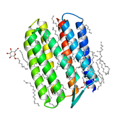
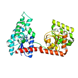
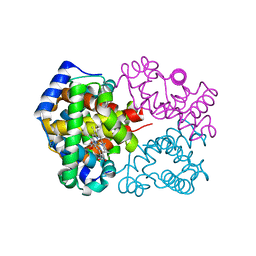
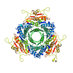
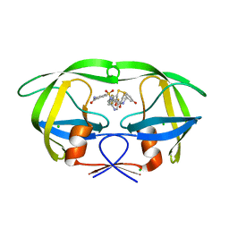
2R7M
 
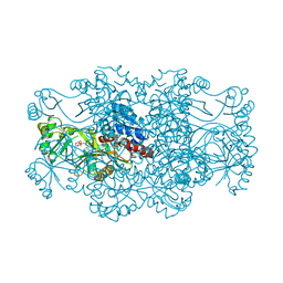 | | Crystal structure of FAICAR synthetase (PurP) from M. jannaschii complexed with AMP | | 分子名称: | 5-formaminoimidazole-4-carboxamide-1-(beta)-D-ribofuranosyl 5'-monophosphate synthetase, ADENOSINE MONOPHOSPHATE, CHLORIDE ION, ... | | 著者 | Zhang, Y, White, R.H, Ealick, S.E. | | 登録日 | 2007-09-09 | | 公開日 | 2007-12-04 | | 最終更新日 | 2024-02-21 | | 実験手法 | X-RAY DIFFRACTION (2.3 Å) | | 主引用文献 | Crystal structure and function of 5-formaminoimidazole-4-carboxamide ribonucleotide synthetase from Methanocaldococcus jannaschii.
Biochemistry, 47, 2008
|
|
3QSL
 | | Structure of CAE31940 from Bordetella bronchiseptica RB50 | | 分子名称: | CITRIC ACID, Putative exported protein | | 著者 | Bajor, J, Kagan, O, Chruszcz, M, Savchenko, A, Joachimiak, A, Minor, W, Midwest Center for Structural Genomics (MCSG) | | 登録日 | 2011-02-21 | | 公開日 | 2011-03-23 | | 最終更新日 | 2023-12-06 | | 実験手法 | X-RAY DIFFRACTION (2 Å) | | 主引用文献 | The crystal structure of pyrimidine/thiamin biosynthesis precursor-like domain-containing protein CAE31940 from proteobacterium Bordetella bronchiseptica RB50, and evolutionary insight into the NMT1/THI5 family.
J Struct Funct Genomics, 15, 2014
|
|
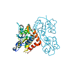
3QO0
 | |
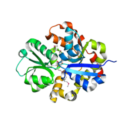
2R82
 | | Pyruvate phosphate dikinase (PPDK) triple mutant R219E/E271R/S262D adapts a second conformational state | | 分子名称: | Pyruvate, phosphate dikinase, SULFATE ION | | 著者 | Lim, K, Read, R.J, Chen, C.C, Herzberg, O. | | 登録日 | 2007-09-10 | | 公開日 | 2008-01-01 | | 最終更新日 | 2023-08-30 | | 実験手法 | X-RAY DIFFRACTION (3.6 Å) | | 主引用文献 | Swiveling domain mechanism in pyruvate phosphate dikinase.
Biochemistry, 46, 2007
|
|
3EFG
 | | Structure of SlyX protein from Xanthomonas campestris pv. campestris str. ATCC 33913 | | 分子名称: | 1,2-ETHANEDIOL, Protein slyX homolog | | 著者 | Cuff, M.E, Xu, X, Cui, H, Savchenko, A, Edwards, A, Joachimiak, A, Midwest Center for Structural Genomics (MCSG) | | 登録日 | 2008-09-08 | | 公開日 | 2008-12-09 | | 最終更新日 | 2024-02-21 | | 実験手法 | X-RAY DIFFRACTION (2 Å) | | 主引用文献 | Structure of SlyX protein from Xanthomonas campestris pv. campestris str. ATCC 33913
TO BE PUBLISHED
|
|
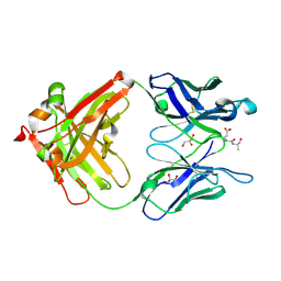
3QAP
 | | Crystal structure of Natronomonas pharaonis sensory rhodopsin II in the ground state | | 分子名称: | 2,3-DI-PHYTANYL-GLYCEROL, EICOSANE, RETINAL, ... | | 著者 | Gushchin, I, Reshetnyak, A, Borshchevskiy, V, Ishchenko, A, Round, E, Grudinin, S, Engelhard, M, Buldt, G, Gordeliy, V. | | 登録日 | 2011-01-11 | | 公開日 | 2011-09-14 | | 最終更新日 | 2024-02-21 | | 実験手法 | X-RAY DIFFRACTION (1.9 Å) | | 主引用文献 | Active State of Sensory Rhodopsin II: Structural Determinants for Signal Transfer and Proton Pumping.
J.Mol.Biol., 412, 2011
|
|
2RI7
 | |
2RIJ
 | | Crystal structure of a putative 2,3,4,5-tetrahydropyridine-2-carboxylate n-succinyltransferase (cj1605c, dapd) from campylobacter jejuni at 1.90 A resolution | | 分子名称: | CHLORIDE ION, CITRIC ACID, GLYCEROL, ... | | 著者 | Joint Center for Structural Genomics (JCSG) | | 登録日 | 2007-10-11 | | 公開日 | 2007-10-23 | | 最終更新日 | 2023-01-25 | | 実験手法 | X-RAY DIFFRACTION (1.9 Å) | | 主引用文献 | Crystal structure of Putative 2,3,4,5-tetrahydropyridine-2-carboxylate N-succinyltransferase (NP_282733.1) from Campylobacter jejuni at 1.90 A resolution
To be published
|
|
3QN8
 | | HIV-1 protease (mutant Q7K L33I L63I) in complex with a novel inhibitor | | 分子名称: | (4aS,7aS)-1,4-bis(3-hydroxybenzyl)hexahydro-1H-pyrrolo[3,4-b]pyrazine-2,3-dione, CHLORIDE ION, Protease | | 著者 | Lindemann, I, Heine, A, Klebe, G. | | 登録日 | 2011-02-08 | | 公開日 | 2012-02-08 | | 最終更新日 | 2023-11-01 | | 実験手法 | X-RAY DIFFRACTION (1.382 Å) | | 主引用文献 | Novel inhibitors for HIV-1 protease
To be Published
|
|
2R68
 | |
3QQ7
 | |
3E7D
 | |
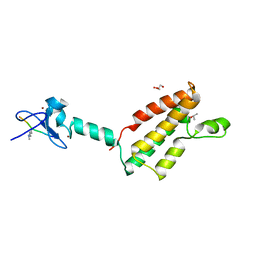
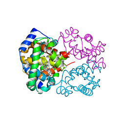
2R7L
 | | Crystal structure of FAICAR synthetase (PurP) from M. jannaschii complexed with ATP and AICAR | | 分子名称: | 5-formaminoimidazole-4-carboxamide-1-(beta)-D-ribofuranosyl 5'-monophosphate synthetase, ADENOSINE-5'-TRIPHOSPHATE, AMINOIMIDAZOLE 4-CARBOXAMIDE RIBONUCLEOTIDE, ... | | 著者 | Zhang, Y, White, R.H, Ealick, S.E. | | 登録日 | 2007-09-09 | | 公開日 | 2007-12-04 | | 最終更新日 | 2024-02-21 | | 実験手法 | X-RAY DIFFRACTION (2.1 Å) | | 主引用文献 | Crystal structure and function of 5-formaminoimidazole-4-carboxamide ribonucleotide synthetase from Methanocaldococcus jannaschii.
Biochemistry, 47, 2008
|
|
3QJC
 | |
3ECJ
 | |
3QS3
 | |
3QJB
 | |
3EI4
 | |
3EK6
 | |
3EN3
 | |
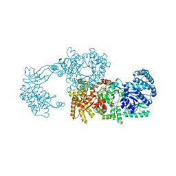
2R7N
 | | Crystal structure of FAICAR synthetase (PurP) from M. jannaschii complexed with ADP and FAICAR | | 分子名称: | 5-(formylamino)-1-(5-O-phosphono-beta-D-ribofuranosyl)-1H-imidazole-4-carboxamide, 5-formaminoimidazole-4-carboxamide-1-(beta)-D-ribofuranosyl 5'-monophosphate synthetase, ADENOSINE-5'-DIPHOSPHATE, ... | | 著者 | Zhang, Y, White, R.H, Ealick, S.E. | | 登録日 | 2007-09-09 | | 公開日 | 2007-12-04 | | 最終更新日 | 2024-02-21 | | 実験手法 | X-RAY DIFFRACTION (2.4 Å) | | 主引用文献 | Crystal structure and function of 5-formaminoimidazole-4-carboxamide ribonucleotide synthetase from Methanocaldococcus jannaschii.
Biochemistry, 47, 2008
|
|
3QNZ
 | | Orthorhombic form of IgG1 Fab fragment (in complex with antigenic tubulin peptide) sharing same Fv as IgA | | 分子名称: | Fab fragment of IMMUNOGLOBULIN G1 HEAVY CHAIN, Fab fragment of IMMUNOGLOBULIN G1 LIGHT CHAIN, GLYCEROL, ... | | 著者 | Trajtenberg, F, Correa, A, Buschiazzo, A. | | 登録日 | 2011-02-09 | | 公開日 | 2012-02-15 | | 最終更新日 | 2023-11-01 | | 実験手法 | X-RAY DIFFRACTION (2.2 Å) | | 主引用文献 | Structure of a human IgA1 Fab fragment at 1.55 angstrom resolution: potential effect of the constant domains on antigen-affinity modulation
Acta Crystallogr.,Sect.D, 69, 2013
|
|
3QPJ
 | |
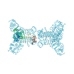
2RCJ
 | | Solution structure of human Immunoglobulin M | | 分子名称: | IgA1 heavy chain, IgA1 light chain, J chain | | 著者 | Perkins, S.J, Nealis, A.S, Sutton, B.J, Feinstein, A. | | 登録日 | 2007-09-20 | | 公開日 | 2008-01-22 | | 最終更新日 | 2024-02-21 | | 実験手法 | SOLUTION SCATTERING | | 主引用文献 | Solution structure of human and mouse immunoglobulin M by synchrotron X-ray scattering and molecular graphics modelling. A possible mechanism for complement activation.
J.Mol.Biol., 221, 1991
|
|
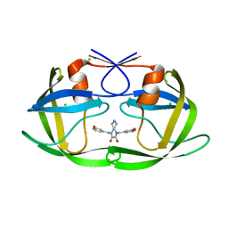
3ETP
 | | The crystal structure of the ligand-binding domain of the EphB2 receptor at 2.0 A resolution | | 分子名称: | Ephrin type-B receptor 2 | | 著者 | Goldgur, Y, Paavilainen, S, Nikolov, D.B, Himanen, J.P. | | 登録日 | 2008-10-08 | | 公開日 | 2008-10-21 | | 最終更新日 | 2023-11-29 | | 実験手法 | X-RAY DIFFRACTION (2 Å) | | 主引用文献 | Structure of the ligand-binding domain of the EphB2 receptor at 2 A resolution.
Acta Crystallogr.,Sect.F, 65, 2009
|
|


