1EDV
 
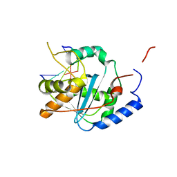
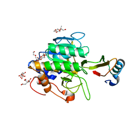
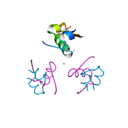
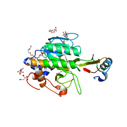
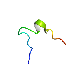 | | SOLUTION STRUCTURE OF 2ND INTRADISKAL LOOP OF BOVINE RHODOPSIN (RESIDUES 172-205) | | 分子名称: | RHODOPSIN | | 著者 | Yeagle, P.L, Salloum, A, Chopra, A, Bhawsar, N, Ali, L. | | 登録日 | 2000-01-28 | | 公開日 | 2000-08-09 | | 最終更新日 | 2024-05-22 | | 実験手法 | SOLUTION NMR | | 主引用文献 | Structures of the intradiskal loops and amino terminus of the G-protein receptor, rhodopsin.
J.Pept.Res., 55, 2000
|
|
1P5H
 | |
3O42
 | |
1SB2
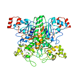
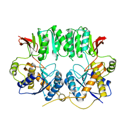
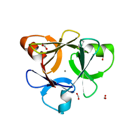
 | | High resolution Structure determination of rhodocetin | | 分子名称: | Rhodocetin alpha subunit, Rhodocetin beta subunit | | 著者 | Paaventhan, P, Kong, C.G, Joseph, J.S, Chung, M.C.M, Kolatkar, P.R. | | 登録日 | 2004-02-10 | | 公開日 | 2005-02-01 | | 最終更新日 | 2011-07-13 | | 実験手法 | X-RAY DIFFRACTION (1.9 Å) | | 主引用文献 | Structure of rhodocetin reveals noncovalently bound heterodimer interface
Protein Sci., 14, 2005
|
|
3IDB
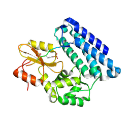
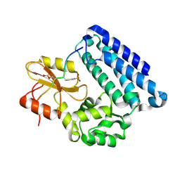
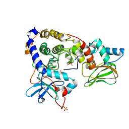 | | Crystal structure of (108-268)RIIb:C holoenzyme of cAMP-dependent protein kinase | | 分子名称: | MANGANESE (II) ION, PHOSPHOAMINOPHOSPHONIC ACID-ADENYLATE ESTER, cAMP-dependent protein kinase catalytic subunit alpha, ... | | 著者 | Brown, S.H.J, Wu, J, Kim, C, Alberto, K, Taylor, S.S. | | 登録日 | 2009-07-20 | | 公開日 | 2009-09-29 | | 最終更新日 | 2023-09-06 | | 実験手法 | X-RAY DIFFRACTION (1.62 Å) | | 主引用文献 | Novel isoform-specific interfaces revealed by PKA RIIbeta holoenzyme structures.
J.Mol.Biol., 393, 2009
|
|
2N63
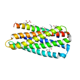
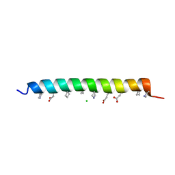
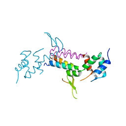
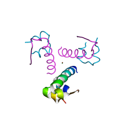
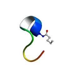 | | Structure of C4VG16KRKP | | 分子名称: | PENTANOIC ACID, antimicrobial peptide C4VG16KRKP | | 著者 | Bhunia, A, Datta, A. | | 登録日 | 2015-08-11 | | 公開日 | 2016-03-23 | | 最終更新日 | 2024-01-24 | | 実験手法 | SOLUTION NMR | | 主引用文献 | Designing potent antimicrobial peptides by disulphide linked dimerization and N-terminal lipidation to increase antimicrobial activity and membrane perturbation: Structural insights into lipopolysaccharide binding.
J Colloid Interface Sci, 461, 2016
|
|
3O2J
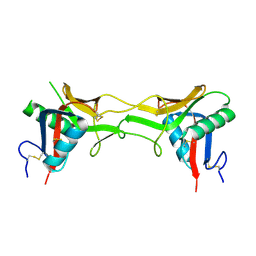
 | | Structure of the GluA2 NTD-dimer interface mutant, N54A | | 分子名称: | 2-acetamido-2-deoxy-beta-D-glucopyranose, Glutamate receptor 2 | | 著者 | Rossmann, M, Sukumaran, M, Penn, A.C, Veprintsev, D.B, Greger, I.H. | | 登録日 | 2010-07-22 | | 公開日 | 2011-03-09 | | 最終更新日 | 2023-11-01 | | 実験手法 | X-RAY DIFFRACTION (1.95 Å) | | 主引用文献 | Subunit-selective N-terminal domain associations organize the formation of AMPA receptor heteromers
Embo J., 30, 2011
|
|
3IDC
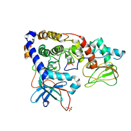 | | Crystal structure of (102-265)RIIb:C holoenzyme of cAMP-dependent protein kinase | | 分子名称: | MANGANESE (II) ION, PHOSPHOAMINOPHOSPHONIC ACID-ADENYLATE ESTER, cAMP-dependent protein kinase catalytic subunit alpha, ... | | 著者 | Brown, S.H.J, Wu, J, Kim, C, Alberto, K, Taylor, S.S. | | 登録日 | 2009-07-20 | | 公開日 | 2009-09-29 | | 最終更新日 | 2023-09-06 | | 実験手法 | X-RAY DIFFRACTION (2.7 Å) | | 主引用文献 | Novel isoform-specific interfaces revealed by PKA RIIbeta holoenzyme structures.
J.Mol.Biol., 393, 2009
|
|
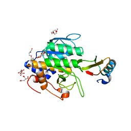
1NMU
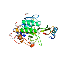
 | | MBP-L30 | | 分子名称: | 60S ribosomal protein L30, alpha-D-glucopyranose-(1-4)-alpha-D-glucopyranose-(1-4)-alpha-D-glucopyranose-(1-4)-alpha-D-glucopyranose, maltose-binding periplasmic protein | | 著者 | Chao, J.A, Prasad, G.S, White, S.A, Stout, C.D, Williamson, J.R. | | 登録日 | 2003-01-10 | | 公開日 | 2003-02-18 | | 最終更新日 | 2024-05-22 | | 実験手法 | X-RAY DIFFRACTION (2.31 Å) | | 主引用文献 | Inherent Protein Structural Flexibility at the RNA-binding Interface of L30e
J.Mol.Biol., 326, 2003
|
|
1P5R
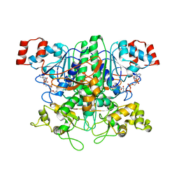 | | Formyl-CoA Transferase in complex with Coenzyme A | | 分子名称: | COENZYME A, Formyl-coenzyme A transferase | | 著者 | Ricagno, S, Jonsson, S, Richards, N, Lindqvist, Y. | | 登録日 | 2003-04-28 | | 公開日 | 2003-07-29 | | 最終更新日 | 2023-08-16 | | 実験手法 | X-RAY DIFFRACTION (2.5 Å) | | 主引用文献 | Formyl-CoA Transferase encloses the CoA binding site at the interface of an interlocked dimer
Embo J., 22, 2003
|
|
3N6V
 | |
4GV3
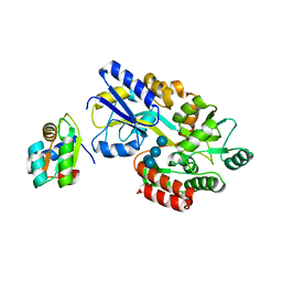
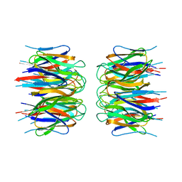
 | | Structures of Lassa and Tacaribe viral nucleoproteins with or without 5 triphosphate dsRNA substrate reveal a unique 3 -5 exoribonuclease mechanism to suppress type I interferon production | | 分子名称: | MANGANESE (II) ION, Nucleoprotein, RNA (5'-R(*(GTP)P*GP*GP*C)-3'), ... | | 著者 | Jiang, X, Huang, Q, Wang, W, Dong, H, Ly, H, Liang, Y, Dong, C. | | 登録日 | 2012-08-30 | | 公開日 | 2013-05-01 | | 最終更新日 | 2024-02-28 | | 実験手法 | X-RAY DIFFRACTION (1.68 Å) | | 主引用文献 | Structures of Arenaviral Nucleoproteins with Triphosphate dsRNA Reveal a Unique Mechanism of Immune Suppression.
J.Biol.Chem., 288, 2013
|
|
3OB1
 | |
3O3Y
 | |
4GV6
 | | Structures of Lassa and Tacaribe viral nucleoproteins with or without 5 triphosphate dsRNA substrate reveal a unique 3 -5 exoribonuclease mechanism to suppress type I interferon production | | 分子名称: | MANGANESE (II) ION, Nucleoprotein, RNA (5'-R(*(GTP)P*GP*GP*C)-3'), ... | | 著者 | Jiang, X, Huang, Q, Wang, W, Dong, H, Ly, H, Liang, Y, Dong, C. | | 登録日 | 2012-08-30 | | 公開日 | 2013-05-01 | | 最終更新日 | 2024-02-28 | | 実験手法 | X-RAY DIFFRACTION (1.98 Å) | | 主引用文献 | Structures of Arenaviral Nucleoproteins with Triphosphate dsRNA Reveal a Unique Mechanism of Immune Suppression.
J.Biol.Chem., 288, 2013
|
|
1TO1
 | | crystal structure of the complex of subtilisin BPN' with chymotrypsin inhibitor 2 Y61A mutant | | 分子名称: | CALCIUM ION, CITRIC ACID, PENTAETHYLENE GLYCOL, ... | | 著者 | Radisky, E.S, Kwan, G, Karen Lu, C.J, Koshland Jr, D.E. | | 登録日 | 2004-06-11 | | 公開日 | 2004-11-09 | | 最終更新日 | 2023-08-23 | | 実験手法 | X-RAY DIFFRACTION (1.68 Å) | | 主引用文献 | Binding, Proteolytic, and Crystallographic Analyses of Mutations at the Protease-Inhibitor Interface of the Subtilisin BPN'/Chymotrypsin Inhibitor 2 Complex(,).
Biochemistry, 43, 2004
|
|
1TO2
 | | crystal structure of the complex of subtilisin BPN' with chymotrypsin inhibitor 2 M59K, in pH 9 cryosoak | | 分子名称: | CALCIUM ION, CITRIC ACID, POLYETHYLENE GLYCOL (N=34), ... | | 著者 | Radisky, E.S, Kwan, G, Karen Lu, C.J, Koshland Jr, D.E. | | 登録日 | 2004-06-11 | | 公開日 | 2004-11-09 | | 最終更新日 | 2023-08-23 | | 実験手法 | X-RAY DIFFRACTION (1.3 Å) | | 主引用文献 | Binding, Proteolytic, and Crystallographic Analyses of Mutations at the Protease-Inhibitor Interface of the Subtilisin BPN'/Chymotrypsin Inhibitor 2 Complex(,).
Biochemistry, 43, 2004
|
|
3KMV
 | | Crystal structure of CBM42A from Clostridium thermocellum | | 分子名称: | ACETATE ION, Alpha-L-arabinofuranosidase B, CALCIUM ION, ... | | 著者 | Santos-Silva, T, Alves, V.D, Prates, J.A.M, Fontes, C.M.G.A, Romao, M.J. | | 登録日 | 2009-11-11 | | 公開日 | 2010-08-04 | | 最終更新日 | 2023-09-06 | | 実験手法 | X-RAY DIFFRACTION (1.8 Å) | | 主引用文献 | Family 42 carbohydrate-binding modules display multiple arabinoxylan-binding interfaces presenting different ligand affinities.
Biochim.Biophys.Acta, 1804, 2010
|
|
3OB2
 | |
3MHV
 | | Crystal Structure of Vps4 and Vta1 | | 分子名称: | Vacuolar protein sorting-associated protein 4, Vacuolar protein sorting-associated protein VTA1 | | 著者 | Yang, D, Hurley, J.H. | | 登録日 | 2010-04-09 | | 公開日 | 2010-10-06 | | 最終更新日 | 2023-11-01 | | 実験手法 | X-RAY DIFFRACTION (3.1 Å) | | 主引用文献 | Structural role of the Vps4-Vta1 interface in ESCRT-III recycling
Structure, 18, 2010
|
|
2P1B
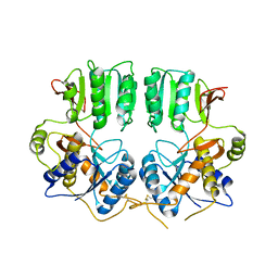
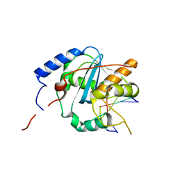
 | | Crystal structure of human nucleophosmin-core | | 分子名称: | Nucleophosmin | | 著者 | Lee, H.H, Kim, H.S, Kang, J.Y, Lee, B.I, Ha, J.Y, Yoon, H.J, Lim, S.O, Jung, G, Suh, S.W. | | 登録日 | 2007-03-03 | | 公開日 | 2007-03-27 | | 最終更新日 | 2023-10-25 | | 実験手法 | X-RAY DIFFRACTION (2.75 Å) | | 主引用文献 | Crystal structure of human nucleophosmin-core reveals plasticity of the pentamer-pentamer interface
Proteins, 69, 2007
|
|
1TM4
 | | crystal structure of the complex of subtilsin BPN'with chymotrypsin inhibitor 2 M59G mutant | | 分子名称: | CALCIUM ION, CITRIC ACID, PENTAETHYLENE GLYCOL, ... | | 著者 | Radisky, E.S, Kwan, G, Karen Lu, C.J, Koshland Jr, D.E. | | 登録日 | 2004-06-10 | | 公開日 | 2004-11-09 | | 最終更新日 | 2023-08-23 | | 実験手法 | X-RAY DIFFRACTION (1.7 Å) | | 主引用文献 | Binding, Proteolytic, and Crystallographic Analyses of Mutations at the Protease-Inhibitor Interface of the Subtilisin BPN'/Chymotrypsin Inhibitor 2 Complex(,).
Biochemistry, 43, 2004
|
|
3P2X
 | | Insulin fibrillation is the Janus face of induced fit. A chiaral clamp stabilizes the native state at the expense of activity | | 分子名称: | CHLORIDE ION, Insulin, PHENOL, ... | | 著者 | Hua, Q.X, Wan, Z.L, Huang, K, Hu, S.Q, Phillip, N.F, Jia, W.H, Whittingham, J, Dodson, G.G, Katsoyannis, P.G, Weiss, M.A. | | 登録日 | 2010-10-04 | | 公開日 | 2011-11-23 | | 最終更新日 | 2023-09-06 | | 実験手法 | X-RAY DIFFRACTION (2 Å) | | 主引用文献 | Insulin fibrillation is the Janus face of induced fit. A chiral clamp stabilizes the native state at the expense of activity
To be Published
|
|
3P33
 | | Insulin fibrillation is the Janus face of induced fit. A chiral clamp stabilizes the native state at the expense of activity | | 分子名称: | CHLORIDE ION, Insulin, PHENOL, ... | | 著者 | Hua, Q.X, Wan, Z.L, Huang, K, Hu, S.Q, Phillip, N.F, Jia, W.H, Whittingham, J, Dodson, G.G, Katsoyannis, P.G, Weiss, M.A. | | 登録日 | 2010-10-04 | | 公開日 | 2011-11-23 | | 最終更新日 | 2023-09-06 | | 実験手法 | X-RAY DIFFRACTION (2.3 Å) | | 主引用文献 | Insulin fibrillation is the Janus face of induced fit. A chiral clamp stabilizes the native state at the expense of activity
To be Published
|
|
1TM5
 | | crystal structure of the complex of subtilisin BPN' with chymotrypsin inhibitor 2 M59A mutant | | 分子名称: | CALCIUM ION, CITRIC ACID, PENTAETHYLENE GLYCOL, ... | | 著者 | Radisky, E.S, Kwan, G, Karen Lu, C.J, Koshland Jr, D.E. | | 登録日 | 2004-06-10 | | 公開日 | 2004-11-09 | | 最終更新日 | 2023-08-23 | | 実験手法 | X-RAY DIFFRACTION (1.45 Å) | | 主引用文献 | Binding, Proteolytic, and Crystallographic Analyses of Mutations at the Protease-Inhibitor Interface of the Subtilisin BPN'/Chymotrypsin Inhibitor 2 Complex(,).
Biochemistry, 43, 2004
|
|


