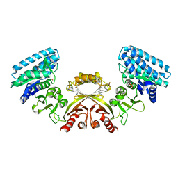
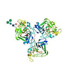
2J0T
 
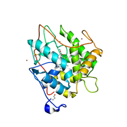
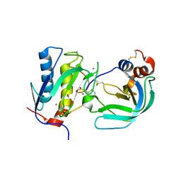 | | Crystal Structure of the Catalytic Domain of MMP-1 in Complex with the Inhibitory Domain of TIMP-1 | | 分子名称: | CALCIUM ION, INTERSTITIAL COLLAGENASE, METALLOPROTEINASE INHIBITOR 1, ... | | 著者 | Iyer, S, Wei, S, Brew, K, Acharya, K.R. | | 登録日 | 2006-08-04 | | 公開日 | 2006-10-18 | | 最終更新日 | 2023-12-13 | | 実験手法 | X-RAY DIFFRACTION (2.54 Å) | | 主引用文献 | Crystal Structure of the Catalytic Domain of Matrix Metalloproteinase-1 in Complex with the Inhibitory Domain of Tissue Inhibitor of Metalloproteinase-1.
J.Biol.Chem., 282, 2007
|
|
2J0U
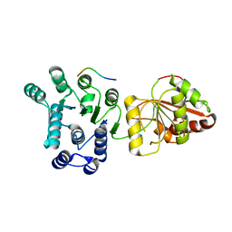 | | The crystal structure of eIF4AIII-Barentsz complex at 3.0 A resolution | | 分子名称: | ATP-DEPENDENT RNA HELICASE DDX48, PROTEIN CASC3 | | 著者 | Bono, F, Ebert, J, Lorentzen, E, Conti, E. | | 登録日 | 2006-08-04 | | 公開日 | 2006-09-06 | | 最終更新日 | 2023-12-13 | | 実験手法 | X-RAY DIFFRACTION (3 Å) | | 主引用文献 | The Crystal Structure of the Exon Junction Complex Reveals How It Mantains a Stable Grip on Mrna
Cell(Cambridge,Mass.), 126, 2006
|
|
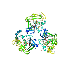
2J0V
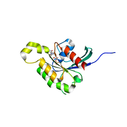 | | The crystal structure of Arabidopsis thaliana RAC7-ROP9: the first RAS superfamily GTPase from the plant kingdom | | 分子名称: | GUANOSINE-5'-DIPHOSPHATE, MAGNESIUM ION, RAC-LIKE GTP-BINDING PROTEIN ARAC7 | | 著者 | Sormo, C.G, Leiros, I, Brembu, T, Winge, P, Os, V, Bones, A.M. | | 登録日 | 2006-08-07 | | 公開日 | 2006-10-04 | | 最終更新日 | 2023-12-13 | | 実験手法 | X-RAY DIFFRACTION (1.78 Å) | | 主引用文献 | The Crystal Structure of Arabidopsis Thaliana Rac7/Rop9: The First Ras Superfamily Gtpase from the Plant Kingdom.
Phytochemistry, 67, 2006
|
|
2J0W
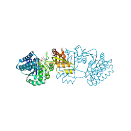 | | Crystal structure of E. coli aspartokinase III in complex with aspartate and ADP (R-state) | | 分子名称: | ADENOSINE-5'-DIPHOSPHATE, ASPARTIC ACID, CHLORIDE ION, ... | | 著者 | Kotaka, M, Ren, J, Lockyer, M, Hawkins, A.R, Stammers, D.K. | | 登録日 | 2006-08-07 | | 公開日 | 2006-08-10 | | 最終更新日 | 2023-12-13 | | 実験手法 | X-RAY DIFFRACTION (2.5 Å) | | 主引用文献 | Structures of R- and T-State Escherichia Coli Aspartokinase III: Mechanisms of the Allosteric Transition and Inhibition by Lysine.
J.Biol.Chem., 281, 2006
|
|
2J0X
 | | CRYSTAL STRUCTURE OF E. COLI ASPARTOKINASE III IN COMPLEX WITH LYSINE AND ASPARTATE (T-STATE) | | 分子名称: | ASPARTIC ACID, LYSINE, LYSINE-SENSITIVE ASPARTOKINASE 3, ... | | 著者 | Kotaka, M, Ren, J, Lockyer, M, Hawkins, A.R, Stammers, D.K. | | 登録日 | 2006-08-07 | | 公開日 | 2006-08-10 | | 最終更新日 | 2024-05-01 | | 実験手法 | X-RAY DIFFRACTION (2.8 Å) | | 主引用文献 | Structures of R- and T-State Escherichia Coli Aspartokinase III: Mechanisms of the Allosteric Transition and Inhibition by Lysine.
J.Biol.Chem., 281, 2006
|
|
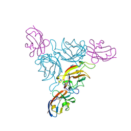
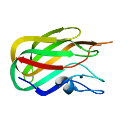
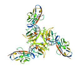
2J0Y
 | | L-ficolin complexed to b-1,3-D-glucan | | 分子名称: | 2-acetamido-2-deoxy-beta-D-glucopyranose, ACETATE ION, CALCIUM ION, ... | | 著者 | Garlatti, V, Gaboriaud, C. | | 登録日 | 2006-08-08 | | 公開日 | 2007-01-23 | | 最終更新日 | 2020-07-29 | | 実験手法 | X-RAY DIFFRACTION (2.35 Å) | | 主引用文献 | Structural Insights Into the Innate Immune Recognition Specificities of L- and H-Ficolins.
Embo J., 26, 2007
|
|
2J0Z
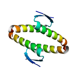 | | p53 tetramerization domain wild type | | 分子名称: | CELLULAR TUMOR ANTIGEN P53 | | 著者 | Carbajo, R.J, Mora, P, Sanchez del Pino, M.M, Perez-Paya, E, Pineda-Lucena, A. | | 登録日 | 2006-08-08 | | 公開日 | 2007-08-28 | | 最終更新日 | 2024-05-15 | | 実験手法 | SOLUTION NMR | | 主引用文献 | Solvent-exposed residues located in the beta-sheet modulate the stability of the tetramerization domain of p53--a structural and combinatorial approach.
Proteins, 71, 2008
|
|
2J10
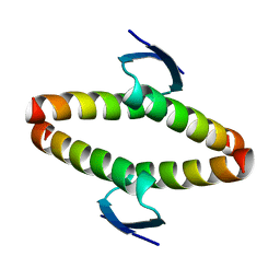 | | p53 tetramerization domain mutant T329F Q331K | | 分子名称: | CELLULAR TUMOR ANTIGEN P53 | | 著者 | Carbajo, R.J, Mora, P, Sanchez del Pino, M.M, Perez-Paya, E, Pineda-Lucena, A. | | 登録日 | 2006-08-08 | | 公開日 | 2007-08-28 | | 最終更新日 | 2024-05-15 | | 実験手法 | SOLUTION NMR | | 主引用文献 | Solvent-exposed residues located in the beta-sheet modulate the stability of the tetramerization domain of p53--a structural and combinatorial approach.
Proteins, 71, 2008
|
|
2J11
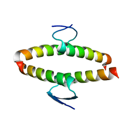 | | p53 tetramerization domain mutant Y327S T329G Q331G | | 分子名称: | CELLULAR TUMOR ANTIGEN P53 | | 著者 | Carbajo, R.J, Mora, P, Sanchez del Pino, M.M, Perez-Paya, E, Pineda-Lucena, A. | | 登録日 | 2006-08-08 | | 公開日 | 2007-08-28 | | 最終更新日 | 2024-05-15 | | 実験手法 | SOLUTION NMR | | 主引用文献 | Solvent-exposed residues located in the beta-sheet modulate the stability of the tetramerization domain of p53--a structural and combinatorial approach.
Proteins, 71, 2008
|
|
2J12
 | | Ad37 fibre head in complex with CAR D1 | | 分子名称: | CALCIUM ION, COXSACKIEVIRUS AND ADENOVIRUS RECEPTOR, FIBER PROTEIN | | 著者 | Seiradake, E, Lortat-Jacob, H, Billet, O, Kremer, E.J, Cusack, S. | | 登録日 | 2006-08-08 | | 公開日 | 2006-08-29 | | 最終更新日 | 2023-12-13 | | 実験手法 | X-RAY DIFFRACTION (1.5 Å) | | 主引用文献 | Structural and Mutational Analysis of Human Ad37 and Canine Adenovirus 2 Fiber Heads in Complex with the D1 Domain of Coxsackie and Adenovirus Receptor.
J.Biol.Chem., 281, 2006
|
|
2J13
 | | Structure of a family 4 carbohydrate esterase from Bacillus anthracis | | 分子名称: | ACETATE ION, CACODYLATE ION, POLYSACCHARIDE DEACETYLASE, ... | | 著者 | Gloster, T.M, Oberbarnscheidt, L, Taylor, E.J, Davies, G.J. | | 登録日 | 2006-08-08 | | 公開日 | 2006-10-03 | | 最終更新日 | 2023-12-13 | | 実験手法 | X-RAY DIFFRACTION (1.7 Å) | | 主引用文献 | Structure of a Carbohydrate Esterase from Bacillus Anthracis.
Proteins: Struct., Funct., Bioinf., 66, 2007
|
|
2J14
 | | 3,4,5-Trisubstituted Isoxazoles as Novel PPARdelta Agonists: Part2 | | 分子名称: | (3-{4-[2-(2,4-DICHLORO-PHENOXY)-ETHYLCARBAMOYL]-5-PHENYL-ISOXAZOL-3-YL}-PHENYL)-ACETIC ACID, PEROXISOME PROLIFERATOR-ACTIVATED RECEPTOR DELTA | | 著者 | Epple, R, Azimioara, M, Russo, R, Xie, Y, Wang, X, Cow, C, Wityak, J, Karanewsky, D, Bursulaya, B, Kreusch, A, Tuntland, T, Gerken, A, Iskandar, M, Saez, E, Seidel, H.M, Tian, S.S. | | 登録日 | 2006-08-08 | | 公開日 | 2006-09-06 | | 最終更新日 | 2023-12-13 | | 実験手法 | X-RAY DIFFRACTION (2.8 Å) | | 主引用文献 | 3,4,5-Trisubstituted Isoxazoles as Novel Ppardelta Agonists. Part 2
Bioorg.Med.Chem.Lett., 16, 2006
|
|
2J15
 | | Cyclic MrIA: An exceptionally stable and potent cyclic conotoxin with a novel topological fold that targets the norepinephrine transporter. | | 分子名称: | MAI126P | | 著者 | Lovelace, E.S, Armishaw, C.J, Colgrave, M.L, Walstrom, M.E, Alewood, P.F, Daly, N.L, Craik, D.J. | | 登録日 | 2006-08-09 | | 公開日 | 2006-11-01 | | 最終更新日 | 2018-05-09 | | 実験手法 | SOLUTION NMR | | 主引用文献 | Cyclic MrIA: a stable and potent cyclic conotoxin with a novel topological fold that targets the norepinephrine transporter.
J. Med. Chem., 49, 2006
|
|
2J16
 | | Apo & Sulphate bound forms of SDP-1 | | 分子名称: | MAGNESIUM ION, SULFATE ION, TYROSINE-PROTEIN PHOSPHATASE YIL113W | | 著者 | Briggs, D.C, McDonald, N.Q. | | 登録日 | 2006-08-09 | | 公開日 | 2007-05-22 | | 最終更新日 | 2024-05-01 | | 実験手法 | X-RAY DIFFRACTION (2.7 Å) | | 主引用文献 | Redox-mediated substrate recognition by Sdp1 defines a new group of tyrosine phosphatases.
Nature, 447, 2007
|
|
2J17
 | | pTyr bound form of SDP-1 | | 分子名称: | MAGNESIUM ION, O-PHOSPHOTYROSINE, TYROSINE-PROTEIN PHOSPHATASE YIL113W | | 著者 | Briggs, D.C, McDonald, N.Q. | | 登録日 | 2006-08-09 | | 公開日 | 2007-05-22 | | 最終更新日 | 2023-12-13 | | 実験手法 | X-RAY DIFFRACTION (2.84 Å) | | 主引用文献 | Redox-mediated substrate recognition by Sdp1 defines a new group of tyrosine phosphatases.
Nature, 447, 2007
|
|
2J18
 | | Chloroperoxidase mixture of ferric and ferrous states (low dose data set) | | 分子名称: | 2-acetamido-2-deoxy-beta-D-glucopyranose, 2-acetamido-2-deoxy-beta-D-glucopyranose-(1-4)-2-acetamido-2-deoxy-beta-D-glucopyranose, BROMIDE ION, ... | | 著者 | Beitlich, T, Kuhnel, K, Schulze-Briese, C, Shoeman, R.L, Schlichting, I. | | 登録日 | 2006-08-09 | | 公開日 | 2006-12-18 | | 最終更新日 | 2023-12-13 | | 実験手法 | X-RAY DIFFRACTION (1.75 Å) | | 主引用文献 | Cryoradiolytic Reduction of Crystalline Heme Proteins: Analysis by Uv-Vis Spectroscopy and X-Ray Crystallography
J.Synchrotron Radiat., 14, 2007
|
|
2J19
 | | Ferrous Chloroperoxidase (high dose data set) | | 分子名称: | 2-acetamido-2-deoxy-beta-D-glucopyranose, 2-acetamido-2-deoxy-beta-D-glucopyranose-(1-4)-2-acetamido-2-deoxy-beta-D-glucopyranose, BROMIDE ION, ... | | 著者 | Beitlich, T, Kuhnel, K, Schulze-Briese, C, Shoeman, R.L, Schlichting, I. | | 登録日 | 2006-08-09 | | 公開日 | 2006-12-18 | | 最終更新日 | 2023-12-13 | | 実験手法 | X-RAY DIFFRACTION (1.75 Å) | | 主引用文献 | Cryoradiolytic Reduction of Crystalline Heme Proteins: Analysis by Uv-Vis Spectroscopy and X-Ray Crystallography
J.Synchrotron Radiat., 14, 2007
|
|
2J1A
 | |
2J1D
 | | Crystallization of hDaam1 C-terminal Fragment | | 分子名称: | DISHEVELED-ASSOCIATED ACTIVATOR OF MORPHOGENESIS 1, GLYCEROL, PHOSPHATE ION | | 著者 | Lu, J, Meng, W, Poy, F, Eck, M.J. | | 登録日 | 2006-08-10 | | 公開日 | 2007-05-15 | | 最終更新日 | 2024-05-08 | | 実験手法 | X-RAY DIFFRACTION (2.25 Å) | | 主引用文献 | Structure of the Fh2 Domain of Daam1: Implications for Formin Regulation of Actin Assembly.
J.Mol.Biol., 369, 2007
|
|
2J1E
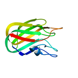 | |
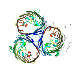
2J1G
 | | L-ficolin complexed to N-acetyl-cystein | | 分子名称: | 2-acetamido-2-deoxy-beta-D-glucopyranose-(1-4)-2-acetamido-2-deoxy-beta-D-glucopyranose, ACETATE ION, CALCIUM ION, ... | | 著者 | Garlatti, V, Gaboriaud, C. | | 登録日 | 2006-08-11 | | 公開日 | 2007-01-23 | | 最終更新日 | 2023-12-13 | | 実験手法 | X-RAY DIFFRACTION (1.95 Å) | | 主引用文献 | Structural Insights Into the Innate Immune Recognition Specificities of L- and H-Ficolins.
Embo J., 26, 2007
|
|
2J1K
 | | CAV-2 fibre head in complex with CAR D1 | | 分子名称: | COXSACKIEVIRUS AND ADENOVIRUS RECEPTOR, FIBER PROTEIN | | 著者 | Seiradake, E, Lortat-Jacob, H, Billet, O, Kremer, E.J, Cusack, S. | | 登録日 | 2006-08-14 | | 公開日 | 2006-09-05 | | 最終更新日 | 2023-12-13 | | 実験手法 | X-RAY DIFFRACTION (2.3 Å) | | 主引用文献 | Structural and Mutational Analysis of Human Ad37 and Canine Adenovirus 2 Fiber Heads in Complex with the D1 Domain of Coxsackie and Adenovirus Receptor.
J.Biol.Chem., 281, 2006
|
|
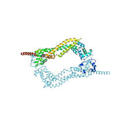
2J1L
 | | Crystal Structure of Human Rho-related GTP-binding protein RhoD | | 分子名称: | GUANOSINE-5'-DIPHOSPHATE, MAGNESIUM ION, RHO-RELATED GTP-BINDING PROTEIN RHOD | | 著者 | Pike, A.C.W, Johansson, C, Gileadi, C, Niesen, F.H, Sobott, F, Schoch, G, Elkins, J, Smee, C, Gorrec, F, Watt, S, Bray, J, Turnbull, A.P, von Delft, F, Arrowsmith, C, Edwards, A, Weigelt, J, Sundstrom, M, Doyle, D. | | 登録日 | 2006-08-14 | | 公開日 | 2006-09-18 | | 最終更新日 | 2023-12-13 | | 実験手法 | X-RAY DIFFRACTION (2.5 Å) | | 主引用文献 | Crystal Structure of Human Rho-Related GTP-Binding Protein Rhod
To be Published
|
|
2J1M
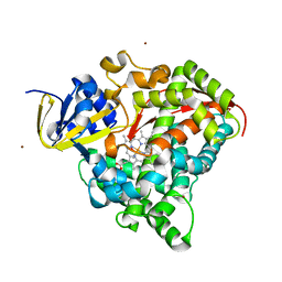 | | P450 BM3 Heme domain in complex with DMSO | | 分子名称: | CYTOCHROME P450 102, DIMETHYL SULFOXIDE, PROTOPORPHYRIN IX CONTAINING FE, ... | | 著者 | Kuper, J, Tuck-Seng, W, Roccatano, D, Wilmanns, M, Schwaneberg, U. | | 登録日 | 2006-08-14 | | 公開日 | 2007-05-15 | | 最終更新日 | 2023-12-13 | | 実験手法 | X-RAY DIFFRACTION (1.7 Å) | | 主引用文献 | Understanding a Mechanism of Organic Cosolvent Inactivation in Heme Monooxygenase P450 Bm-3.
J.Am.Chem.Soc., 129, 2007
|
|
2J1N
 | | osmoporin OmpC | | 分子名称: | CHLORIDE ION, DODECANE, MAGNESIUM ION, ... | | 著者 | Basle, A, Storici, P, Rummel, G, Rosenbusch, J.P, Schirmer, T. | | 登録日 | 2006-08-15 | | 公開日 | 2006-09-06 | | 最終更新日 | 2023-12-13 | | 実験手法 | X-RAY DIFFRACTION (2 Å) | | 主引用文献 | Crystal Structure of Osmoporin Ompc from E. Coli at 2.0 A.
J.Mol.Biol., 362, 2006
|
|


