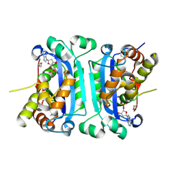
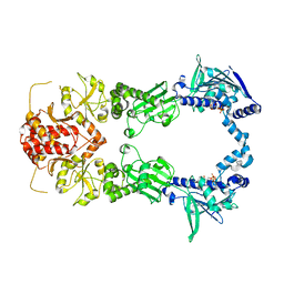
2IO8
 
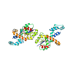
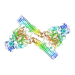
 | | E. coli Bifunctional glutathionylspermidine synthetase/amidase Incomplex with Mg2+ and ADP | | 分子名称: | ADENOSINE-5'-DIPHOSPHATE, Bifunctional glutathionylspermidine synthetase/amidase, CYSTEINE, ... | | 著者 | Pai, C.H, Chiang, B.Y, Ko, T.P, Chou, C.C, Chong, C.M, Yen, F.J, Coward, J.K, Wang, A.H.-J, Lin, C.H. | | 登録日 | 2006-10-10 | | 公開日 | 2006-12-12 | | 最終更新日 | 2024-03-13 | | 実験手法 | X-RAY DIFFRACTION (2.1 Å) | | 主引用文献 | Dual binding sites for translocation catalysis by Escherichia coli glutathionylspermidine synthetase
Embo J., 25, 2006
|
|
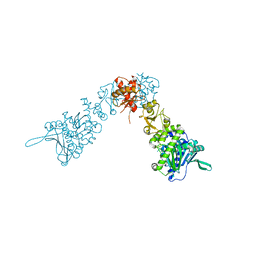
2IO9
 | | E. coli Bifunctional glutathionylspermidine synthetase/amidase Incomplex with Mg2+ ,GSH and ADP | | 分子名称: | ADENOSINE-5'-DIPHOSPHATE, Bifunctional glutathionylspermidine synthetase/amidase, GLUTATHIONE, ... | | 著者 | Pai, C.H, Chiang, B.Y, Ko, T.P, Chou, C.C, Chong, C.M, Yen, F.J, Coward, J.K, Wang, A.H.-J, Lin, C.H. | | 登録日 | 2006-10-10 | | 公開日 | 2006-12-12 | | 最終更新日 | 2011-07-13 | | 実験手法 | X-RAY DIFFRACTION (2.2 Å) | | 主引用文献 | Dual binding sites for translocation catalysis by Escherichia coli glutathionylspermidine synthetase
Embo J., 25, 2006
|
|
2IOA
 | | E. coli Bifunctional glutathionylspermidine synthetase/amidase Incomplex with Mg2+ and ADP and phosphinate inhibitor | | 分子名称: | ADENOSINE-5'-DIPHOSPHATE, Bifunctional glutathionylspermidine synthetase/amidase, D-GAMMA-GLUTAMYL-N-{[(R)-{4-[(4-AMINOBUTYL)AMINO]BUTYL}(PHOSPHONOOXY)PHOSPHORYL]METHYL}-D-ALANINAMIDE, ... | | 著者 | Pai, C.H, Chiang, B.Y, Ko, T.P, Chou, C.C, Chong, C.M, Yen, F.J, Coward, J.K, Wang, A.H.-J, Lin, C.H. | | 登録日 | 2006-10-10 | | 公開日 | 2006-12-12 | | 最終更新日 | 2024-03-13 | | 実験手法 | X-RAY DIFFRACTION (2.8 Å) | | 主引用文献 | Dual binding sites for translocation catalysis by Escherichia coli glutathionylspermidine synthetase
Embo J., 25, 2006
|
|
2IOB
 | | E. coli Bifunctional glutathionylspermidine synthetase/amidase Apo protein | | 分子名称: | Bifunctional glutathionylspermidine synthetase/amidase | | 著者 | Pai, C.H, Chiang, B.Y, Ko, T.P, Chou, C.C, Chong, C.M, Yen, F.J, Coward, J.K, Wang, A.H.-J, Lin, C.H. | | 登録日 | 2006-10-10 | | 公開日 | 2006-12-12 | | 最終更新日 | 2024-03-13 | | 実験手法 | X-RAY DIFFRACTION (2.2 Å) | | 主引用文献 | Dual binding sites for translocation catalysis by Escherichia coli glutathionylspermidine synthetase
Embo J., 25, 2006
|
|
2IOC
 | |
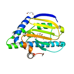
2IOD
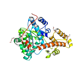
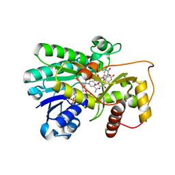 | | Binding of two substrate analogue molecules to dihydroflavonol-4-reductase alters the functional geometry of the catalytic site | | 分子名称: | 3,5,7-TRIHYDROXY-2-(3,4,5-TRIHYDROXYPHENYL)-4H-CHROMEN-4-ONE, Dihydroflavonol 4-reductase, NADP NICOTINAMIDE-ADENINE-DINUCLEOTIDE PHOSPHATE | | 著者 | Petit, P, Langlois d'Estaintot, B, Granier, T, Gallois, B. | | 登録日 | 2006-10-10 | | 公開日 | 2007-09-11 | | 最終更新日 | 2023-10-25 | | 実験手法 | X-RAY DIFFRACTION (2.06 Å) | | 主引用文献 | Binding of two substrate analogue molecules to dihydroflavonol-4-reductase alters the functional geometry of the catalytic site
To be Published
|
|
2IOF
 | | Crystal structure of phosphonoacetaldehyde hydrolase with sodium borohydride-reduced substrate intermediate | | 分子名称: | MAGNESIUM ION, PHOSPHATE ION, Phosphonoacetaldehyde hydrolase | | 著者 | Allen, K.A, Lahiri, S.D, Zhang, G, Dunaway-Mariano, D. | | 登録日 | 2006-10-10 | | 公開日 | 2007-07-17 | | 最終更新日 | 2019-07-24 | | 実験手法 | X-RAY DIFFRACTION (2.5 Å) | | 主引用文献 | Diversification of function in the haloacid dehalogenase enzyme superfamily: The role of the cap domain in hydrolytic phosphoruscarbon bond cleavage.
Bioorg.Chem., 34, 2006
|
|
2IOG
 | |
2IOH
 | | Crystal structure of phosphonoacetaldehyde hydrolase with a K53R mutation | | 分子名称: | MAGNESIUM ION, PHOSPHATE ION, Phosphonoacetaldehyde hydrolase | | 著者 | Allen, K.A, Lahiri, S.D, Zhang, G, Dunaway-Mariano, D, Peisach, E. | | 登録日 | 2006-10-10 | | 公開日 | 2007-08-07 | | 最終更新日 | 2024-02-21 | | 実験手法 | X-RAY DIFFRACTION (2.9 Å) | | 主引用文献 | Diversification of function in the haloacid dehalogenase enzyme superfamily: The role of the cap domain in hydrolytic phosphoruscarbon bond cleavage.
Bioorg.Chem., 34, 2006
|
|
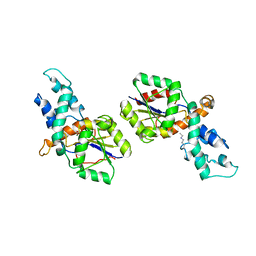
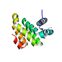
2IOI
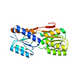
 | | Crystal structure of the mouse p53 core domain at 1.55 A | | 分子名称: | 2-AMINO-2-HYDROXYMETHYL-PROPANE-1,3-DIOL, Cellular tumor antigen p53, ZINC ION | | 著者 | Ho, W.C, Luo, C, Zhao, K, Chai, X, Fitzgerald, M.X, Marmorstein, R. | | 登録日 | 2006-10-10 | | 公開日 | 2006-12-05 | | 最終更新日 | 2024-02-21 | | 実験手法 | X-RAY DIFFRACTION (1.55 Å) | | 主引用文献 | High-resolution structure of the p53 core domain: implications for binding small-molecule stabilizing compounds.
Acta Crystallogr.,Sect.D, 62, 2006
|
|
2IOJ
 | |
2IOK
 | |
2IOL
 | |
2IOM
 | | Mouse p53 core domain soaked with 2-propanol | | 分子名称: | 2-AMINO-2-HYDROXYMETHYL-PROPANE-1,3-DIOL, Cellular tumor antigen p53, ISOPROPYL ALCOHOL, ... | | 著者 | Ho, W.C, Luo, C, Zhao, K, Chai, X, Fitzgerald, M.X, Marmorstein, R. | | 登録日 | 2006-10-10 | | 公開日 | 2006-12-05 | | 最終更新日 | 2024-02-21 | | 実験手法 | X-RAY DIFFRACTION (2 Å) | | 主引用文献 | High-resolution structure of the p53 core domain: implications for binding small-molecule stabilizing compounds.
ACTA CRYSTALLOGR.,SECT.D, 62, 2006
|
|
2ION
 | |
2IOO
 | | Crystal structure of the mouse p53 core domain | | 分子名称: | Cellular tumor antigen p53, ZINC ION | | 著者 | Ho, W.C, Luo, C, Zhao, K, Chai, X, Fitzgerald, M.X, Marmorstein, R. | | 登録日 | 2006-10-10 | | 公開日 | 2006-12-05 | | 最終更新日 | 2024-02-21 | | 実験手法 | X-RAY DIFFRACTION (2.02 Å) | | 主引用文献 | High-resolution structure of the p53 core domain: implications for binding small-molecule stabilizing compounds.
Acta Crystallogr.,Sect.D, 62, 2006
|
|
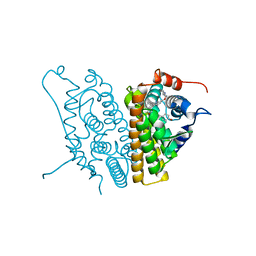
2IOP
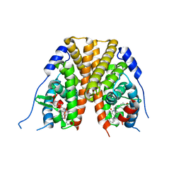
 | | Crystal Structure of Full-length HtpG, the Escherichia coli Hsp90, Bound to ADP | | 分子名称: | ADENOSINE-5'-DIPHOSPHATE, Chaperone protein htpG | | 著者 | Shiau, A.K, Harris, S.F, Agard, D.A. | | 登録日 | 2006-10-10 | | 公開日 | 2006-11-21 | | 最終更新日 | 2024-02-21 | | 実験手法 | X-RAY DIFFRACTION (3.55 Å) | | 主引用文献 | Structural Analysis of E. coli hsp90 reveals dramatic nucleotide-dependent conformational rearrangements.
Cell(Cambridge,Mass.), 127, 2006
|
|
2IOQ
 | |
2IOR
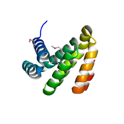
 | | Crystal Structure of the N-terminal Domain of HtpG, the Escherichia coli Hsp90, Bound to ADP | | 分子名称: | ADENOSINE-5'-DIPHOSPHATE, Chaperone protein htpG, HEXANE-1,6-DIOL, ... | | 著者 | Shiau, A.K, Harris, S.F, Agard, D.A. | | 登録日 | 2006-10-10 | | 公開日 | 2006-11-21 | | 最終更新日 | 2023-08-30 | | 実験手法 | X-RAY DIFFRACTION (1.65 Å) | | 主引用文献 | Structural Analysis of E. coli hsp90 reveals dramatic nucleotide-dependent conformational rearrangements.
Cell(Cambridge,Mass.), 127, 2006
|
|
2IOS
 | |
2IOT
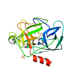 | | Clavulanic Acid bound to Elastase | | 分子名称: | Elastase-1, N-(3-OXOPROPYL)GLYCINE, SULFATE ION | | 著者 | Farady, C, Navia, M.A. | | 登録日 | 2006-10-10 | | 公開日 | 2008-04-08 | | 最終更新日 | 2023-08-30 | | 実験手法 | X-RAY DIFFRACTION (1.6 Å) | | 主引用文献 | Elastase Inhibition by Clavulanic Acid, and Inhibitor of Bacterial b-lactamases: Mechanistic and Structural Studies
To be Published
|
|
2IOU
 | |
2IOV
 | | Bright-state structure of the reversibly switchable fluorescent protein Dronpa | | 分子名称: | Fluorescent protein Dronpa | | 著者 | Stiel, A.C, Trowitzsch, S, Weber, G, Andresen, M, Eggeling, C, Hell, S.W, Jakobs, S, Wahl, M.C. | | 登録日 | 2006-10-11 | | 公開日 | 2006-12-05 | | 最終更新日 | 2023-11-15 | | 実験手法 | X-RAY DIFFRACTION (1.8 Å) | | 主引用文献 | 1.8 A bright-state structure of the reversibly switchable fluorescent protein Dronpa guides the generation of fast switching variants
Biochem.J., 402, 2007
|
|
2IOY
 | |
2IP1
 | |


