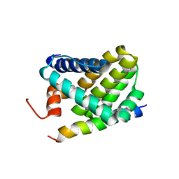
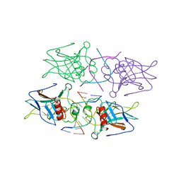
2UWQ
 
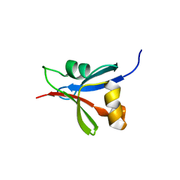 | |
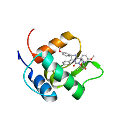
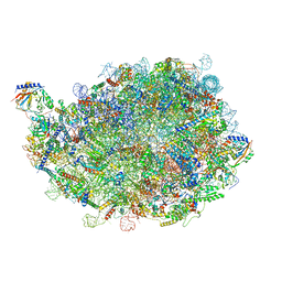
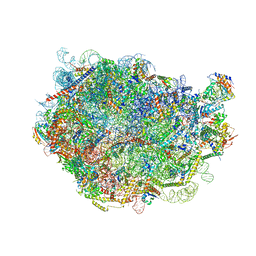
6R7F
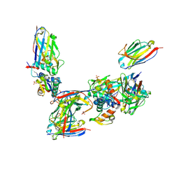
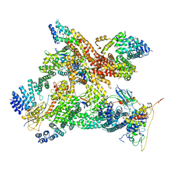 | | Structural basis of Cullin-2 RING E3 ligase regulation by the COP9 signalosome | | 分子名称: | COP9 signalosome complex subunit 1, COP9 signalosome complex subunit 2, COP9 signalosome complex subunit 3, ... | | 著者 | Faull, S.V, Lau, A.M.C, Martens, C, Ahdash, Z, Yebenes, H, Schmidt, C, Beuron, F, Cronin, N.B, Morris, E.P, Politis, A. | | 登録日 | 2019-03-28 | | 公開日 | 2019-08-28 | | 最終更新日 | 2024-05-22 | | 実験手法 | ELECTRON MICROSCOPY (8.2 Å) | | 主引用文献 | Structural basis of Cullin 2 RING E3 ligase regulation by the COP9 signalosome.
Nat Commun, 10, 2019
|
|
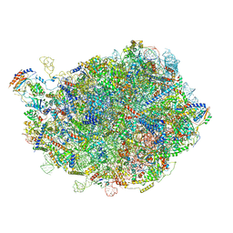
6R6H
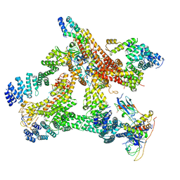 | | Structural basis of Cullin-2 RING E3 ligase regulation by the COP9 signalosome | | 分子名称: | COP9 signalosome complex subunit 1, COP9 signalosome complex subunit 2, COP9 signalosome complex subunit 3, ... | | 著者 | Morris, E.P, Faull, S.V, Lau, A.M.C, Politis, A, Beuron, F, Cronin, N. | | 登録日 | 2019-03-27 | | 公開日 | 2019-08-28 | | 最終更新日 | 2024-05-22 | | 実験手法 | ELECTRON MICROSCOPY (8.4 Å) | | 主引用文献 | Structural basis of Cullin 2 RING E3 ligase regulation by the COP9 signalosome.
Nat Commun, 10, 2019
|
|
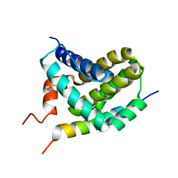
2FHI
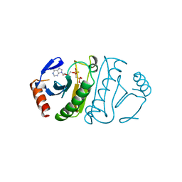 | | SUBSTRATE ANALOG (IB2) COMPLEX WITH THE HIS 96 ASN SUBSTITUTION OF THE FRAGILE HISTIDINE TRIAD PROTEIN, FHIT | | 分子名称: | FRAGILE HISTIDINE TRIAD PROTEIN, P1-P2-METHYLENE-P3-THIO-DIADENOSINE TRIPHOSPHATE | | 著者 | Pace, H.C, Garrison, P.N, Robinson, A.K, Barnes, L.D, Draganescu, A, Rosler, A, Blackburn, G.M, Siprashvili, Z, Croce, C.M, Heubner, K, Brenner, C. | | 登録日 | 1998-04-01 | | 公開日 | 1998-06-17 | | 最終更新日 | 2024-05-29 | | 実験手法 | X-RAY DIFFRACTION (2.6 Å) | | 主引用文献 | Genetic, biochemical, and crystallographic characterization of Fhit-substrate complexes as the active signaling form of Fhit.
Proc.Natl.Acad.Sci.USA, 95, 1998
|
|
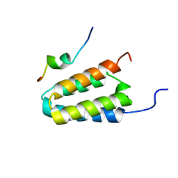
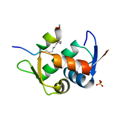
2ROC
 | | Solution structure of Mcl-1 Complexed with Puma | | 分子名称: | Bcl-2-binding component 3, Induced myeloid leukemia cell differentiation protein Mcl-1 homolog | | 著者 | Day, C.L, Smits, C, Fan, F.C, Lee, E.F, Fairlie, W.D, Hinds, M.G. | | 登録日 | 2008-03-17 | | 公開日 | 2008-07-08 | | 最終更新日 | 2024-05-29 | | 実験手法 | SOLUTION NMR | | 主引用文献 | Structure of the BH3 Domains from the p53-Inducible BH3-Only Proteins Noxa and Puma in Complex with Mcl-1
J.Mol.Biol., 380, 2008
|
|
2N06
 | | Mdmx-298 | | 分子名称: | 4-[[(4S,5R)-5-(4-chlorophenyl)-4-(3-methoxyphenyl)-2-(4-methoxy-2-propan-2-yloxy-phenyl)-4,5-dihydroimidazol-1-yl]carbonyl]piperazin-2-one, Protein Mdm4 | | 著者 | Grace, C.R, Kriwacki, R.W. | | 登録日 | 2015-03-04 | | 公開日 | 2016-01-27 | | 最終更新日 | 2024-05-01 | | 実験手法 | SOLUTION NMR | | 主引用文献 | Monitoring Ligand-Induced Protein Ordering in Drug Discovery.
J.Mol.Biol., 428, 2016
|
|
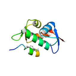
2ROD
 | | Solution Structure of MCL-1 Complexed with NoxaA | | 分子名称: | Induced myeloid leukemia cell differentiation protein Mcl-1 homolog, Noxa | | 著者 | Day, C.L, Smits, C, Fan, F.C, Lee, E.F, Fairlie, W.D, Hinds, M.G. | | 登録日 | 2008-03-17 | | 公開日 | 2008-07-08 | | 最終更新日 | 2024-05-29 | | 実験手法 | SOLUTION NMR | | 主引用文献 | Structure of the BH3 Domains from the p53-Inducible BH3-Only Proteins Noxa and Puma in Complex with Mcl-1
J.Mol.Biol., 380, 2008
|
|
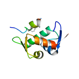
2KZU
 | | DAXX helical bundle (DHB) domain / Rassf1C complex | | 分子名称: | Death-associated protein 6, Ras association (RalGDS/AF-6) domain family 1 | | 著者 | Escobar-Cabrera, E, Lau, D.K.W, Giovinazzi, S, Ishov, A.M, McIntosh, L.P. | | 登録日 | 2010-06-25 | | 公開日 | 2010-12-15 | | 最終更新日 | 2024-05-15 | | 実験手法 | SOLUTION NMR | | 主引用文献 | Structural Characterization of the DAXX N-Terminal Helical Bundle Domain and Its Complex with Rassf1C.
Structure, 18, 2010
|
|
2Y9U
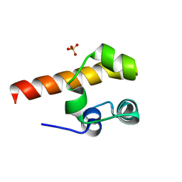 | | Structural basis of p63a SAM domain mutants involved in AEC syndrome | | 分子名称: | SULFATE ION, TUMOR PROTEIN 63 | | 著者 | Sathyamurthy, A, Freund, S.M.V, Johnson, C.M, Allen, M.D. | | 登録日 | 2011-02-16 | | 公開日 | 2011-08-03 | | 最終更新日 | 2024-05-08 | | 実験手法 | X-RAY DIFFRACTION (1.6 Å) | | 主引用文献 | Structural Basis of P63Alpha Sam Domain Mutants Involved in Aec Syndrome.
FEBS J., 278, 2011
|
|
6V4E
 | |
6V4G
 | |
6V4H
 | |
6V4F
 | |
4G83
 | |
8INE
 | |
8INF
 | |
8FLB
 | |
8FLA
 | |
1TTV
 | | NMR Structure of a Complex Between MDM2 and a Small Molecule Inhibitor | | 分子名称: | 1-{[4,5-BIS(4-CHLOROPHENYL)-2-(2-ISOPROPOXY-4-METHOXYPHENYL)-4,5-DIHYDRO-1H-IMIDAZOL-1-YL]CARBONYL}PIPERAZINE, Ubiquitin-protein ligase E3 Mdm2 | | 著者 | Fry, D.C, Emerson, S.D, Palme, S, Vu, B.T, Liu, C.M, Podlaski, F. | | 登録日 | 2004-06-23 | | 公開日 | 2005-01-04 | | 最終更新日 | 2024-05-22 | | 実験手法 | SOLUTION NMR | | 主引用文献 | NMR structure of a complex between MDM2 and a small molecule inhibitor.
J.Biomol.Nmr, 30, 2004
|
|
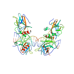
3VD0
 | | structure of p73 DNA binding domain tetramer modulates p73 transactivation | | 分子名称: | DNA (5'-D(*CP*AP*GP*GP*CP*AP*TP*GP*CP*CP*TP*G)-3'), Tumor protein p73, ZINC ION | | 著者 | Ethayathulla, A.S, Tse, P.W, Nguyen, S, Viadiu, H. | | 登録日 | 2012-01-04 | | 公開日 | 2012-04-18 | | 最終更新日 | 2023-09-13 | | 実験手法 | X-RAY DIFFRACTION (2.95 Å) | | 主引用文献 | Structure of p73 DNA-binding domain tetramer modulates p73 transactivation.
Proc.Natl.Acad.Sci.USA, 109, 2012
|
|
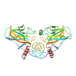
3VD1
 | | structure of p73 DNA binding domain tetramer modulates p73 transactivation | | 分子名称: | DNA (5'-D(*CP*GP*GP*GP*CP*AP*TP*GP*CP*CP*CP*G)-3'), Tumor protein p73, ZINC ION | | 著者 | Ethayathulla, A.S, Tse, P.W, Nguyen, S, Viadiu, H. | | 登録日 | 2012-01-04 | | 公開日 | 2012-04-18 | | 最終更新日 | 2023-09-13 | | 実験手法 | X-RAY DIFFRACTION (2.95 Å) | | 主引用文献 | Structure of p73 DNA-binding domain tetramer modulates p73 transactivation.
Proc.Natl.Acad.Sci.USA, 109, 2012
|
|
3VD2
 | | structure of p73 DNA binding domain tetramer modulates p73 transactivation | | 分子名称: | DNA (5'-D(*AP*TP*GP*GP*AP*CP*AP*TP*GP*TP*CP*CP*AP*T)-3'), Tumor protein p73, ZINC ION | | 著者 | Ethayathulla, A.S, Tse, P.W, Nguyen, S, Viadiu, H. | | 登録日 | 2012-01-04 | | 公開日 | 2012-04-18 | | 最終更新日 | 2023-11-29 | | 実験手法 | X-RAY DIFFRACTION (4 Å) | | 主引用文献 | Structure of p73 DNA-binding domain tetramer modulates p73 transactivation.
Proc.Natl.Acad.Sci.USA, 109, 2012
|
|
5KBD
 | |
2VYR
 | | Structure of human MDM4 N-terminal domain bound to a single domain antibody | | 分子名称: | HUMAN SINGLE DOMAIN ANTIBODY, MDM4 PROTEIN, SULFATE ION | | 著者 | Yu, G.W, Vaysburd, M, Allen, M.D, Settanni, G, Fersht, A.R. | | 登録日 | 2008-07-28 | | 公開日 | 2008-11-25 | | 最終更新日 | 2013-03-06 | | 実験手法 | X-RAY DIFFRACTION (2 Å) | | 主引用文献 | Structure of Human Mdm4 N-Terminal Domain Bound to a Single-Domain Antibody.
J.Mol.Biol., 385, 2009
|
|
5VK1
 | |


