3FOO
 
 | |
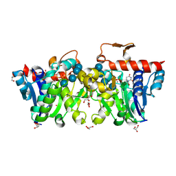
3ASV
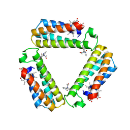
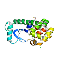
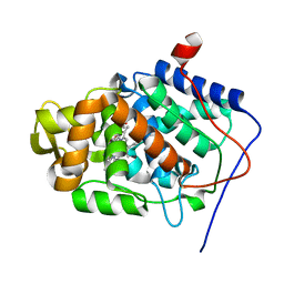
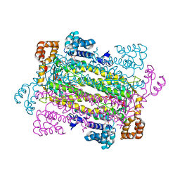
 | | The Closed form of serine dehydrogenase complexed with NADP+ | | 分子名称: | NADP NICOTINAMIDE-ADENINE-DINUCLEOTIDE PHOSPHATE, PHOSPHATE ION, Short-chain dehydrogenase/reductase SDR | | 著者 | Yamazawa, R, Nakajima, Y, Yoshimoto, T, Ito, K. | | 登録日 | 2010-12-21 | | 公開日 | 2011-10-12 | | 最終更新日 | 2023-11-01 | | 実験手法 | X-RAY DIFFRACTION (2.7 Å) | | 主引用文献 | Crystal structure of serine dehydrogenase from Escherichia coli: important role of the C-terminal region for closed-complex formation.
J.Biochem., 149, 2011
|
|
1KG8
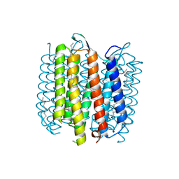
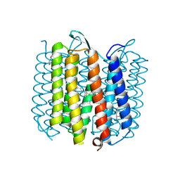 | | X-ray structure of an early-M intermediate of bacteriorhodopsin | | 分子名称: | 1-[2,6,10.14-TETRAMETHYL-HEXADECAN-16-YL]-2-[2,10,14-TRIMETHYLHEXADECAN-16-YL]GLYCEROL, RETINAL, bacteriorhodopsin | | 著者 | Facciotti, M.T, Rouhani, S, Burkard, F.T, Betancourt, F.M, Downing, K.H, Rose, R.B, McDermott, G, Glaeser, R.M. | | 登録日 | 2001-11-26 | | 公開日 | 2001-12-05 | | 最終更新日 | 2023-08-16 | | 実験手法 | X-RAY DIFFRACTION (2 Å) | | 主引用文献 | Structure of an early intermediate in the M-state phase of the bacteriorhodopsin photocycle.
Biophys.J., 81, 2001
|
|
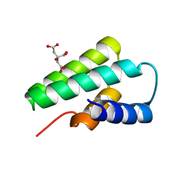
2PPX
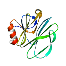
 | | Crystal structure of a HTH XRE-family like protein from Agrobacterium tumefaciens | | 分子名称: | GLYCEROL, SULFATE ION, Uncharacterized protein Atu1735 | | 著者 | Cuff, M.E, Skarina, T, Onopriyenko, O, Edwards, A, Savchenko, A, Joachimiak, A, Midwest Center for Structural Genomics (MCSG) | | 登録日 | 2007-04-30 | | 公開日 | 2007-05-29 | | 最終更新日 | 2011-07-13 | | 実験手法 | X-RAY DIFFRACTION (2 Å) | | 主引用文献 | Structure of a HTH XRE-family like protein from Agrobacterium tumefaciens.
TO BE PUBLISHED
|
|
1KGM
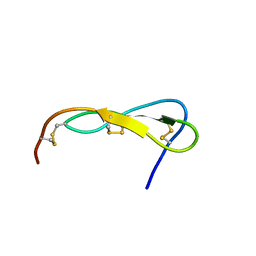 | | SOLUTION STRUCTURE OF THE SMALL SERINE PROTEASE INHIBITOR SGCI | | 分子名称: | SERINE PROTEASE INHIBITOR I | | 著者 | Gaspari, Z, Patthy, A, Graf, L, Perczel, A. | | 登録日 | 2001-11-28 | | 公開日 | 2001-12-12 | | 最終更新日 | 2022-02-23 | | 実験手法 | SOLUTION NMR | | 主引用文献 | Comparative structure analysis of proteinase inhibitors from the desert locust, Schistocerca gregaria.
Eur.J.Biochem., 269, 2002
|
|
1KNI
 | | Stabilizing Disulfide Bridge Mutant of T4 Lysozyme | | 分子名称: | BETA-MERCAPTOETHANOL, CHLORIDE ION, LYSOZYME | | 著者 | Jacobson, R.H, Matsumura, M, Faber, H.R, Matthews, B.W. | | 登録日 | 2001-12-18 | | 公開日 | 2001-12-28 | | 最終更新日 | 2024-04-03 | | 実験手法 | X-RAY DIFFRACTION (1.7 Å) | | 主引用文献 | Structure of a stabilizing disulfide bridge mutant that closes the active-site cleft of T4 lysozyme.
Protein Sci., 1, 1992
|
|
3FQ4
 | |
1KOZ
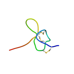 | | SOLUTION STRUCTURE OF OMEGA-GRAMMOTOXIN SIA | | 分子名称: | Voltage-dependent Channel Inhibitor | | 著者 | Takeuchi, K, Park, E.J, Lee, C.W, Kim, J.I, Takahashi, H, Swartz, K.J, Shimada, I. | | 登録日 | 2001-12-25 | | 公開日 | 2002-08-28 | | 最終更新日 | 2022-02-23 | | 実験手法 | SOLUTION NMR | | 主引用文献 | Solution structure of omega-grammotoxin SIA, a gating modifier of P/Q and N-type Ca(2+) channel.
J.Mol.Biol., 321, 2002
|
|
1KPU
 | |
3U2U
 | | Crystal Structure of Human Glycogenin-1 (GYG1) complexed with manganese, UDP and maltotetraose | | 分子名称: | GLYCEROL, Glycogenin-1, MANGANESE (II) ION, ... | | 著者 | Chaikuad, A, Froese, D.S, Krysztofinska, E, von Delft, F, Weigelt, J, Arrowsmith, C.H, Edwards, A.M, Bountra, C, Oppermann, U, Yue, W.W, Structural Genomics Consortium (SGC) | | 登録日 | 2011-10-04 | | 公開日 | 2011-11-02 | | 最終更新日 | 2023-09-13 | | 実験手法 | X-RAY DIFFRACTION (1.45 Å) | | 主引用文献 | Conformational plasticity of glycogenin and its maltosaccharide substrate during glycogen biogenesis.
Proc.Natl.Acad.Sci.USA, 108, 2011
|
|
1KKL
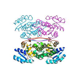
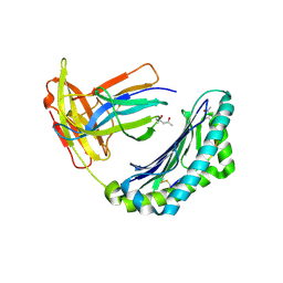
 | | L.casei HprK/P in complex with B.subtilis HPr | | 分子名称: | CALCIUM ION, HprK protein, PHOSPHOCARRIER PROTEIN HPR | | 著者 | Fieulaine, S, Morera, S, Poncet, S, Galinier, A, Janin, J, Deutscher, J, Nessler, S. | | 登録日 | 2001-12-10 | | 公開日 | 2002-08-28 | | 最終更新日 | 2023-08-16 | | 実験手法 | X-RAY DIFFRACTION (2.8 Å) | | 主引用文献 | X-ray structure of a bifunctional protein kinase in complex with its protein substrate HPr.
Proc.Natl.Acad.Sci.USA, 99, 2002
|
|
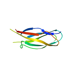
3F2E
 | | Crystal structure of Yellowstone SIRV coat protein C-terminus | | 分子名称: | CITRIC ACID, SIRV coat protein | | 著者 | Taurog, R.E, Szymczyna, B.R, Williamson, J.R, Johnson, J.E. | | 登録日 | 2008-10-29 | | 公開日 | 2009-04-21 | | 最終更新日 | 2024-04-03 | | 実験手法 | X-RAY DIFFRACTION (1.668 Å) | | 主引用文献 | Synergy of NMR, computation, and X-ray crystallography for structural biology.
Structure, 17, 2009
|
|
3UBQ
 | | Influenza hemagglutinin from the 2009 pandemic in complex with ligand 3SLN | | 分子名称: | 2-acetamido-2-deoxy-beta-D-glucopyranose, 2-acetamido-2-deoxy-beta-D-glucopyranose-(1-4)-2-acetamido-2-deoxy-beta-D-glucopyranose, N-acetyl-alpha-neuraminic acid-(2-3)-beta-D-galactopyranose, ... | | 著者 | Xu, R, Wilson, I.A. | | 登録日 | 2011-10-24 | | 公開日 | 2011-11-23 | | 最終更新日 | 2023-09-13 | | 実験手法 | X-RAY DIFFRACTION (2 Å) | | 主引用文献 | Structural Characterization of the Hemagglutinin Receptor Specificity from the 2009 H1N1 Influenza Pandemic.
J.Virol., 86, 2012
|
|
1KRJ
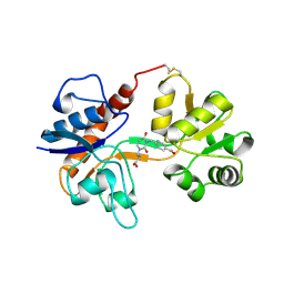
 | | Engineering Calcium-binding site into Cytochrome c Peroxidase (CcP) | | 分子名称: | Cytochrome c Peroxidase, POTASSIUM ION, PROTOPORPHYRIN IX CONTAINING FE | | 著者 | Bonagura, C.A, Bhaskar, B, Sundaramoorthy, M, Poulos, T.L. | | 登録日 | 2002-01-09 | | 公開日 | 2002-01-23 | | 最終更新日 | 2024-04-03 | | 実験手法 | X-RAY DIFFRACTION (2 Å) | | 主引用文献 | Conversion of an engineered potassium-binding site into a calcium-selective site in cytochrome c peroxidase.
J.Biol.Chem., 274, 1999
|
|
3GLF
 | |
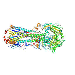
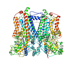
2QCU
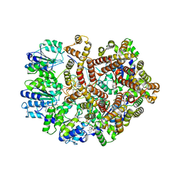
 | | Crystal structure of Glycerol-3-phosphate Dehydrogenase from Escherichia coli | | 分子名称: | 1,2-ETHANEDIOL, Aerobic glycerol-3-phosphate dehydrogenase, FLAVIN-ADENINE DINUCLEOTIDE, ... | | 著者 | Yeh, J.I, Chinte, U, Du, S. | | 登録日 | 2007-06-19 | | 公開日 | 2008-04-15 | | 最終更新日 | 2024-02-21 | | 実験手法 | X-RAY DIFFRACTION (1.75 Å) | | 主引用文献 | Structure of glycerol-3-phosphate dehydrogenase, an essential monotopic membrane enzyme involved in respiration and metabolism.
Proc.Natl.Acad.Sci.Usa, 105, 2008
|
|
3GVD
 | | Crystal Structure of Serine Acetyltransferase CysE from Yersinia pestis | | 分子名称: | 1-METHOXY-2-[2-(2-METHOXY-ETHOXY]-ETHANE, ACETIC ACID, CYSTEINE, ... | | 著者 | Kim, Y, Zhou, M, Peterson, S, Anderson, W.F, Joachimiak, A, Center for Structural Genomics of Infectious Diseases (CSGID) | | 登録日 | 2009-03-30 | | 公開日 | 2009-05-12 | | 最終更新日 | 2023-09-06 | | 実験手法 | X-RAY DIFFRACTION (2.4 Å) | | 主引用文献 | Crystal Structure of Serine Acetyltransferase CysE from Yersinia pestis
To be Published
|
|
3H06
 | |
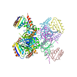
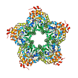
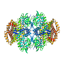
2QGA
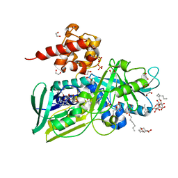
 | | Plasmodium vivax adenylosuccinate lyase Pv003765 with AMP bound | | 分子名称: | ADENOSINE MONOPHOSPHATE, Adenylosuccinate lyase, CALCIUM ION, ... | | 著者 | Lunin, V.V, Wernimont, A.K, Lew, J, Kozieradzki, I, Bochkarev, A, Arrowsmith, C.H, Sundstrom, M, Weigelt, J, Edwards, A.E, Hui, R, Hills, T, Altamentova, S, Structural Genomics Consortium (SGC) | | 登録日 | 2007-06-28 | | 公開日 | 2007-07-24 | | 最終更新日 | 2023-08-30 | | 実験手法 | X-RAY DIFFRACTION (2.01 Å) | | 主引用文献 | Plasmodium vivax adenylosuccinate lyase Pv003765 with AMP bound
To be Published
|
|
2QJI
 | | M. jannaschii ADH synthase complexed with dihydroxyacetone phosphate and glycerol | | 分子名称: | 1,3-DIHYDROXYACETONEPHOSPHATE, GLYCEROL, Putative aldolase MJ0400 | | 著者 | Ealick, S.E, Morar, M. | | 登録日 | 2007-07-07 | | 公開日 | 2007-10-30 | | 最終更新日 | 2011-07-13 | | 実験手法 | X-RAY DIFFRACTION (2.8 Å) | | 主引用文献 | Structure of 2-amino-3,7-dideoxy-D-threo-hept-6-ulosonic acid synthase, a catalyst in the archaeal pathway for the biosynthesis of aromatic amino acids.
Biochemistry, 46, 2007
|
|
1KG9
 | | Structure of a "mock-trapped" early-M intermediate of bacteriorhosopsin | | 分子名称: | 1-[2,6,10.14-TETRAMETHYL-HEXADECAN-16-YL]-2-[2,10,14-TRIMETHYLHEXADECAN-16-YL]GLYCEROL, RETINAL, bacteriorhodopsin | | 著者 | Facciotti, M.T, Rouhani, S, Burkard, F.T, Betancourt, F.M, Downing, K.H, Rose, R.B, McDermott, G, Glaeser, R.M. | | 登録日 | 2001-11-26 | | 公開日 | 2001-12-05 | | 最終更新日 | 2023-08-16 | | 実験手法 | X-RAY DIFFRACTION (1.81 Å) | | 主引用文献 | Structure of an early intermediate in the M-state phase of the bacteriorhodopsin photocycle.
Biophys.J., 81, 2001
|
|
1KRI
 | |
3C3Z
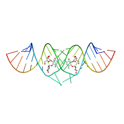 | | Crystal structure of HIV-1 subtype F DIS extended duplex RNA bound to ribostamycin | | 分子名称: | HIV-1 subtype F genomic RNA, RIBOSTAMYCIN | | 著者 | Freisz, S, Lang, K, Micura, R, Dumas, P, Ennifar, E. | | 登録日 | 2008-01-29 | | 公開日 | 2008-05-06 | | 最終更新日 | 2024-03-13 | | 実験手法 | X-RAY DIFFRACTION (1.5 Å) | | 主引用文献 | Binding of aminoglycoside antibiotics to the duplex form of the HIV-1 genomic RNA dimerization initiation site.
Angew.Chem.Int.Ed.Engl., 47, 2008
|
|
3U33
 | |
2QJK
 | | Crystal Structure Analysis of mutant rhodobacter sphaeroides bc1 with stigmatellin and antimycin | | 分子名称: | (1R)-2-{[(R)-(2-AMINOETHOXY)(HYDROXY)PHOSPHORYL]OXY}-1-[(DODECANOYLOXY)METHYL]ETHYL (9Z)-OCTADEC-9-ENOATE, (2R,3S,6S,7R,8R)-3-{[3-(FORMYLAMINO)-2-HYDROXYBENZOYL]AMINO}-8-HEXYL-2,6-DIMETHYL-4,9-DIOXO-1,5-DIOXONAN-7-YL (2S)-2-METHYLBUTANOATE, 2-O-octyl-beta-D-glucopyranose, ... | | 著者 | Esser, L. | | 登録日 | 2007-07-07 | | 公開日 | 2007-12-25 | | 最終更新日 | 2023-08-30 | | 実験手法 | X-RAY DIFFRACTION (3.1 Å) | | 主引用文献 | Inhibitor-complexed structures of the cytochrome bc1 from the photosynthetic bacterium Rhodobacter sphaeroides.
J.Biol.Chem., 283, 2008
|
|


