3ULH
 
 | |
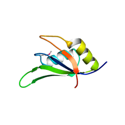
4A8X
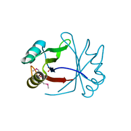
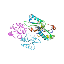
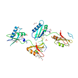
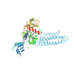
 | | Structure of the core ASAP complex | | 分子名称: | HISTONE DEACETYLASE COMPLEX SUBUNIT SAP18, HOOK-LIKE, ISOFORM A, ... | | 著者 | Murachelli, A.G, Ebert, J, Basquin, C, Le Hir, H, Conti, E. | | 登録日 | 2011-11-21 | | 公開日 | 2012-03-07 | | 最終更新日 | 2023-12-20 | | 実験手法 | X-RAY DIFFRACTION (1.9 Å) | | 主引用文献 | The Structure of the Asap Core Complex Reveals the Existence of a Pinin-Containing Psap Complex
Nat.Struct.Mol.Biol., 19, 2012
|
|
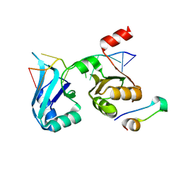
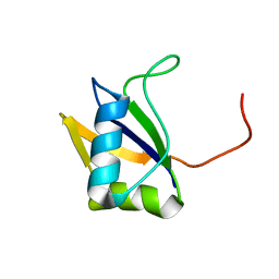
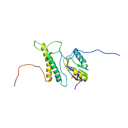
2RS8
 | | Solution structure of the N-terminal RNA recognition motif of NonO | | 分子名称: | Non-POU domain-containing octamer-binding protein | | 著者 | Nagata, T, Muto, Y, Inoue, M, Kigawa, T, Terada, T, Shirouzu, M, Yokoyama, S, RIKEN Structural Genomics/Proteomics Initiative (RSGI) | | 登録日 | 2011-11-29 | | 公開日 | 2012-12-19 | | 最終更新日 | 2024-05-15 | | 実験手法 | SOLUTION NMR | | 主引用文献 | Solution structure of the N-terminal RNA recognition motif of NonO
To be Published
|
|
3UWT
 | |
3V4M
 | |
3VAG
 | | Structure of U2AF65 variant with BrU3C2 DNA | | 分子名称: | 1,4-DIETHYLENE DIOXIDE, DNA 5'-D(*U*CP*(BRU)P*UP*UP*UP*U)-3', GLYCEROL, ... | | 著者 | Jenkins, J.L, Kielkopf, C.L. | | 登録日 | 2011-12-29 | | 公開日 | 2013-02-13 | | 最終更新日 | 2024-02-28 | | 実験手法 | X-RAY DIFFRACTION (2.19 Å) | | 主引用文献 | U2AF65 adapts to diverse pre-mRNA splice sites through conformational selection of specific and promiscuous RNA recognition motifs.
Nucleic Acids Res., 41, 2013
|
|
3VAH
 | | Structure of U2AF65 variant with BrU3C4 DNA | | 分子名称: | 1,4-DIETHYLENE DIOXIDE, DNA 5'-D(P*UP*UP*(BRU)P*CP*UP*UP*U)-3', GLYCEROL, ... | | 著者 | Jenkins, J.L, Kielkopf, C.L. | | 登録日 | 2011-12-29 | | 公開日 | 2013-02-13 | | 最終更新日 | 2024-02-28 | | 実験手法 | X-RAY DIFFRACTION (2.5 Å) | | 主引用文献 | U2AF65 adapts to diverse pre-mRNA splice sites through conformational selection of specific and promiscuous RNA recognition motifs.
Nucleic Acids Res., 41, 2013
|
|
3VAL
 | | Structure of U2AF65 variant with BrU5C1 DNA | | 分子名称: | 1,4-DIETHYLENE DIOXIDE, DNA (5'-D(*C*UP*UP*UP*(BRU)P*UP*U)-3'), SULFATE ION, ... | | 著者 | Jenkins, J.L, Frato, K.H, Kielkopf, C.L. | | 登録日 | 2011-12-29 | | 公開日 | 2013-02-13 | | 最終更新日 | 2023-09-13 | | 実験手法 | X-RAY DIFFRACTION (2.5 Å) | | 主引用文献 | U2AF65 adapts to diverse pre-mRNA splice sites through conformational selection of specific and promiscuous RNA recognition motifs.
Nucleic Acids Res., 41, 2013
|
|
3VAK
 | | Structure of U2AF65 variant with BrU5 DNA | | 分子名称: | 1,4-DIETHYLENE DIOXIDE, DNA (5'-D(*UP*UP*UP*UP*(BRU)P*UP*U)-3'), GLYCEROL, ... | | 著者 | Jenkins, J.L, Kielkopf, C.L. | | 登録日 | 2011-12-29 | | 公開日 | 2013-02-13 | | 最終更新日 | 2017-08-16 | | 実験手法 | X-RAY DIFFRACTION (2.17 Å) | | 主引用文献 | U2AF65 adapts to diverse pre-mRNA splice sites through conformational selection of specific and promiscuous RNA recognition motifs.
Nucleic Acids Res., 41, 2013
|
|
3VAF
 | | Structure of U2AF65 variant with BrU3 DNA | | 分子名称: | 1,4-DIETHYLENE DIOXIDE, DNA 5'-D(*UP*UP*(BRU)P*(BRU)P*UP*UP*U)-3', GLYCEROL, ... | | 著者 | Jenkins, J.L, Kielkopf, C.L. | | 登録日 | 2011-12-29 | | 公開日 | 2013-02-13 | | 最終更新日 | 2023-09-13 | | 実験手法 | X-RAY DIFFRACTION (2.49 Å) | | 主引用文献 | U2AF65 adapts to diverse pre-mRNA splice sites through conformational selection of specific and promiscuous RNA recognition motifs.
Nucleic Acids Res., 41, 2013
|
|
3VAM
 | | Structure of U2AF65 variant with BrU5C2 DNA | | 分子名称: | 1,4-DIETHYLENE DIOXIDE, DNA (5'-D(*UP*CP*UP*UP*(BRU)P*UP*U)-3'), GLYCEROL, ... | | 著者 | Jenkins, J.L, Kielkopf, C.L. | | 登録日 | 2011-12-29 | | 公開日 | 2013-02-13 | | 最終更新日 | 2024-02-28 | | 実験手法 | X-RAY DIFFRACTION (2.4 Å) | | 主引用文献 | U2AF65 adapts to diverse pre-mRNA splice sites through conformational selection of specific and promiscuous RNA recognition motifs.
Nucleic Acids Res., 41, 2013
|
|
3VAJ
 | | Structure of U2AF65 variant with BrU5C6 DNA | | 分子名称: | 1,4-DIETHYLENE DIOXIDE, DNA (5'-D(*UP*UP*UP*UP*(BRU)P*CP*U)-3'), GLYCEROL, ... | | 著者 | Jenkins, J.L, Kielkopf, C.L. | | 登録日 | 2011-12-29 | | 公開日 | 2013-02-13 | | 最終更新日 | 2024-02-28 | | 実験手法 | X-RAY DIFFRACTION (1.9 Å) | | 主引用文献 | U2AF65 adapts to diverse pre-mRNA splice sites through conformational selection of specific and promiscuous RNA recognition motifs.
Nucleic Acids Res., 41, 2013
|
|
3VAI
 | | Structure of U2AF65 variant with BrU3C5 DNA | | 分子名称: | 1,4-DIETHYLENE DIOXIDE, DNA 5'-D(*UP*UP*(BRU)P*UP*CP*UP*U)-3', GLYCEROL, ... | | 著者 | Jenkins, J.L, Frato, K.H, Kielkopf, C.L. | | 登録日 | 2011-12-29 | | 公開日 | 2013-02-13 | | 最終更新日 | 2024-02-28 | | 実験手法 | X-RAY DIFFRACTION (2.2 Å) | | 主引用文献 | U2AF65 adapts to diverse pre-mRNA splice sites through conformational selection of specific and promiscuous RNA recognition motifs.
Nucleic Acids Res., 41, 2013
|
|
3VF0
 | | Raver1 in complex with metavinculin L954 deletion mutant | | 分子名称: | 4-(2-HYDROXYETHYL)-1-PIPERAZINE ETHANESULFONIC ACID, GLYCEROL, Ribonucleoprotein PTB-binding 1, ... | | 著者 | Lee, J.H, Vonrhein, C, Bricogne, G, Izard, T. | | 登録日 | 2012-01-09 | | 公開日 | 2012-07-25 | | 最終更新日 | 2012-09-19 | | 実験手法 | X-RAY DIFFRACTION (2.54 Å) | | 主引用文献 | The Metavinculin Tail Domain Directs Constitutive Interactions with Raver1 and vinculin RNA.
J.Mol.Biol., 422, 2012
|
|
4ED5
 | | Crystal structure of the two N-terminal RRM domains of HuR complexed with RNA | | 分子名称: | 1,2-ETHANEDIOL, 1-METHOXY-2-(2-METHOXYETHOXY)ETHANE, 5'-R(*A*UP*UP*UP*UP*UP*AP*UP*UP*UP*U)-3', ... | | 著者 | Wang, H, Zeng, F, Liu, Q, Niu, L, Teng, M, Li, X. | | 登録日 | 2012-03-27 | | 公開日 | 2012-05-23 | | 最終更新日 | 2024-03-20 | | 実験手法 | X-RAY DIFFRACTION (2 Å) | | 主引用文献 | The structure of the ARE-binding domains of Hu antigen R (HuR) undergoes conformational changes during RNA binding.
Acta Crystallogr.,Sect.D, 69, 2013
|
|
4EGL
 | | Crystal structure of two tandem RNA recognition motifs of Human antigen R | | 分子名称: | ELAV-like protein 1, GLYCEROL, SULFATE ION | | 著者 | Wang, H, Zeng, F, Liu, H, Teng, M, Li, X. | | 登録日 | 2012-03-31 | | 公開日 | 2012-05-30 | | 最終更新日 | 2023-11-08 | | 実験手法 | X-RAY DIFFRACTION (2.9 Å) | | 主引用文献 | Crystal structure of two tandem RNA recognition motifs of Human antigen R
To be Published
|
|
4F02
 | |
4F26
 | |
4F25
 | |
4FXW
 | | Structure of phosphorylated SF1 complex with U2AF65-UHM domain | | 分子名称: | SULFATE ION, Splicing factor 1, Splicing factor U2AF 65 kDa subunit | | 著者 | Wang, W, Bauer, W.J, Wedekind, J.E, Kielkopf, C.L. | | 登録日 | 2012-07-03 | | 公開日 | 2013-01-16 | | 最終更新日 | 2017-11-15 | | 実験手法 | X-RAY DIFFRACTION (2.29 Å) | | 主引用文献 | Structure of Phosphorylated SF1 Bound to U2AF(65) in an Essential Splicing Factor Complex.
Structure, 21, 2013
|
|
4FXV
 | |
2LXI
 | |
2LXU
 | | Solution NMR Structure of the eukaryotic RNA recognition motif, RRM1, from the heterogeneous nuclear ribonucleoprotein H from Homo sapiens, Northeast Structural Genomics Consortium (NESG) Target HR8614A | | 分子名称: | Heterogeneous nuclear ribonucleoprotein H | | 著者 | Ramelot, T.A, Yang, Y, Pederson, K, Shastry, R, Kohan, E, Janjua, H, Xiao, R, Acton, T.B, Everett, J.K, Prestegard, J.H, Montelione, G.T, Kennedy, M.A, Northeast Structural Genomics Consortium (NESG) | | 登録日 | 2012-08-31 | | 公開日 | 2012-10-31 | | 最終更新日 | 2024-05-15 | | 実験手法 | SOLUTION NMR | | 主引用文献 | Solution NMR Structure of the eukaryotic RNA recognition motif, RRM1, from the heterogeneous nuclear ribonucleoprotein H from Homo sapiens, Northeast Structural Genomics Consortium (NESG) Target HR8614A
To be Published
|
|
2LYV
 | |
2M0G
 | | Structure, phosphorylation and U2AF65 binding of the Nterminal Domain of splicing factor 1 during 3 splice site Recognition | | 分子名称: | Splicing factor 1, Splicing factor U2AF 65 kDa subunit | | 著者 | Madl, T, Sattler, M, Zhang, Y, Bagdiul, I, Kern, T, Kang, H, Zou, P, Maeusbacher, N, Sieber, S.A, Kraemer, A. | | 登録日 | 2012-10-25 | | 公開日 | 2013-01-30 | | 最終更新日 | 2024-05-01 | | 実験手法 | SOLUTION NMR | | 主引用文献 | Structure, phosphorylation and U2AF65 binding of the N-terminal domain of splicing factor 1 during 3'-splice site recognition.
Nucleic Acids Res., 41, 2013
|
|


