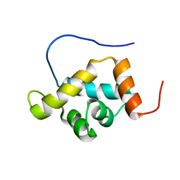
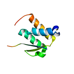
2DO5
 
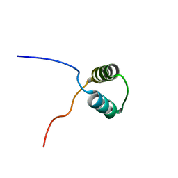 | | Solution structure of the SAP domain of human splicing factor 3B subunit 2 | | 分子名称: | Splicing factor 3B subunit 2 | | 著者 | Suzuki, S, Muto, Y, Inoue, M, Kigawa, T, Terada, T, Shirouzu, M, Yokoyama, S, RIKEN Structural Genomics/Proteomics Initiative (RSGI) | | 登録日 | 2006-04-27 | | 公開日 | 2007-04-17 | | 最終更新日 | 2024-05-29 | | 実験手法 | SOLUTION NMR | | 主引用文献 | Solution structure of the SAP domain of human splicing factor 3B subunit 2
To be Published
|
|
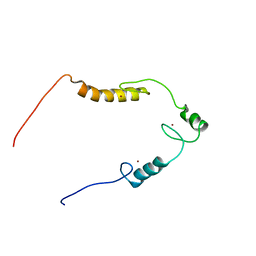
2DN5
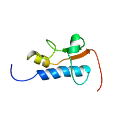 | | Solution Structure of RSGI RUH-057, a GTF2I domain in human cDNA | | 分子名称: | General transcription factor II-I repeat domain-containing protein 1 | | 著者 | Doi-Katayama, Y, Hirota, H, Hayashi, F, Yokoyama, S, RIKEN Structural Genomics/Proteomics Initiative (RSGI) | | 登録日 | 2006-04-25 | | 公開日 | 2006-10-25 | | 最終更新日 | 2024-05-29 | | 実験手法 | SOLUTION NMR | | 主引用文献 | Solution Structure of RSGI RUH-057, a GTF2I domain in human cDNA
To be Published
|
|
2ND1
 | |
2DHS
 | | Solution Structure of Nucleic Acid Binding Protein CUGBP1ab and its Binding Study with DNA and RNA | | 分子名称: | CUG triplet repeat RNA-binding protein 1 | | 著者 | Xia, Y.L, Jun, K.Y, Zhu, Q, Han, X.G, Zhang, H, Timchenko, L, Swanson, M, Gao, X.L. | | 登録日 | 2006-03-25 | | 公開日 | 2007-04-24 | | 最終更新日 | 2024-05-01 | | 実験手法 | SOLUTION NMR | | 主引用文献 | Solution Structure of Nucleic Acid Binding Protein CUGBP1ab and its Binding Study with DNA and RNA
To be Published
|
|
2DW4
 | |
2FBV
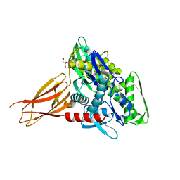
 | | WRN exonuclease, Mn complex | | 分子名称: | MANGANESE (II) ION, Werner syndrome helicase | | 著者 | Perry, J.J. | | 登録日 | 2005-12-10 | | 公開日 | 2006-04-25 | | 最終更新日 | 2023-08-30 | | 実験手法 | X-RAY DIFFRACTION (2.4 Å) | | 主引用文献 | WRN exonuclease structure and molecular mechanism imply an editing role in DNA end processing.
Nat.Struct.Mol.Biol., 13, 2006
|
|
2FBT
 | | WRN exonuclease | | 分子名称: | ACETIC ACID, Werner syndrome helicase | | 著者 | Perry, J.J. | | 登録日 | 2005-12-10 | | 公開日 | 2006-04-25 | | 最終更新日 | 2024-04-03 | | 実験手法 | X-RAY DIFFRACTION (2.05 Å) | | 主引用文献 | WRN exonuclease structure and molecular mechanism imply an editing role in DNA end processing.
Nat.Struct.Mol.Biol., 13, 2006
|
|
2CR9
 | |
2CSF
 | |
2NML
 | | Crystal structure of HEF2/ERH at 1.55 A resolution | | 分子名称: | Enhancer of rudimentary homolog | | 著者 | Jin, T.C, Guo, F, Serebriiskii, I.G, Howard, A.J, Zhang, Y.Z. | | 登録日 | 2006-10-21 | | 公開日 | 2006-10-31 | | 最終更新日 | 2023-12-27 | | 実験手法 | X-RAY DIFFRACTION (1.55 Å) | | 主引用文献 | A 1.55 A resolution X-ray crystal structure of HEF2/ERH and insights into its transcriptional and cell-cycle interaction networks.
Proteins, 68, 2007
|
|
2CSH
 | | Solution structure of tandem repeat of the zf-C2H2 domains of human zinc finger protein 297B | | 分子名称: | ZINC ION, Zinc finger protein 297B | | 著者 | Inoue, K, Saitoh, K, Hayashi, F, Kigawa, T, Yokoyama, S, RIKEN Structural Genomics/Proteomics Initiative (RSGI) | | 登録日 | 2005-05-21 | | 公開日 | 2005-11-21 | | 最終更新日 | 2024-05-29 | | 実験手法 | SOLUTION NMR | | 主引用文献 | Solution structure of tandem repeat of the zf-C2H2 domains of human zinc finger protein 297B
To be Published
|
|
2ND0
 | |
2N80
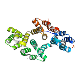
 | | p75NTR DD:RhoGDI | | 分子名称: | Rho GDP-dissociation inhibitor 1, Tumor necrosis factor receptor superfamily member 16 | | 著者 | Lin, Z, Ibanez, C.F. | | 登録日 | 2015-09-30 | | 公開日 | 2015-12-23 | | 最終更新日 | 2024-05-01 | | 実験手法 | SOLUTION NMR | | 主引用文献 | Structural basis of death domain signaling in the p75 neurotrophin receptor
Elife, 4, 2015
|
|
2NCZ
 | |
2QZV
 | | Draft Crystal Structure of the Vault Shell at 9 Angstroms Resolution | | 分子名称: | Major vault protein | | 著者 | Anderson, D.H, Kickhoefer, V.A, Sievers, S.A, Rome, L.H, Eisenberg, D. | | 登録日 | 2007-08-17 | | 公開日 | 2007-12-04 | | 最終更新日 | 2024-04-03 | | 実験手法 | X-RAY DIFFRACTION (9 Å) | | 主引用文献 | Draft crystal structure of the vault shell at 9-A resolution.
Plos Biol., 5, 2007
|
|
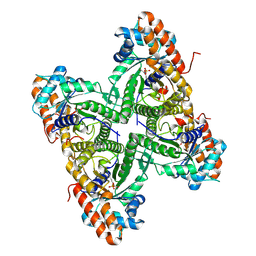
2G76
 | | Crystal structure of human 3-phosphoglycerate dehydrogenase | | 分子名称: | D-3-phosphoglycerate dehydrogenase, D-MALATE, NICOTINAMIDE-ADENINE-DINUCLEOTIDE | | 著者 | Turnbull, A.P, Salah, E, Savitsky, P, Gileadi, O, von Delft, F, Edwards, A, Arrowsmith, C, Weigelt, J, Sundstrom, M, Oppermann, U, Structural Genomics Consortium (SGC) | | 登録日 | 2006-02-27 | | 公開日 | 2006-03-21 | | 最終更新日 | 2023-08-30 | | 実験手法 | X-RAY DIFFRACTION (1.7 Å) | | 主引用文献 | Crystal structure of human 3-phosphoglycerate dehydrogenase
To be Published
|
|
2FMM
 | | Crystal Structure of EMSY-HP1 complex | | 分子名称: | Chromobox protein homolog 1, Protein EMSY, SULFATE ION | | 著者 | Huang, Y. | | 登録日 | 2006-01-09 | | 公開日 | 2006-05-23 | | 最終更新日 | 2024-02-14 | | 実験手法 | X-RAY DIFFRACTION (1.8 Å) | | 主引用文献 | Crystal structure of the HP1-EMSY complex reveals an unusual mode of HP1 binding.
Structure, 14, 2006
|
|
2QQS
 | |
2QUT
 | |
2RAN
 | | RAT ANNEXIN V CRYSTAL STRUCTURE: CA2+-INDUCED CONFORMATIONAL CHANGES | | 分子名称: | ANNEXIN V, CALCIUM ION, SULFATE ION | | 著者 | Concha, N.O, Head, J.F, Kaetzel, M.A, Dedman, J.R, Seaton, B.A. | | 登録日 | 1994-09-01 | | 公開日 | 1994-11-30 | | 最終更新日 | 2024-02-21 | | 実験手法 | X-RAY DIFFRACTION (1.89 Å) | | 主引用文献 | Rat annexin V crystal structure: Ca(2+)-induced conformational changes.
Science, 261, 1993
|
|
2G9A
 | |
6EIN
 | | Crystal structure of KDM5B in complex with S49365a. | | 分子名称: | 1,2-ETHANEDIOL, 8-[4-[2-(4-propanoylpiperazin-1-yl)ethyl]pyrazol-1-yl]-3~{H}-pyrido[3,4-d]pyrimidin-4-one, Lysine-specific demethylase 5B,Lysine-specific demethylase 5B, ... | | 著者 | Srikannathasan, V, Newman, J.A, Szykowska, A, Wright, M, Ruda, G.F, Vazquez-Rodriguez, S.A, Kupinska, K, Strain-Damerell, C, Burgess-Brown, N.A, Arrowsmith, C.H, Edwards, A, Bountra, C, Oppermann, U, Huber, K, von Delft, F. | | 登録日 | 2017-09-19 | | 公開日 | 2018-05-02 | | 最終更新日 | 2024-01-17 | | 実験手法 | X-RAY DIFFRACTION (2.11 Å) | | 主引用文献 | Crystal structure of KDM5B in complex with S49365a.
To Be Published
|
|
6E93
 | | Crystal Structure of ZBTB38 C-terminal Zinc Fingers 6-9 in complex with methylated DNA | | 分子名称: | DNA (5'-D(*GP*CP*AP*CP*TP*CP*AP*TP*(DCM)P*GP*GP*(DCM)P*GP*CP*AP*GP*AP*C)-3'), DNA (5'-D(*GP*TP*CP*TP*GP*(DCM)P*GP*CP*(DCM)P*GP*AP*TP*GP*AP*GP*TP*GP*C)-3'), ZINC ION, ... | | 著者 | Hudson, N.O, Whitby, F.G, Buck-Koehntop, B.A. | | 登録日 | 2018-07-31 | | 公開日 | 2018-11-07 | | 最終更新日 | 2024-03-13 | | 実験手法 | X-RAY DIFFRACTION (1.747 Å) | | 主引用文献 | Structural insights into methylated DNA recognition by the C-terminal zinc fingers of the DNA reader protein ZBTB38.
J. Biol. Chem., 293, 2018
|
|
6EOC
 | | Crystal structure of AMPylated GRP78 in apo form (Crystal form 2) | | 分子名称: | 78 kDa glucose-regulated protein, CITRATE ANION, SULFATE ION | | 著者 | Yan, Y, Preissler, S, Ron, D, Read, R.J. | | 登録日 | 2017-10-09 | | 公開日 | 2017-11-01 | | 最終更新日 | 2024-01-17 | | 実験手法 | X-RAY DIFFRACTION (1.67 Å) | | 主引用文献 | AMPylation targets the rate-limiting step of BiP's ATPase cycle for its functional inactivation.
Elife, 6, 2017
|
|
6EHT
 | | Modulation of PCNA sliding surface by p15PAF suggests a suppressive mechanism for cisplatin-induced DNA lesion bypass by pol eta holoenzyme | | 分子名称: | DNA (5'-D(P*AP*TP*AP*CP*GP*AP*TP*GP*GP*G)-3'), DNA (5'-D(P*CP*CP*CP*AP*TP*CP*GP*TP*AP*T)-3'), PCNA-associated factor, ... | | 著者 | De March, M, Barrera-Vilarmau, S, Mentegari, E, Merino, N, Bressan, E, Maga, G, Crehuet, R, Onesti, S, Blanco, F.J, De Biasio, A. | | 登録日 | 2017-09-15 | | 公開日 | 2018-08-08 | | 最終更新日 | 2024-01-17 | | 実験手法 | X-RAY DIFFRACTION (3.2 Å) | | 主引用文献 | p15PAF binding to PCNA modulates the DNA sliding surface.
Nucleic Acids Res., 46, 2018
|
|


