2CST
 
 | |
2CSU
 | |
2CSV
 | |
2CSW
 | |
2CSX
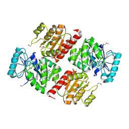
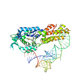
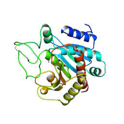
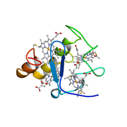
 | | Crystal structure of Aquifex aeolicus methionyl-tRNA synthetase complexed with tRNA(Met) | | 分子名称: | Methionyl-tRNA synthetase, RNA (75-MER) | | 著者 | Nakanishi, K, Ogiso, Y, Nakama, T, Fukai, S, Nureki, O, RIKEN Structural Genomics/Proteomics Initiative (RSGI) | | 登録日 | 2005-05-23 | | 公開日 | 2005-09-20 | | 最終更新日 | 2024-03-13 | | 実験手法 | X-RAY DIFFRACTION (2.7 Å) | | 主引用文献 | Structural basis for anticodon recognition by methionyl-tRNA synthetase.
Nat.Struct.Mol.Biol., 12, 2005
|
|
2CSY
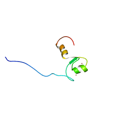
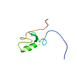
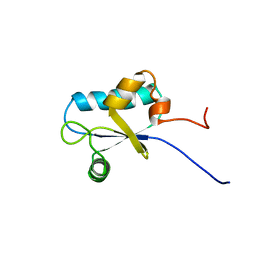
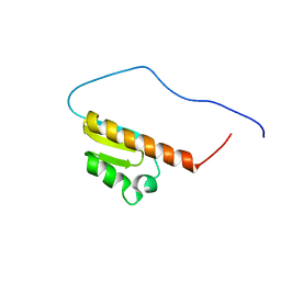
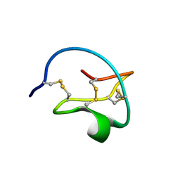
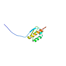
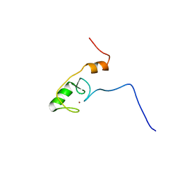 | | Solution structure of the RING domain of the Zinc finger protein 183-like 1 | | 分子名称: | ZINC ION, Zinc finger protein 183-like 1 | | 著者 | Miyamoto, K, Sato, M, Tomizawa, T, Saito, K, Koshiba, S, Inoue, M, Kigawa, T, Yokoyama, S, RIKEN Structural Genomics/Proteomics Initiative (RSGI) | | 登録日 | 2005-05-23 | | 公開日 | 2005-11-23 | | 最終更新日 | 2024-05-29 | | 実験手法 | SOLUTION NMR | | 主引用文献 | Solution structure of the RING domain of the Zinc finger protein 183-like 1
To be Published
|
|
2CSZ
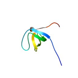
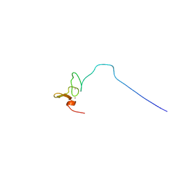 | | Solution Structure of the RING domain of the Synaptotagmin-like protein 4 | | 分子名称: | Synaptotagmin-like protein 4, ZINC ION | | 著者 | Miyamoto, K, Sato, M, Koshiba, S, Inoue, M, Kigawa, T, Yokoyama, S, RIKEN Structural Genomics/Proteomics Initiative (RSGI) | | 登録日 | 2005-05-23 | | 公開日 | 2005-11-23 | | 最終更新日 | 2024-05-29 | | 実験手法 | SOLUTION NMR | | 主引用文献 | Solution Structure of the RING domain of the Synaptotagmin-like protein 4
To be Published
|
|
2CT0
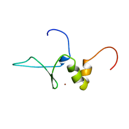
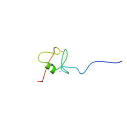 | | Solution structure of the RING domain of the Non-SMC element 1 protein | | 分子名称: | Non-SMC element 1 homolog, ZINC ION | | 著者 | Miyamoto, K, Sato, M, Koshiba, S, Inoue, M, Kigawa, T, Yokoyama, S, RIKEN Structural Genomics/Proteomics Initiative (RSGI) | | 登録日 | 2005-05-23 | | 公開日 | 2005-11-23 | | 最終更新日 | 2024-05-29 | | 実験手法 | SOLUTION NMR | | 主引用文献 | Solution structure of the RING domain of the Non-SMC element 1 protein
To be Published
|
|
2CT1
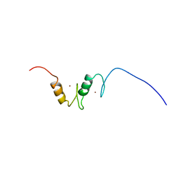 | | Solution Structure of the zinc finger domain of Transcriptional repressor CTCF protein | | 分子名称: | Transcriptional repressor CTCF, ZINC ION | | 著者 | Miyamoto, K, Koshiba, S, Inoue, M, Kigawa, T, Yokoyama, S, RIKEN Structural Genomics/Proteomics Initiative (RSGI) | | 登録日 | 2005-05-23 | | 公開日 | 2005-11-23 | | 最終更新日 | 2024-05-29 | | 実験手法 | SOLUTION NMR | | 主引用文献 | Solution Structure of the zinc finger domain of Transcriptional repressor CTCF protein
To be Published
|
|
2CT2
 | | Solution Structure of the RING domain of the Tripartite motif protein 32 | | 分子名称: | Tripartite motif protein 32, ZINC ION | | 著者 | Miyamoto, K, Tochio, N, Sato, M, Koshiba, S, Inoue, M, Kigawa, T, Yokoyama, S, RIKEN Structural Genomics/Proteomics Initiative (RSGI) | | 登録日 | 2005-05-23 | | 公開日 | 2005-11-23 | | 最終更新日 | 2024-05-29 | | 実験手法 | SOLUTION NMR | | 主引用文献 | Solution Structure of the RING domain of the Tripartite motif protein 32
To be Published
|
|
2CT3
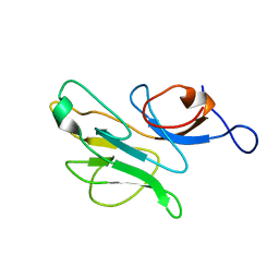
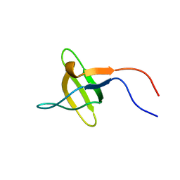
 | | Solution Structure of the SH3 domain of the Vinexin protein | | 分子名称: | Vinexin | | 著者 | Miyamoto, K, Tomizawa, T, Koshiba, S, Inoue, M, Kigawa, T, Yokoyama, S, RIKEN Structural Genomics/Proteomics Initiative (RSGI) | | 登録日 | 2005-05-23 | | 公開日 | 2005-11-23 | | 最終更新日 | 2024-05-29 | | 実験手法 | SOLUTION NMR | | 主引用文献 | Solution Structure of the SH3 domain of the Vinexin protein
To be Published
|
|
2CT4
 | | Solution Strutcure of the SH3 domain of the Cdc42-interacting protein 4 | | 分子名称: | Cdc42-interacting protein 4 | | 著者 | Miyamoto, K, Tomizawa, T, Koshiba, S, Inoue, M, Kigawa, T, Yokoyama, S, RIKEN Structural Genomics/Proteomics Initiative (RSGI) | | 登録日 | 2005-05-23 | | 公開日 | 2005-11-23 | | 最終更新日 | 2024-05-29 | | 実験手法 | SOLUTION NMR | | 主引用文献 | Solution Strutcure of the SH3 domain of the Cdc42-interacting protein 4
To be Published
|
|
2CT5
 | | Solution Structure of the zinc finger BED domain of the zinc finger BED domain containing protein 1 | | 分子名称: | ZINC ION, Zinc finger BED domain containing protein 1 | | 著者 | Miyamoto, K, Tomizawa, T, Koshiba, S, Inoue, M, Kigawa, T, Yokoyama, S, RIKEN Structural Genomics/Proteomics Initiative (RSGI) | | 登録日 | 2005-05-23 | | 公開日 | 2005-11-23 | | 最終更新日 | 2024-05-29 | | 実験手法 | SOLUTION NMR | | 主引用文献 | Solution Structure of the zinc finger BED domain of the zinc finger BED domain containing protein 1
To be Published
|
|
2CT6
 | | solution structure of the sh3 domain-binding glutamic acid-rich-like protein 2 | | 分子名称: | sh3 domain-binding glutamic acid-rich-like protein 2 | | 著者 | Miyamoto, K, Koshiba, S, Inoue, M, Kigawa, T, Yokoyama, S, RIKEN Structural Genomics/Proteomics Initiative (RSGI) | | 登録日 | 2005-05-23 | | 公開日 | 2005-11-23 | | 最終更新日 | 2024-05-29 | | 実験手法 | SOLUTION NMR | | 主引用文献 | solution structure of the sh3 domain-binding glutamic acid-rich-like protein 2
To be Published
|
|
2CT7
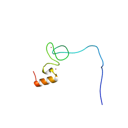 | | Solution Structure of the IBR domain of the RING finger protein 31 protein | | 分子名称: | RING finger protein 31, ZINC ION | | 著者 | Miyamoto, K, Koshiba, S, Tomizawa, T, Inoue, M, Kigawa, T, Yokoyama, S, RIKEN Structural Genomics/Proteomics Initiative (RSGI) | | 登録日 | 2005-05-23 | | 公開日 | 2005-11-23 | | 最終更新日 | 2024-05-29 | | 実験手法 | SOLUTION NMR | | 主引用文献 | Solution Structure of the IBR domain of the RING finger protein 31 protein
To be Published
|
|
2CT8
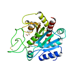
 | | Crystal structure of Aquifex aeolicus methionyl-tRNA synthetase complexed with tRNA(Met) and methionyl-adenylate anologue | | 分子名称: | 5'-O-[(L-METHIONYL)-SULPHAMOYL]ADENOSINE, Methionyl-tRNA synthetase, RNA (74-MER) | | 著者 | Nakanishi, K, Ogiso, Y, Nakama, T, Fukai, S, Nureki, O, RIKEN Structural Genomics/Proteomics Initiative (RSGI) | | 登録日 | 2005-05-23 | | 公開日 | 2005-09-20 | | 最終更新日 | 2024-03-13 | | 実験手法 | X-RAY DIFFRACTION (2.7 Å) | | 主引用文献 | Structural basis for anticodon recognition by methionyl-tRNA synthetase.
Nat.Struct.Mol.Biol., 12, 2005
|
|
2CT9
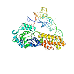
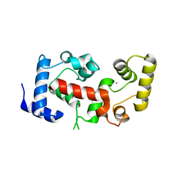 | | The crystal structure of calcineurin B homologous proein 1 (CHP1) | | 分子名称: | CALCIUM ION, Calcium-binding protein p22 | | 著者 | Naoe, Y, Arita, K, Hashimoto, H, Kanazawa, H, Sato, M, Shimizu, T. | | 登録日 | 2005-05-23 | | 公開日 | 2005-07-05 | | 最終更新日 | 2024-03-13 | | 実験手法 | X-RAY DIFFRACTION (2.2 Å) | | 主引用文献 | Structural characterization of calcineurin B homologous protein 1
J.Biol.Chem., 280, 2005
|
|
2CTB
 | | THE HIGH RESOLUTION CRYSTAL STRUCTURE OF THE COMPLEX BETWEEN CARBOXYPEPTIDASE A AND L-PHENYL LACTATE | | 分子名称: | CARBOXYPEPTIDASE A, ZINC ION | | 著者 | Teplyakov, A, Wilson, K.S, Orioli, P, Mangani, S. | | 登録日 | 1993-04-02 | | 公開日 | 1994-01-31 | | 最終更新日 | 2017-11-29 | | 実験手法 | X-RAY DIFFRACTION (1.5 Å) | | 主引用文献 | High-resolution structure of the complex between carboxypeptidase A and L-phenyl lactate.
Acta Crystallogr.,Sect.D, 49, 1993
|
|
2CTC
 | | THE HIGH RESOLUTION CRYSTAL STRUCTURE OF THE COMPLEX BETWEEN CARBOXYPEPTIDASE A AND L-PHENYL LACTATE | | 分子名称: | ALPHA-HYDROXY-BETA-PHENYL-PROPIONIC ACID, CARBOXYPEPTIDASE A, ZINC ION | | 著者 | Teplyakov, A, Wilson, K.S, Orioli, P, Mangani, S. | | 登録日 | 1993-04-02 | | 公開日 | 1994-01-31 | | 最終更新日 | 2017-11-29 | | 実験手法 | X-RAY DIFFRACTION (1.4 Å) | | 主引用文献 | High-resolution structure of the complex between carboxypeptidase A and L-phenyl lactate.
Acta Crystallogr.,Sect.D, 49, 1993
|
|
2CTD
 | | Solution structure of two zf-C2H2 domains from human Zinc finger protein 512 | | 分子名称: | ZINC ION, Zinc finger protein 512 | | 著者 | Tomizawa, T, Kigawa, T, Koshiba, S, Inoue, M, Yokoyama, S, RIKEN Structural Genomics/Proteomics Initiative (RSGI) | | 登録日 | 2005-05-24 | | 公開日 | 2005-11-24 | | 最終更新日 | 2024-05-29 | | 実験手法 | SOLUTION NMR | | 主引用文献 | Solution structure of two zf-C2H2 domains from human Zinc finger protein 512
To be Published
|
|
2CTE
 | | Solution structure of the 1st KH type I domain from human Vigilin | | 分子名称: | Vigilin | | 著者 | Tomizawa, T, Kigawa, T, Koshiba, S, Inoue, M, Yokoyama, S, RIKEN Structural Genomics/Proteomics Initiative (RSGI) | | 登録日 | 2005-05-24 | | 公開日 | 2005-11-24 | | 最終更新日 | 2024-05-29 | | 実験手法 | SOLUTION NMR | | 主引用文献 | Solution structure of the 1st KH type I domain from human Vigilin
To be Published
|
|
2CTF
 | | Solution structure of the 4th KH type I domain from human Vigilin | | 分子名称: | Vigilin | | 著者 | Tomizawa, T, Kigawa, T, Koshiba, S, Inoue, M, Yokoyama, S, RIKEN Structural Genomics/Proteomics Initiative (RSGI) | | 登録日 | 2005-05-24 | | 公開日 | 2005-11-24 | | 最終更新日 | 2024-05-29 | | 実験手法 | SOLUTION NMR | | 主引用文献 | Solution structure of the 4th KH type I domain from human Vigilin
To be Published
|
|
2CTH
 | | CYTOCHROME C3 FROM DESULFOVIBRIO VULGARIS HILDENBOROUGH | | 分子名称: | CYTOCHROME C3, PROTOPORPHYRIN IX CONTAINING FE | | 著者 | Simoes, P, Matias, P.M, Morais, J, Wilson, K, Dauter, Z, Carrondo, M.A. | | 登録日 | 1997-06-18 | | 公開日 | 1997-12-24 | | 最終更新日 | 2023-08-09 | | 実験手法 | X-RAY DIFFRACTION (1.67 Å) | | 主引用文献 | Refinement of the Three-Dimensional Structures of Cytochromes C3 from Desulfovibrio Vulgaris Hildenborough at 1.67 Angstrom Resolution and from Desulfovibrio Desulfuricans Atcc 27774 at 1.6 Angstrom Resolution
Inorg.Chim.Acta., 273, 1998
|
|
2CTI
 | |
2CTJ
 | | Solution structure of the 8th KH type I domain from human Vigilin | | 分子名称: | Vigilin | | 著者 | Tomizawa, T, Kigawa, T, Koshiba, S, Inoue, M, Yokoyama, S, RIKEN Structural Genomics/Proteomics Initiative (RSGI) | | 登録日 | 2005-05-24 | | 公開日 | 2005-11-24 | | 最終更新日 | 2024-05-29 | | 実験手法 | SOLUTION NMR | | 主引用文献 | Solution structure of the 8th KH type I domain from human Vigilin
To be Published
|
|


