1K9D
 
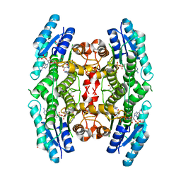
 | | The 1.7 A crystal structure of alpha-D-glucuronidase, a family-67 glycoside hydrolase from Bacillus stearothermophilus T-1 | | 分子名称: | GLYCEROL, alpha-D-glucuronidase | | 著者 | Golan, G, Shallom, D, Teplitsky, A, Zaide, G, Shulami, S, Baasov, T, Stojanoff, V, Thompson, A, Shoham, Y, Shoham, G. | | 登録日 | 2001-10-29 | | 公開日 | 2002-10-29 | | 最終更新日 | 2024-04-03 | | 実験手法 | X-RAY DIFFRACTION (1.7 Å) | | 主引用文献 | Crystal Structures of Geobacillus stearothermophilus {alpha}-Glucuronidase Complexed with Its Substrate and Products: MECHANISTIC IMPLICATIONS.
J.Biol.Chem., 279, 2004
|
|
6F7K
 | | Crystal structure of Dettilon tailspike protein (gp208) | | 分子名称: | 1,2-ETHANEDIOL, Tailspike, alpha-D-galactopyranose-(1-6)-beta-D-mannopyranose-(1-4)-alpha-L-rhamnopyranose-(1-3)-alpha-D-galactopyranose, ... | | 著者 | Roske, Y, Heinemann, U. | | 登録日 | 2017-12-11 | | 公開日 | 2018-12-19 | | 最終更新日 | 2024-01-17 | | 実験手法 | X-RAY DIFFRACTION (2.1 Å) | | 主引用文献 | Time-resolved DNA release from an O-antigen-specificSalmonellabacteriophage with a contractile tail.
J.Biol.Chem., 294, 2019
|
|
5QQY
 | | PanDDA analysis group deposition -- Crystal Structure of human ALAS2A in complex with Z2856434899 | | 分子名称: | 1-[(2,5-dihydrothiophen-3-yl)methyl]piperidin-4-ol, 5-aminolevulinate synthase, erythroid-specific, ... | | 著者 | Bezerra, G.A, Foster, W, Bailey, H, Shrestha, L, Krojer, T, Talon, R, Brandao-Neto, J, Douangamath, A, Nicola, B.B, von Delft, F, Arrowsmith, C.H, Edwards, A, Bountra, C, Brennan, P.E, Yue, W.W. | | 登録日 | 2019-05-22 | | 公開日 | 2019-08-07 | | 最終更新日 | 2024-03-06 | | 実験手法 | X-RAY DIFFRACTION (1.49 Å) | | 主引用文献 | PanDDA analysis group deposition
To Be Published
|
|
4Q1D
 | | Human dCK C4S-S74E mutant in complex with UDP and the inhibitor 9 {2-{[(1R)-1-{2-[3-(2-fluoroethoxy)-4-methoxyphenyl]-5-propyl-1,3-thiazol-4-yl}ethyl]sulfanyl}pyrimidine-4,6-diamine} | | 分子名称: | (R)-2-((1-(2-(3-(2-fluoroethoxy)-4-methoxyphenyl)-5-propylthiazol-4-yl)ethyl)thio)pyrimidine-4,6-diamine, Deoxycytidine kinase, URIDINE-5'-DIPHOSPHATE | | 著者 | Nomme, J, Lavie, A. | | 登録日 | 2014-04-03 | | 公開日 | 2015-02-18 | | 最終更新日 | 2023-09-20 | | 実験手法 | X-RAY DIFFRACTION (2 Å) | | 主引用文献 | Structure-guided development of deoxycytidine kinase inhibitors with nanomolar affinity and improved metabolic stability.
J.Med.Chem., 57, 2014
|
|
2CXE
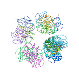 | | Crystal structure of octameric ribulose-1,5-bisphosphate carboxylase/oxygenase (Rubisco) from Pyrococcus horikoshii OT3 (form-2 crystal) | | 分子名称: | Ribulose bisphosphate carboxylase | | 著者 | Mizohata, E, Mishima, C, Akasaka, R, Uda, H, Terada, T, Shirouzu, M, Yokoyama, S, RIKEN Structural Genomics/Proteomics Initiative (RSGI) | | 登録日 | 2005-06-28 | | 公開日 | 2005-12-28 | | 最終更新日 | 2024-03-13 | | 実験手法 | X-RAY DIFFRACTION (3 Å) | | 主引用文献 | Crystal structure of octameric ribulose-1,5-bisphosphate carboxylase/oxygenase (Rubisco) from Pyrococcus horikoshii OT3 (form-2 crystal)
To be Published
|
|
1NXY
 | | Crystal Structure of the complex between M182T mutant of TEM-1 and a boronic acid inhibitor (SM2) | | 分子名称: | (1R)-1-(2-THIENYLACETYLAMINO)-1-(3-CARBOXYPHENYL)METHYLBORONIC ACID, Beta-lactamase TEM, POTASSIUM ION | | 著者 | Wang, X, Minasov, G, Blazquez, J, Caselli, E, Prati, F, Shoichet, B.K. | | 登録日 | 2003-02-11 | | 公開日 | 2003-08-26 | | 最終更新日 | 2024-10-30 | | 実験手法 | X-RAY DIFFRACTION (1.6 Å) | | 主引用文献 | Recognition and Resistance in TEM beta-lactamase
Biochemistry, 42, 2003
|
|
5QON
 | | PanDDA analysis group deposition -- Crystal Structure of DCP2 (NUDT20) in complex with FMOPL000446a | | 分子名称: | 1,2-ETHANEDIOL, ACETATE ION, DCP2 (NUDT20), ... | | 著者 | Nelson, E.R, Velupillai, S, Talon, R, Collins, P.M, Krojer, T, Wang, D, Brandao-Neto, J, Douangamath, A, Burgess-Brown, N, Arrowsmith, C.H, Bountra, C, Huber, K, von Delft, F. | | 登録日 | 2019-02-22 | | 公開日 | 2019-05-08 | | 最終更新日 | 2024-03-06 | | 実験手法 | X-RAY DIFFRACTION (1.8 Å) | | 主引用文献 | PanDDA analysis group deposition
To Be Published
|
|
1C1D
 | | L-PHENYLALANINE DEHYDROGENASE STRUCTURE IN TERNARY COMPLEX WITH NADH AND L-PHENYLALANINE | | 分子名称: | 1,4-DIHYDRONICOTINAMIDE ADENINE DINUCLEOTIDE, ISOPROPYL ALCOHOL, L-PHENYLALANINE DEHYDROGENASE, ... | | 著者 | Vanhooke, J.L, Thoden, J.B. | | 登録日 | 1999-07-21 | | 公開日 | 2000-08-30 | | 最終更新日 | 2024-02-07 | | 実験手法 | X-RAY DIFFRACTION (1.25 Å) | | 主引用文献 | Rhodococcus L-phenylalanine dehydrogenase: kinetics, mechanism, and structural basis for catalytic specificity.
Biochemistry, 39, 2000
|
|
6FF7
 | | human Bact spliceosome core structure | | 分子名称: | 116 kDa U5 small nuclear ribonucleoprotein component, ADENOSINE-5'-DIPHOSPHATE, BUD13 homolog, ... | | 著者 | Haselbach, D, Komarov, I, Agafonov, D, Hartmuth, K, Graf, B, Kastner, B, Luehrmann, R, Stark, H. | | 登録日 | 2018-01-03 | | 公開日 | 2019-03-13 | | 最終更新日 | 2024-05-08 | | 実験手法 | ELECTRON MICROSCOPY (4.5 Å) | | 主引用文献 | Structure and Conformational Dynamics of the Human Spliceosomal BactComplex.
Cell, 172, 2018
|
|
5QPC
 | | Crystal Structure of DCP2 (NUDT20) after initial refinement with no ligand modelled (structure $n) | | 分子名称: | 1,2-ETHANEDIOL, ACETATE ION, DCP2 (NUDT20), ... | | 著者 | Nelson, E.R, Velupillai, S, Talon, R, Collins, P.M, Krojer, T, Wang, D, Brandao-Neto, J, Douangamath, A, Burgess-Brown, N, Arrowsmith, C.H, Bountra, C, Huber, K, von Delft, F. | | 登録日 | 2019-02-22 | | 公開日 | 2019-05-08 | | 最終更新日 | 2024-03-06 | | 実験手法 | X-RAY DIFFRACTION (1.66 Å) | | 主引用文献 | PanDDA analysis group deposition of ground-state model
To Be Published
|
|
1K5W
 | | THREE-DIMENSIONAL STRUCTURE OF THE SYNAPTOTAGMIN 1 C2B-DOMAIN: SYNAPTOTAGMIN 1 AS A PHOSPHOLIPID BINDING MACHINE | | 分子名称: | CALCIUM ION, Synaptotagmin I | | 著者 | Fernandez, I, Arac, D, Ubach, J, Gerber, S.H, Shin, O, Gao, Y, Anderson, R.G.W, Sudhof, T.C, Rizo, J. | | 登録日 | 2001-10-12 | | 公開日 | 2002-01-23 | | 最終更新日 | 2024-05-22 | | 実験手法 | SOLUTION NMR | | 主引用文献 | Three-dimensional structure of the synaptotagmin 1 C2B-domain: synaptotagmin 1 as a phospholipid binding machine.
Neuron, 32, 2001
|
|
3B06
 | | Crystal structure of Sulfolobus shibatae isopentenyl diphosphate isomerase in complex with reduced FMN and DMAPP. | | 分子名称: | 1-DEOXY-1-(7,8-DIMETHYL-2,4-DIOXO-3,4-DIHYDRO-2H-BENZO[G]PTERIDIN-1-ID-10(5H)-YL)-5-O-PHOSPHONATO-D-RIBITOL, DIMETHYLALLYL DIPHOSPHATE, Isopentenyl-diphosphate delta-isomerase, ... | | 著者 | Unno, H, Nagai, T, Hemmi, H. | | 登録日 | 2011-06-03 | | 公開日 | 2011-11-02 | | 最終更新日 | 2024-03-13 | | 実験手法 | X-RAY DIFFRACTION (2.29 Å) | | 主引用文献 | Covalent modification of reduced flavin mononucleotide in type-2 isopentenyl diphosphate isomerase by active-site-directed inhibitors.
Proc.Natl.Acad.Sci.USA, 108, 2011
|
|
4LSK
 | | Crystal Structure of tRNA Proline (CGG) Bound to Codon CCG-G on the Ribosome | | 分子名称: | 16S rRNA, 23S rRNA, 30S ribosomal protein S10, ... | | 著者 | Maehigashi, T, Dunkle, J.A, Dunham, C.M. | | 登録日 | 2013-07-22 | | 公開日 | 2014-08-06 | | 最終更新日 | 2024-11-06 | | 実験手法 | X-RAY DIFFRACTION (3.48000622 Å) | | 主引用文献 | Structural insights into +1 frameshifting promoted by expanded or modification-deficient anticodon stem loops.
Proc.Natl.Acad.Sci.USA, 111, 2014
|
|
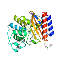
3GN1
 | | Structure of Pteridine Reductase 1 (PTR1) from TRYPANOSOMA BRUCEI in ternary complex with cofactor (NADP+) and inhibitor (DDD00067116) | | 分子名称: | 1H-benzimidazol-2-amine, ACETATE ION, NADP NICOTINAMIDE-ADENINE-DINUCLEOTIDE PHOSPHATE, ... | | 著者 | Tulloch, L.B, Brenk, R, Hunter, W.N. | | 登録日 | 2009-03-16 | | 公開日 | 2009-12-29 | | 最終更新日 | 2025-03-26 | | 実験手法 | X-RAY DIFFRACTION (2 Å) | | 主引用文献 | One scaffold, three binding modes: novel and selective pteridine reductase 1 inhibitors derived from fragment hits discovered by virtual screening.
J.Med.Chem., 52, 2009
|
|
2GCN
 | |
1XB7
 | | X-ray structure of ERRalpha LBD in complex with a PGC-1alpha peptide at 2.5A resolution | | 分子名称: | IODIDE ION, Peroxisome proliferator activated receptor gamma coactivator 1 alpha, Steroid hormone receptor ERR1 | | 著者 | Kallen, J, Schlaeppi, J.M, Bitsch, F, Filipuzzi, I, Schilb, A, Riou, V, Graham, A, Strauss, A, Geiser, M, Fournier, B. | | 登録日 | 2004-08-30 | | 公開日 | 2004-09-14 | | 最終更新日 | 2023-10-25 | | 実験手法 | X-RAY DIFFRACTION (2.5 Å) | | 主引用文献 | Evidence for Ligand-independent Transcriptional Activation of the Human Estrogen-related Receptor {alpha} (ERR{alpha}): CRYSTAL STRUCTURE OF ERR{alpha} LIGAND BINDING DOMAIN IN COMPLEX WITH PEROXISOME PROLIFERATOR-ACTIVATED RECEPTOR COACTIVATOR-1{alpha}
J.Biol.Chem., 279, 2004
|
|
1X81
 | | Farnesyl transferase structure of Jansen compound | | 分子名称: | 6-[(S)-AMINO(4-CHLOROPHENYL)(1-METHYL-1H-IMIDAZOL-5-YL)METHYL]-4-(3-CHLOROPHENYL)-1-METHYLQUINOLIN-2(1H)-ONE, Protein farnesyltransferase beta subunit, Protein farnesyltransferase/geranylgeranyltransferase type I alpha subunit, ... | | 著者 | Li, Q, Claiborne, A, Li, T, Hasvold, L, Stoll, V.S, Muchmore, S, Jakob, C.G, Gu, W, Cohen, J, Hutchins, C, Frost, D, Rosenberg, S.H, Sham, H.L. | | 登録日 | 2004-08-16 | | 公開日 | 2004-12-21 | | 最終更新日 | 2024-02-14 | | 実験手法 | X-RAY DIFFRACTION (3.5 Å) | | 主引用文献 | Design, synthesis, and activity of 4-quinolone and pyridone compounds as nonthiol-containing farnesyltransferase inhibitors.
Bioorg.Med.Chem.Lett., 14, 2004
|
|
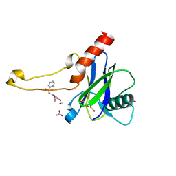
3SC1
 | | Novel Isoquinolone PDK1 Inhibitors Discovered through Fragment-Based Lead Discovery | | 分子名称: | 3-phosphoinositide-dependent protein kinase 1, 6-[2-(hydroxymethyl)phenyl]isoquinolin-1(2H)-one, GLYCEROL, ... | | 著者 | Greasley, S.E, Ferre, R.-A, Krauss, M, Cronin, C. | | 登録日 | 2011-06-06 | | 公開日 | 2011-10-26 | | 最終更新日 | 2024-11-06 | | 実験手法 | X-RAY DIFFRACTION (2.7 Å) | | 主引用文献 | Novel isoquinolone PDK1 inhibitors discovered through fragment-based lead discovery.
J Comput Aided Mol Des, 25, 2011
|
|
5QOU
 | | PanDDA analysis group deposition -- Crystal Structure of DCP2 (NUDT20) in complex with Z296300542 | | 分子名称: | 1,2-ETHANEDIOL, ACETATE ION, DCP2 (NUDT20), ... | | 著者 | Nelson, E.R, Velupillai, S, Talon, R, Collins, P.M, Krojer, T, Wang, D, Brandao-Neto, J, Douangamath, A, Burgess-Brown, N, Arrowsmith, C.H, Bountra, C, Huber, K, von Delft, F. | | 登録日 | 2019-02-22 | | 公開日 | 2019-05-08 | | 最終更新日 | 2024-03-06 | | 実験手法 | X-RAY DIFFRACTION (2.19 Å) | | 主引用文献 | PanDDA analysis group deposition
To Be Published
|
|
9H0W
 | | Human Carbonic Anhydrase II in complex with B-Thujaplicin (2-hydroxy-4-(propan-2-yl)cyclohepta-2,4,6-trien-1-one) | | 分子名称: | 2-oxidanyl-4-propan-2-yl-cyclohepta-2,4,6-trien-1-one, Carbonic anhydrase 2, ZINC ION | | 著者 | Baroni, C, Ferraroni, M. | | 登録日 | 2024-10-09 | | 公開日 | 2025-10-29 | | 実験手法 | X-RAY DIFFRACTION (1.44 Å) | | 主引用文献 | O-derivatization of natural tropolone and beta-thujaplicin leading to effective inhibitors of human carbonic anhydrases IX and XII.
Eur.J.Med.Chem., 290, 2025
|
|
2G95
 | |
2GCP
 | |
2WHK
 | |
2GCH
 | |
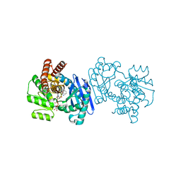
3WEJ
 | | Crystal structure of the human squalene synthase F288A mutant in complex with presqualene pyrophosphate | | 分子名称: | MAGNESIUM ION, Squalene synthase, {(1R,2R,3R)-2-[(3E)-4,8-dimethylnona-3,7-dien-1-yl]-2-methyl-3-[(1E,5E)-2,6,10-trimethylundeca-1,5,9-trien-1-yl]cyclopropyl}methyl trihydrogen diphosphate | | 著者 | Liu, C.I, Jeng, W.Y, Wang, A.H.J. | | 登録日 | 2013-07-07 | | 公開日 | 2014-02-12 | | 最終更新日 | 2023-11-08 | | 実験手法 | X-RAY DIFFRACTION (2 Å) | | 主引用文献 | Structural insights into the catalytic mechanism of human squalene synthase.
Acta Crystallogr.,Sect.D, 70, 2014
|
|


