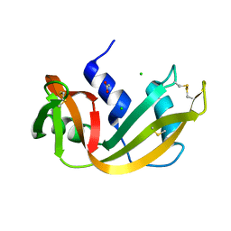
2P47
 
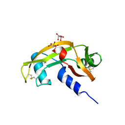
 | | Complex of a camelid single-domain vhh antibody fragment with RNASE A at 2.5A resolution: SE5B-TRI crystal form with five se-met sites (L4M, M34, M51, F68M, M83) in vhh scaffold. | | 分子名称: | ANTIBODY CAB-RN05, Ribonuclease pancreatic | | 著者 | Tereshko, V, Uysal, S, Koide, A, Margalef, K, Koide, S, Kossiakoff, A.A. | | 登録日 | 2007-03-11 | | 公開日 | 2008-03-11 | | 最終更新日 | 2023-11-15 | | 実験手法 | X-RAY DIFFRACTION (2.5 Å) | | 主引用文献 | Toward chaperone-assisted crystallography: protein engineering enhancement of crystal packing and X-ray phasing capabilities of a camelid single-domain antibody (VHH) scaffold
Protein Sci., 17, 2008
|
|
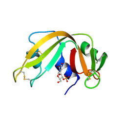
2QCA
 | | A New Crystal Form of Bovine Pancreatic RNase A in Complex with 2'-Deoxyguanosine-5'-monophosphate | | 分子名称: | 2'-DEOXYGUANOSINE-5'-MONOPHOSPHATE, Ribonuclease pancreatic | | 著者 | Larson, S.B, Day, J.S, Cudney, R, McPherson, A, Center for High-Throughput Structural Biology (CHTSB) | | 登録日 | 2007-06-19 | | 公開日 | 2007-07-03 | | 最終更新日 | 2023-08-30 | | 実験手法 | X-RAY DIFFRACTION (1.33 Å) | | 主引用文献 | A new crystal form of bovine pancreatic RNase A in complex with 2'-deoxyguanosine-5'-monophosphate.
Acta Crystallogr.,Sect.F, 63, 2007
|
|
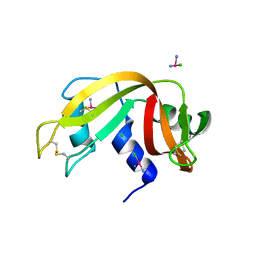
2P44
 | | Complex of a camelid single-domain vhh antibody fragment with RNASE A at 1.8A resolution: SE5A-mono-1 crystal form with five se-met sites (M34, M51, F68M, M83, L86M) in vhh scaffold | | 分子名称: | ANTIBODY CAB-RN05, Ribonuclease pancreatic | | 著者 | Tereshko, V, Uysal, S, Koide, A, Margalef, K, Koide, S, Kossiakoff, A.A. | | 登録日 | 2007-03-11 | | 公開日 | 2008-03-11 | | 最終更新日 | 2011-07-13 | | 実験手法 | X-RAY DIFFRACTION (1.8 Å) | | 主引用文献 | Toward chaperone-assisted crystallography: protein engineering enhancement of crystal packing and X-ray phasing capabilities of a camelid single-domain antibody (VHH) scaffold
Protein Sci., 17, 2008
|
|
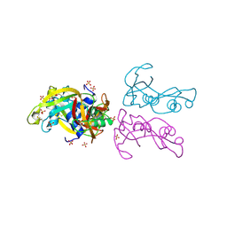
2P43
 | | Complex of a camelid single-domain vhh antibody fragment with RNASE A at 1.65A resolution: SE3-mono-1 crystal form with three se-met sites (M34, M51, M83) in vhh scaffold | | 分子名称: | ANTIBODY CAB-RN05, Ribonuclease pancreatic | | 著者 | Tereshko, V, Uysal, S, Koide, A, Margalef, K, Koide, S, Kossiakoff, A.A. | | 登録日 | 2007-03-11 | | 公開日 | 2008-03-11 | | 最終更新日 | 2011-07-13 | | 実験手法 | X-RAY DIFFRACTION (1.65 Å) | | 主引用文献 | Toward chaperone-assisted crystallography: protein engineering enhancement of crystal packing and X-ray phasing capabilities of a camelid single-domain antibody (VHH) scaffold
Protein Sci., 17, 2008
|
|
2P7S
 | |
2P42
 | | Complex of a camelid single-domain vhh antibody fragment with RNASE A at 1.8A resolution: SE3-mono-2 crystal form with three se-met sites (M34, M51, M83) in vhh scaffold | | 分子名称: | ANTIBODY CAB-RN05, MAGNESIUM ION, Ribonuclease pancreatic | | 著者 | Tereshko, V, Uysal, S, Koide, A, Margalef, K, Koide, S, Kossiakoff, A.A. | | 登録日 | 2007-03-11 | | 公開日 | 2008-03-11 | | 最終更新日 | 2011-07-13 | | 実験手法 | X-RAY DIFFRACTION (1.8 Å) | | 主引用文献 | Toward chaperone-assisted crystallography: protein engineering enhancement of crystal packing and X-ray phasing capabilities of a camelid single-domain antibody (VHH) scaffold
Protein Sci., 17, 2008
|
|
4OKF
 | |
4OXB
 | |
4OOH
 | |
4OWZ
 | |
4OT4
 | |
4QFI
 | | The crystal structure of rat angiogenin-heparin complex | | 分子名称: | ACETIC ACID, Angiogenin, ZINC ION | | 著者 | Yeo, K.J, Hwang, E, Min, K.M, Hwang, K.Y, Jeon, Y.H, Chang, S.I, Cheong, H.K. | | 登録日 | 2014-05-21 | | 公開日 | 2014-08-27 | | 最終更新日 | 2023-11-08 | | 実験手法 | X-RAY DIFFRACTION (1.784 Å) | | 主引用文献 | The crystal structure of rat angiogenin-heparin complex
To be Published
|
|
3ZBW
 | | Crystal Structure of murine Angiogenin-3 | | 分子名称: | ACETIC ACID, ANGIOGENIN-3, SULFATE ION, ... | | 著者 | Iyer, S, Holloway, D.E, Acharya, K.R. | | 登録日 | 2012-11-13 | | 公開日 | 2012-12-26 | | 最終更新日 | 2023-12-20 | | 実験手法 | X-RAY DIFFRACTION (1.801 Å) | | 主引用文献 | Crystal Structures of Murine Angiogenin-2 and -3 - Probing 'Structure - Function' Relationships Amongst Angiogenin Homologues.
FEBS J., 280, 2013
|
|
3ZBV
 | | Crystal Structure of murine Angiogenin-2 | | 分子名称: | ANGIOGENIN-2, GLYCEROL, SULFATE ION, ... | | 著者 | Iyer, S, Holloway, D.E, Acharya, K.R. | | 登録日 | 2012-11-13 | | 公開日 | 2012-12-26 | | 最終更新日 | 2023-12-20 | | 実験手法 | X-RAY DIFFRACTION (1.64 Å) | | 主引用文献 | Crystal Structures of Murine Angiogenin-2 and -3 - Probing 'Structure - Function' Relationships Amongst Angiogenin Homologues.
FEBS J., 280, 2013
|
|
4POU
 | | VHH-metal in Complex with RNase A | | 分子名称: | Ribonuclease pancreatic, VHH Antibody | | 著者 | Fanning, S.W, Walter, R, Horn, J.R. | | 登録日 | 2014-02-26 | | 公開日 | 2014-09-24 | | 最終更新日 | 2014-10-22 | | 実験手法 | X-RAY DIFFRACTION (1.75 Å) | | 主引用文献 | Structural basis of an engineered dual-specific antibody: conformational diversity leads to a hypervariable loop metal-binding site.
Protein Eng.Des.Sel., 27, 2014
|
|
4AHL
 | | P112L - Angiogenin mutants and amyotrophic lateral sclerosis - a biochemical and biological analysis | | 分子名称: | ANGIOGENIN, L(+)-TARTARIC ACID | | 著者 | Thiyagarajan, N, Ferguson, R, Saha, S, Pham, T, Subramanian, V, Acharya, K.R. | | 登録日 | 2012-02-06 | | 公開日 | 2012-10-10 | | 最終更新日 | 2023-12-20 | | 実験手法 | X-RAY DIFFRACTION (2.053 Å) | | 主引用文献 | Structural and Molecular Insights Into the Mechanism of Action of Human Angiogenin-Als Variants in Neurons.
Nat.Commun., 3, 2012
|
|
4SRN
 | | STRUCTURAL CHANGES THAT ACCOMPANY THE REDUCED CATALYTIC EFFICIENCY OF TWO SEMISYNTHETIC RIBONUCLEASE ANALOGS | | 分子名称: | RIBONUCLEASE A, SULFATE ION | | 著者 | deMel, V.S.J, Martin, P.D, Doscher, M.S, Edwards, B.F.P. | | 登録日 | 1991-05-20 | | 公開日 | 1994-12-20 | | 最終更新日 | 2019-08-14 | | 実験手法 | X-RAY DIFFRACTION (2 Å) | | 主引用文献 | Structural changes that accompany the reduced catalytic efficiency of two semisynthetic ribonuclease analogs.
J.Biol.Chem., 267, 1992
|
|
4S0Q
 | |
4A2Y
 | | STRUCTURE OF THE HUMAN EOSINOPHIL CATIONIC PROTEIN IN COMPLEX WITH CITRATE ANIONS | | 分子名称: | (4S)-2-METHYL-2,4-PENTANEDIOL, CITRIC ACID, EOSINOPHIL CATIONIC PROTEIN | | 著者 | Boix, E, Pulido, D, Moussaoui, M, Nogues, V, Russi, S. | | 登録日 | 2011-09-29 | | 公開日 | 2012-06-27 | | 最終更新日 | 2023-12-20 | | 実験手法 | X-RAY DIFFRACTION (1.7 Å) | | 主引用文献 | The Sulfate-Binding Site Structure of the Human Eosinophil Cationic Protein as Revealed by a New Crystal Form.
J.Struct.Biol., 179, 2012
|
|
4RAT
 | |
4RSD
 | | STRUCTURE OF THE D121A VARIANT OF RIBONUCLEASE A | | 分子名称: | ACETATE ION, CHLORIDE ION, RIBONUCLEASE A | | 著者 | Schultz, L.W, Quirk, D.J, Raines, R.T. | | 登録日 | 1998-02-05 | | 公開日 | 1998-07-15 | | 最終更新日 | 2024-10-09 | | 実験手法 | X-RAY DIFFRACTION (1.6 Å) | | 主引用文献 | His...Asp catalytic dyad of ribonuclease A: structure and function of the wild-type, D121N, and D121A enzymes.
Biochemistry, 37, 1998
|
|
4RSK
 | |
4RTE
 | |
4A2O
 | | STRUCTURE OF THE HUMAN EOSINOPHIL CATIONIC PROTEIN IN COMPLEX WITH SULFATE ANIONS | | 分子名称: | EOSINOPHIL CATIONIC PROTEIN, SULFATE ION | | 著者 | Boix, E, Pulido, D, Moussaoui, M, Nogues, V, Russi, S. | | 登録日 | 2011-09-28 | | 公開日 | 2012-06-27 | | 最終更新日 | 2023-12-20 | | 実験手法 | X-RAY DIFFRACTION (1.69 Å) | | 主引用文献 | The Sulfate-Binding Site Structure of the Human Eosinophil Cationic Protein as Revealed by a New Crystal Form.
J.Struct.Biol., 179, 2012
|
|
4AOH
 | | Structural snapshots and functional analysis of human angiogenin variants associated with Amyotrophic Lateral Sclerosis (ALS) | | 分子名称: | ANGIOGENIN, D(-)-TARTARIC ACID, L(+)-TARTARIC ACID | | 著者 | Thiyagarajan, N, Ferguson, R, Subramanian, V, Acharya, K.R. | | 登録日 | 2012-03-27 | | 公開日 | 2012-10-10 | | 最終更新日 | 2024-10-16 | | 実験手法 | X-RAY DIFFRACTION (1.041 Å) | | 主引用文献 | Structural and Molecular Insights Into the Mechanism of Action of Human Angiogenin-Als Variants in Neurons
Nat.Commun., 3, 2012
|
|


