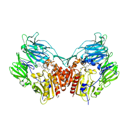
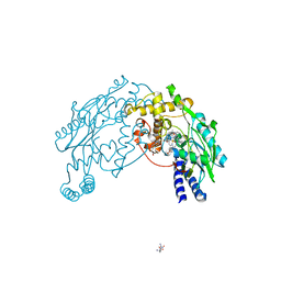
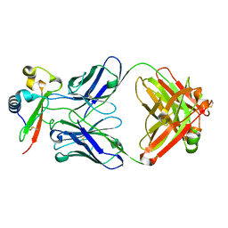
2F38
 
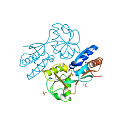
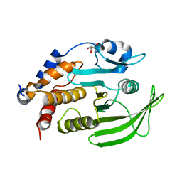
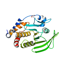
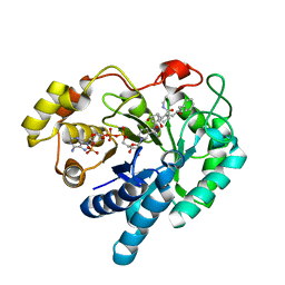 | | Crystal structure of prostaglandin F synathase containing bimatoprost | | 分子名称: | (5Z)-7-{(1R,2R,3R,5S)-3,5-DIHYDROXY-2-[(1E,3S)-3-HYDROXY-5-PHENYLPENT-1-ENYL]CYCLOPENTYL}-N-ETHYLHEPT-5-ENAMIDE, Aldo-keto reductase family 1 member C3, NADP NICOTINAMIDE-ADENINE-DINUCLEOTIDE PHOSPHATE | | 著者 | Komoto, J, Yamada, T, Watanabe, K, Woodward, D.F, Takusagawa, F. | | 登録日 | 2005-11-18 | | 公開日 | 2006-10-31 | | 最終更新日 | 2023-08-23 | | 実験手法 | X-RAY DIFFRACTION (2 Å) | | 主引用文献 | Prostaglandin F2alpha formation from prostaglandin H2 by prostaglandin F synthase (PGFS): crystal structure of PGFS containing bimatoprost.
Biochemistry, 45, 2006
|
|
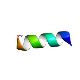
2FBS
 | |
2FJT
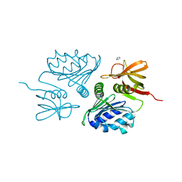
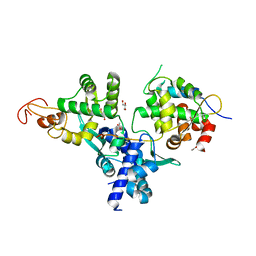
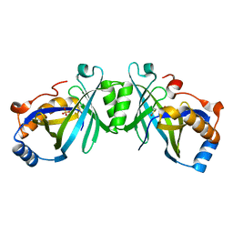 | | Adenylyl cyclase class iv from Yersinia pestis | | 分子名称: | Adenylyl cyclase class IV, SULFATE ION | | 著者 | Gallagher, D.T, Smith, N.N, Kim, S.-K, Reddy, P.T, Robinson, H, Heroux, A. | | 登録日 | 2006-01-03 | | 公開日 | 2006-11-14 | | 最終更新日 | 2024-04-03 | | 実験手法 | X-RAY DIFFRACTION (1.901 Å) | | 主引用文献 | Structure of the class IV adenylyl cyclase reveals a novel fold
J.Mol.Biol., 362, 2006
|
|
2FIK
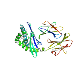 | | Structure of a microbial glycosphingolipid bound to mouse CD1d | | 分子名称: | (2S,3R)-3-HYDROXY-2-(TETRADECANOYLAMINO)OCTADECYL ALPHA-D-GALACTOPYRANOSIDURONIC ACID, 2-acetamido-2-deoxy-beta-D-glucopyranose, 2-acetamido-2-deoxy-beta-D-glucopyranose-(1-4)-2-acetamido-2-deoxy-beta-D-glucopyranose, ... | | 著者 | Wu, D, Zajonc, D.M. | | 登録日 | 2005-12-29 | | 公開日 | 2006-03-21 | | 最終更新日 | 2023-08-30 | | 実験手法 | X-RAY DIFFRACTION (1.8 Å) | | 主引用文献 | Design of natural killer T cell activators: structure and function of a microbial glycosphingolipid bound to mouse CD1d.
Proc.Natl.Acad.Sci.Usa, 103, 2006
|
|
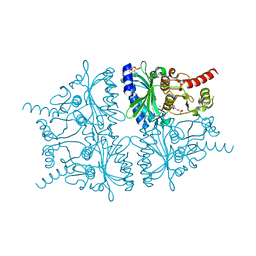
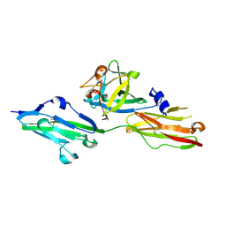
2FJP
 | | Human dipeptidyl peptidase IV/CD26 in complex with an inhibitor | | 分子名称: | 2-acetamido-2-deoxy-beta-D-glucopyranose, 2-acetamido-2-deoxy-beta-D-glucopyranose-(1-4)-2-acetamido-2-deoxy-beta-D-glucopyranose, 6-(4-{(1S,2S)-2-AMINO-1-[(DIMETHYLAMINO)CARBONYL]-3-[(3S)-3-FLUOROPYRROLIDIN-1-YL]-3-OXOPROPYL}PHENYL)-1H-[1,2,4]TRIAZOLO[1,5-A]PYRIDIN-4-IUM, ... | | 著者 | Scapin, G, Patel, S.B, Becker, J.W. | | 登録日 | 2006-01-03 | | 公開日 | 2006-07-04 | | 最終更新日 | 2023-08-30 | | 実験手法 | X-RAY DIFFRACTION (2.4 Å) | | 主引用文献 | (2S,3S)-3-Amino-4-(3,3-difluoropyrrolidin-1-yl)-N,N-dimethyl-4-oxo-2-(4-[1,2,4]triazolo[1,5-a]- pyridin-6-ylphenyl)butanamide: a selective alpha-amino amide dipeptidyl peptidase IV inhibitor for the treatment of type 2 diabetes.
J.Med.Chem., 49, 2006
|
|
2FKW
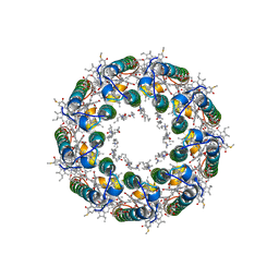 | | Structure of LH2 from Rps. acidophila crystallized in lipidic mesophases | | 分子名称: | BACTERIOCHLOROPHYLL A, LAURYL DIMETHYLAMINE-N-OXIDE, Light-harvesting protein B-800/850, ... | | 著者 | Papiz, M.Z, Cherezov, V, Clogston, J, Caffrey, M. | | 登録日 | 2006-01-05 | | 公開日 | 2006-03-28 | | 最終更新日 | 2023-08-30 | | 実験手法 | X-RAY DIFFRACTION (2.45 Å) | | 主引用文献 | Room to Move: Crystallizing Membrane Proteins in Swollen Lipidic Mesophases
J.Mol.Biol., 357, 2006
|
|
2FL8
 | |
2ES2
 | |
2F3D
 | | Mechanism of displacement of a catalytically essential loop from the active site of fructose-1,6-bisphosphatase | | 分子名称: | 6-O-phosphono-beta-D-fructofuranose, ADENOSINE MONOPHOSPHATE, Fructose-1,6-bisphosphatase 1, ... | | 著者 | Iancu, C.V, Mukund, S, Choe, J.-Y, Fromm, H.J, Honzatko, R.B. | | 登録日 | 2005-11-21 | | 公開日 | 2006-04-25 | | 最終更新日 | 2024-02-14 | | 実験手法 | X-RAY DIFFRACTION (1.83 Å) | | 主引用文献 | Mechanism of displacement of a catalytically essential loop from the active site of mammalian fructose-1,6-bisphosphatase.
Biochemistry, 52, 2013
|
|
2F3J
 | |
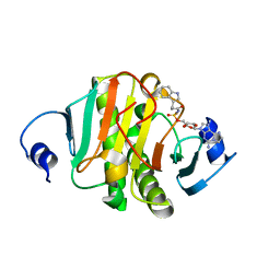
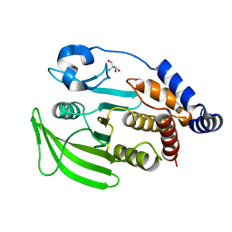
2F5T
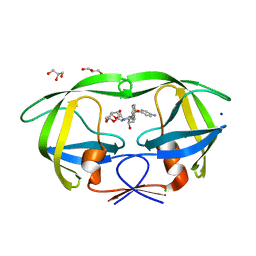
 | | Crystal Structure of the sugar binding domain of the archaeal transcriptional regulator TrmB | | 分子名称: | IMIDAZOLE, alpha-D-glucopyranose-(1-4)-alpha-D-glucopyranose, archaeal transcriptional regulator TrmB | | 著者 | Krug, M, Lee, S.J, Diederichs, K, Boos, W, Welte, W. | | 登録日 | 2005-11-27 | | 公開日 | 2006-02-21 | | 最終更新日 | 2024-02-14 | | 実験手法 | X-RAY DIFFRACTION (1.45 Å) | | 主引用文献 | Crystal Structure of the Sugar Binding Domain of the Archaeal Transcriptional Regulator TrmB
J.Biol.Chem., 281, 2006
|
|
2F7B
 | | CatM effector binding domain | | 分子名称: | CHLORIDE ION, HTH-type transcriptional regulator catM, SULFATE ION | | 著者 | Clark, T, Haddad, S, Ezezika, O, Neidle, E, Momany, C. | | 登録日 | 2005-11-30 | | 公開日 | 2006-10-31 | | 最終更新日 | 2023-08-23 | | 実験手法 | X-RAY DIFFRACTION (1.9 Å) | | 主引用文献 | Distinct Effector-binding Sites Enable Synergistic Transcriptional Activation by BenM, a LysR-type Regulator.
J.Mol.Biol., 367, 2007
|
|
2F81
 | | HIV-1 Protease mutant L90M complexed with inhibitor TMC114 | | 分子名称: | (3R,3AS,6AR)-HEXAHYDROFURO[2,3-B]FURAN-3-YL(1S,2R)-3-[[(4-AMINOPHENYL)SULFONYL](ISOBUTYL)AMINO]-1-BENZYL-2-HYDROXYPROPYLCARBAMATE, CHLORIDE ION, GLYCEROL, ... | | 著者 | Kovalevsky, A.Y, Weber, I.T. | | 登録日 | 2005-12-01 | | 公開日 | 2006-03-07 | | 最終更新日 | 2024-02-14 | | 実験手法 | X-RAY DIFFRACTION (1.25 Å) | | 主引用文献 | Effectiveness of Nonpeptide Clinical Inhibitor TMC-114 on HIV-1 Protease with Highly Drug Resistant Mutations D30N, I50V, and L90M.
J.Med.Chem., 49, 2006
|
|
2FC2
 | |
2FDB
 | |
2FFL
 | |
2FD4
 | |
8PAF
 | |
7GTZ
 | |
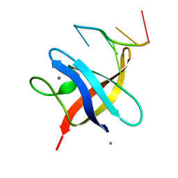
3GSH
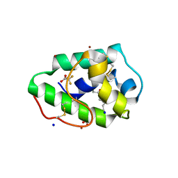 | | Three-dimensional structure of a post translational modified barley LTP1 | | 分子名称: | (12E)-10-oxooctadec-12-enoic acid, Non-specific lipid-transfer protein 1, SODIUM ION, ... | | 著者 | Lascombe, M.B, Prange, T, Bakan, B, Marion, D. | | 登録日 | 2009-03-27 | | 公開日 | 2009-12-15 | | 最終更新日 | 2017-11-01 | | 実験手法 | X-RAY DIFFRACTION (1.8 Å) | | 主引用文献 | The crystal structure of oxylipin-conjugated barley LTP1 highlights the unique plasticity of the hydrophobic cavity of these plant lipid-binding proteins.
Biochem.Biophys.Res.Commun., 390, 2009
|
|
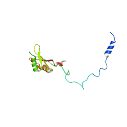
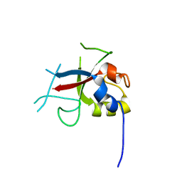
2L33
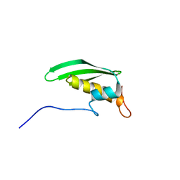 | | Solution NMR Structure of DRBM 2 domain of Interleukin enhancer-binding factor 3 from Homo sapiens, Northeast Structural Genomics Consortium Target HR4527E | | 分子名称: | Interleukin enhancer-binding factor 3 | | 著者 | Liu, G, Janjua, H, Xiao, R, Acton, T.B, Ciccosanti, A, Shastry, R.B, Everett, J, Montelione, G.T, Northeast Structural Genomics Consortium, Northeast Structural Genomics Consortium (NESG) | | 登録日 | 2010-09-03 | | 公開日 | 2010-09-22 | | 最終更新日 | 2024-05-15 | | 実験手法 | SOLUTION NMR | | 主引用文献 | Northeast Structural Genomics Consortium Target HR4527E
To be Published
|
|
7GTW
 | |
7RXP
 | |
7DG2
 | | Nse1-Nse3-Nse4 complex | | 分子名称: | ACETATE ION, GLYCEROL, MAGE domain-containing protein, ... | | 著者 | Cho, Y, Jo, A. | | 登録日 | 2020-11-10 | | 公開日 | 2021-05-26 | | 最終更新日 | 2023-11-29 | | 実験手法 | X-RAY DIFFRACTION (1.7 Å) | | 主引用文献 | Structure Basis for Shaping the Nse4 Protein by the Nse1 and Nse3 Dimer within the Smc5/6 Complex.
J.Mol.Biol., 433, 2021
|
|
7GU6
 | |


