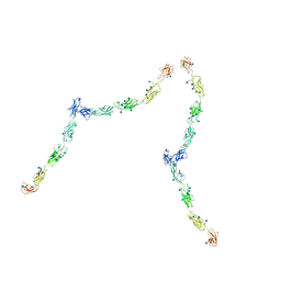
1Q5C
 
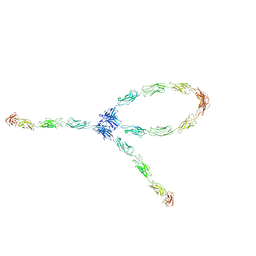 | | S-S-lambda-shaped TRANS and CIS interactions of cadherins model based on fitting C-cadherin (1L3W) to 3D map of desmosomes obtained by electron tomography | | 分子名称: | 2-acetamido-2-deoxy-alpha-D-glucopyranose, 2-acetamido-2-deoxy-beta-D-glucopyranose, CALCIUM ION, ... | | 著者 | He, W, Cowin, P, Stokes, D.L. | | 登録日 | 2003-08-06 | | 公開日 | 2003-10-07 | | 最終更新日 | 2020-07-29 | | 実験手法 | ELECTRON MICROSCOPY (30 Å) | | 主引用文献 | Untangling Desmosomal Knots with Electron Tomography
Science, 302, 2003
|
|
1Q5B
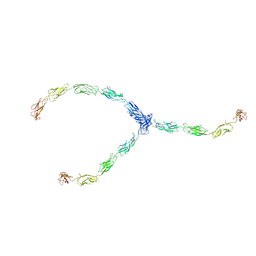 | | lambda-shaped TRANS and CIS interactions of cadherins model based on fitting C-cadherin (1L3W) to 3D map of desmosomes obtained by electron tomography | | 分子名称: | 2-acetamido-2-deoxy-alpha-D-glucopyranose, 2-acetamido-2-deoxy-beta-D-glucopyranose, CALCIUM ION, ... | | 著者 | He, W, Cowin, P, Stokes, D.L. | | 登録日 | 2003-08-06 | | 公開日 | 2003-10-07 | | 最終更新日 | 2020-07-29 | | 実験手法 | ELECTRON MICROSCOPY (30 Å) | | 主引用文献 | Untangling Desmosomal Knots with Electron Tomography
Science, 302, 2003
|
|
1Q5A
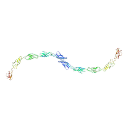 | | S-shaped trans interactions of cadherins model based on fitting C-cadherin (1L3W) to 3D map of desmosomes obtained by electron tomography | | 分子名称: | 2-acetamido-2-deoxy-alpha-D-glucopyranose, 2-acetamido-2-deoxy-beta-D-glucopyranose, CALCIUM ION, ... | | 著者 | He, W, Cowin, P, Stokes, D.L. | | 登録日 | 2003-08-06 | | 公開日 | 2003-10-07 | | 最終更新日 | 2020-07-29 | | 実験手法 | ELECTRON MICROSCOPY (30 Å) | | 主引用文献 | Untangling Desmosomal Knots with Electron Tomography
Science, 302, 2003
|
|
2AIN
 | |
1Q55
 | | W-shaped trans interactions of cadherins model based on fitting C-cadherin (1L3W) to 3D map of desmosomes obtained by electron tomography | | 分子名称: | 2-acetamido-2-deoxy-alpha-D-glucopyranose, 2-acetamido-2-deoxy-beta-D-glucopyranose, CALCIUM ION, ... | | 著者 | He, W, Cowin, P, Stokes, D.L. | | 登録日 | 2003-08-06 | | 公開日 | 2003-10-07 | | 最終更新日 | 2020-07-29 | | 実験手法 | ELECTRON MICROSCOPY (30 Å) | | 主引用文献 | Untangling Desmosomal Knots with Electron Tomography
Science, 302, 2003
|
|
1SJS
 | |
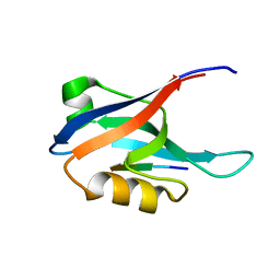
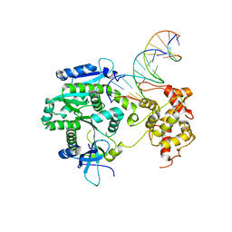
4R11
 | | A conserved phosphorylation switch controls the interaction between cadherin and beta-catenin in vitro and in vivo | | 分子名称: | Cadherin-related hmr-1, IODIDE ION, Protein humpback-2 | | 著者 | Choi, H.-J, Loveless, T, Lynch, A, Bang, I, Hardin, J, Weis, W.I. | | 登録日 | 2014-08-03 | | 公開日 | 2015-04-29 | | 実験手法 | X-RAY DIFFRACTION (2.789 Å) | | 主引用文献 | A Conserved Phosphorylation Switch Controls the Interaction between Cadherin and beta-Catenin In Vitro and In Vivo
Dev.Cell, 33, 2015
|
|
4LYT
 | |
3EX7
 | |
7UUW
 | | Cryogenic electron microscopy 3D map of F-actin bound by the Actin Binding Domain of alpha-catenin ortholog, HMP1 | | 分子名称: | ADENOSINE-5'-DIPHOSPHATE, Actin, alpha skeletal muscle, ... | | 著者 | Rangarajan, E.S, Smith, E.W, Izard, T. | | 登録日 | 2022-04-29 | | 公開日 | 2023-01-18 | | 最終更新日 | 2023-01-25 | | 実験手法 | ELECTRON MICROSCOPY (3.36 Å) | | 主引用文献 | The nematode alpha-catenin ortholog, HMP1, has an extended alpha-helix when bound to actin filaments.
J.Biol.Chem., 299, 2022
|
|
5GNI
 | |
4R10
 | | A conserved phosphorylation switch controls the interaction between cadherin and beta-catenin in vitro and in vivo | | 分子名称: | 1,2-ETHANEDIOL, Cadherin-related hmr-1, Protein humpback-2, ... | | 著者 | Choi, H.-J, Loveless, T, Lynch, A, Bang, I, Hardin, J, Weis, W.I. | | 登録日 | 2014-08-03 | | 公開日 | 2015-04-29 | | 実験手法 | X-RAY DIFFRACTION (2.3 Å) | | 主引用文献 | A Conserved Phosphorylation Switch Controls the Interaction between Cadherin and beta-Catenin In Vitro and In Vivo
Dev.Cell, 33, 2015
|
|
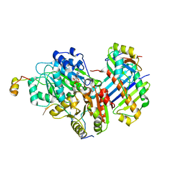
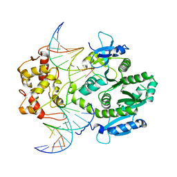
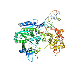
4P0P
 | | Crystal structure of Human Mus81-Eme1 in complex with 5'-flap DNA, and Mg2+ | | 分子名称: | Crossover junction endonuclease EME1, Crossover junction endonuclease MUS81, DNA GAATGTGTGTCTCAATC, ... | | 著者 | Gwon, G.H, Baek, K, Cho, Y. | | 登録日 | 2014-02-22 | | 公開日 | 2014-05-28 | | 最終更新日 | 2023-12-27 | | 実験手法 | X-RAY DIFFRACTION (2.8 Å) | | 主引用文献 | Crystal structures of the structure-selective nuclease Mus81-Eme1 bound to flap DNA substrates.
Embo J., 33, 2014
|
|
4TKD
 | | Sulfolobus solfataricus HJC mutants | | 分子名称: | Holliday junction resolvase Hjc | | 著者 | Bond, C.S. | | 登録日 | 2014-05-26 | | 公開日 | 2015-08-12 | | 最終更新日 | 2023-12-27 | | 実験手法 | X-RAY DIFFRACTION (2.01 Å) | | 主引用文献 | Crystal Unengineering: Reducing the Crystallisability of Sulfolobus solfataricus Hjc
Aust.J.Chem., 67, 2014
|
|
4TKK
 | | Sulfolobus solfataricus HJC mutants | | 分子名称: | Holliday junction resolvase Hjc | | 著者 | Bond, C.S. | | 登録日 | 2014-05-27 | | 公開日 | 2015-08-12 | | 最終更新日 | 2023-12-27 | | 実験手法 | X-RAY DIFFRACTION (2.4 Å) | | 主引用文献 | Crystal Unengineering: Reducing the Crystallisability of Sulfolobus solfataricus Hjc
Aust.J.Chem., 67, 2014
|
|
4P0R
 | | human Mus81-Eme1-3'flap DNA complex | | 分子名称: | Crossover junction endonuclease EME1, Crossover junction endonuclease MUS81, DNA ACGTGCTTACACACAGAGGTTAGGGTGAACTT, ... | | 著者 | Gwon, G.H, Baek, K, Cho, Y. | | 登録日 | 2014-02-22 | | 公開日 | 2014-05-28 | | 最終更新日 | 2023-12-27 | | 実験手法 | X-RAY DIFFRACTION (6.501 Å) | | 主引用文献 | Crystal structures of the structure-selective nuclease Mus81-Eme1 bound to flap DNA substrates.
Embo J., 33, 2014
|
|
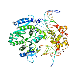
4P0Q
 | | Crystal structure of Human Mus81-Eme1 in complex with 5'-flap DNA | | 分子名称: | Crossover junction endonuclease EME1, Crossover junction endonuclease MUS81, DNA GAATGTGTGTCTCAATC, ... | | 著者 | Gwon, G.H, Baek, K, Cho, Y. | | 登録日 | 2014-02-22 | | 公開日 | 2014-05-28 | | 最終更新日 | 2023-12-27 | | 実験手法 | X-RAY DIFFRACTION (2.851 Å) | | 主引用文献 | Crystal structures of the structure-selective nuclease Mus81-Eme1 bound to flap DNA substrates.
Embo J., 33, 2014
|
|
4P0S
 | | human Mus81-Eme1-3'flap DNA complex | | 分子名称: | Crossover junction endonuclease EME1, Crossover junction endonuclease MUS81, DNA GAATGTGTGTCT, ... | | 著者 | Gwon, G.H, Baek, K, Cho, Y. | | 登録日 | 2014-02-22 | | 公開日 | 2014-05-28 | | 最終更新日 | 2023-12-27 | | 実験手法 | X-RAY DIFFRACTION (6 Å) | | 主引用文献 | Crystal structures of the structure-selective nuclease Mus81-Eme1 bound to flap DNA substrates.
Embo J., 33, 2014
|
|
5FD1
 | |
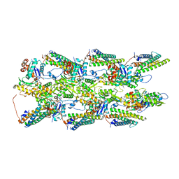
4V4H
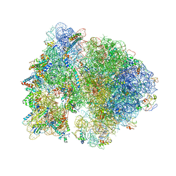 | | Crystal structure of the bacterial ribosome from Escherichia coli in complex with the antibiotic kasugamyin at 3.5A resolution. | | 分子名称: | (1S,2R,3S,4R,5S,6S)-2,3,4,5,6-PENTAHYDROXYCYCLOHEXYL 2-AMINO-4-{[CARBOXY(IMINO)METHYL]AMINO}-2,3,4,6-TETRADEOXY-ALPHA-D-ARABINO-HEXOPYRANOSIDE, 16S RIBOSOMAL RNA, 23S RIBOSOMAL RNA, ... | | 著者 | Schuwirth, B.S, Vila-Sanjurjo, A, Cate, J.H.D. | | 登録日 | 2006-08-04 | | 公開日 | 2014-07-09 | | 最終更新日 | 2023-09-20 | | 実験手法 | X-RAY DIFFRACTION (3.46 Å) | | 主引用文献 | Structural analysis of kasugamycin inhibition of translation.
Nat.Struct.Mol.Biol., 13, 2006
|
|
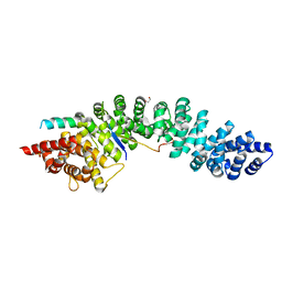
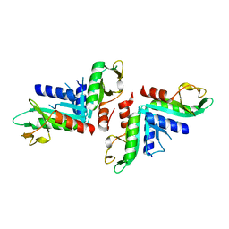
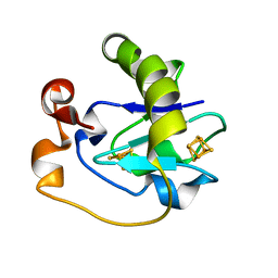
1LM5
 | | Structures of two intermediate filament-binding fragments of desmoplakin reveal a unique repeat motif structure | | 分子名称: | subdomain of Desmoplakin Carboxy-Terminal domain (DPCT) | | 著者 | Choi, H.J, Park-Snyder, S, Pascoe, L.T, Green, K.J, Weis, W.I. | | 登録日 | 2002-04-30 | | 公開日 | 2002-07-31 | | 最終更新日 | 2024-02-14 | | 実験手法 | X-RAY DIFFRACTION (1.8 Å) | | 主引用文献 | Structures of two intermediate filament-binding fragments of desmoplakin reveal a unique repeat motif structure.
Nat.Struct.Biol., 9, 2002
|
|
4URP
 | | The Crystal structure of Nitroreductase from Saccharomyces cerevisiae | | 分子名称: | FATTY ACID REPRESSION MUTANT PROTEIN 2 | | 著者 | Song, H.-N, Woo, E.-J, Bang, S.-Y, Jung, D.-G, Park, S.-G. | | 登録日 | 2014-07-01 | | 公開日 | 2015-04-29 | | 最終更新日 | 2024-05-08 | | 実験手法 | X-RAY DIFFRACTION (2.991 Å) | | 主引用文献 | Crystal Structure of the Fungal Nitroreductase Frm2 from Saccharomyces Cerevisiae.
Protein Sci., 24, 2015
|
|
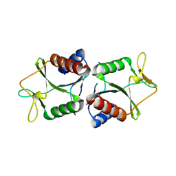
1LM7
 | | Structures of two intermediate filament-binding fragments of desmoplakin reveal a unique repeat motif structure | | 分子名称: | subdomain of Desmoplakin Carboxy-Terminal domain (DPCT) | | 著者 | Choi, H.J, Park-Snyder, S, Pascoe, L.T, Green, K.J, Weis, W.I. | | 登録日 | 2002-04-30 | | 公開日 | 2002-07-31 | | 最終更新日 | 2024-02-14 | | 実験手法 | X-RAY DIFFRACTION (3 Å) | | 主引用文献 | Structures of two intermediate filament-binding fragments of desmoplakin reveal a unique repeat motif structure.
Nat.Struct.Biol., 9, 2002
|
|
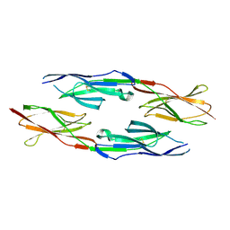
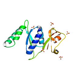
5A6C
 | | Concomitant binding of Afadin to LGN and F-actin directs planar spindle orientation | | 分子名称: | G-PROTEIN-SIGNALING MODULATOR 2, AFADIN, SULFATE ION | | 著者 | Carminati, M, Gallini, S, Pirovano, L, Alfieri, A, Bisi, S, Mapelli, M. | | 登録日 | 2015-06-25 | | 公開日 | 2015-12-30 | | 最終更新日 | 2024-01-10 | | 実験手法 | X-RAY DIFFRACTION (2.901 Å) | | 主引用文献 | Concomitant Binding of Afadin to Lgn and F-Actin Directs Planar Spindle Orientation.
Nat.Struct.Mol.Biol., 23, 2016
|
|
3RF2
 | |


