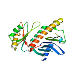
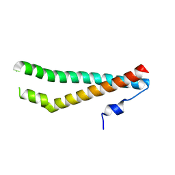
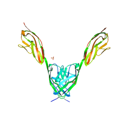
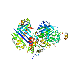
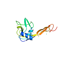
5ZIY
 
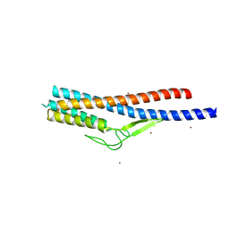 | | Crystal structure of Bacillus cereus FlgL | | 分子名称: | Flagellar hook-associated protein 3, ZINC ION | | 著者 | Hong, H.J, Kim, T.H, Song, W.S, Yoon, S.I. | | 登録日 | 2018-03-18 | | 公開日 | 2018-10-17 | | 最終更新日 | 2024-03-27 | | 実験手法 | X-RAY DIFFRACTION (2.2 Å) | | 主引用文献 | Crystal structure of FlgL and its implications for flagellar assembly
Sci Rep, 8, 2018
|
|
5ZIZ
 | |
5ZJ0
 | |
7X9F
 | | Crystal structure of actinomycin D-echinomycin-d(AGCGCGT/ACGCGCT) complex | | 分子名称: | 2-CARBOXYQUINOXALINE, DNA (5'-D(P*AP*CP*GP*CP*GP*CP*T)-3'), DNA (5'-D(P*AP*GP*CP*GP*CP*GP*T)-3'), ... | | 著者 | Kao, S.H, Satange, R.B, Hou, M.H. | | 登録日 | 2022-03-15 | | 公開日 | 2022-12-14 | | 最終更新日 | 2024-07-10 | | 実験手法 | X-RAY DIFFRACTION (2.96 Å) | | 主引用文献 | Staggered intercalation of DNA duplexes with base-pair modulation by two distinct drug molecules induces asymmetric backbone twisting and structure polymorphism.
Nucleic Acids Res., 50, 2022
|
|
3E17
 | | Crystal structure of the second PDZ domain from human Zona Occludens-2 | | 分子名称: | Tight junction protein ZO-2 | | 著者 | Chen, H, Tong, S.L, Teng, M.K, Niu, L.W. | | 登録日 | 2008-08-01 | | 公開日 | 2009-07-21 | | 最終更新日 | 2023-11-01 | | 実験手法 | X-RAY DIFFRACTION (1.75 Å) | | 主引用文献 | Structure of the second PDZ domain from human zonula occludens 2
Acta Crystallogr.,Sect.F, 65, 2009
|
|
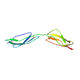
1HL6
 | | A novel mode of RBD-protein recognition in the Y14-mago complex | | 分子名称: | CG8781 PROTEIN, MAGO NASHI PROTEIN | | 著者 | Fribourg, S, Gatfield, D, Yao, W, Izaurralde, E, Conti, E. | | 登録日 | 2003-03-13 | | 公開日 | 2003-05-08 | | 最終更新日 | 2024-05-08 | | 実験手法 | X-RAY DIFFRACTION (2.5 Å) | | 主引用文献 | A Novel Mode of Rbd-Protein Recognition in the Y14-Mago Complex
Nat.Struct.Biol., 10, 2003
|
|
1WAT
 | |
3MJ9
 | |
1WAS
 | |
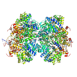
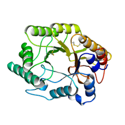
3M34
 | | Crystal structure of transketolase in complex with thiamin diphosphate and calcium ion | | 分子名称: | 1,2-ETHANEDIOL, CALCIUM ION, GLYCEROL, ... | | 著者 | Nocek, B, Makowska-Grzyska, M, Maltseva, N, Grimshaw, S, Joachimiak, A, Anderson, W, Center for Structural Genomics of Infectious Diseases (CSGID) | | 登録日 | 2010-03-08 | | 公開日 | 2010-04-28 | | 最終更新日 | 2023-11-22 | | 実験手法 | X-RAY DIFFRACTION (1.65 Å) | | 主引用文献 | Crystal structure of transketolase in complex with thiamin diphosphate and calcium ion
To be Published
|
|
5N7I
 | |
5N7H
 | |
5N7K
 | |
6VAI
 | |
2IEP
 | |
8EFY
 | |
8LDH
 | |
8U5B
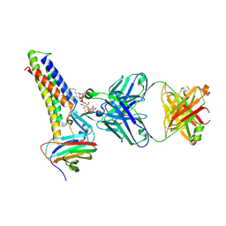 | |
2XB2
 | | Crystal structure of the core Mago-Y14-eIF4AIII-Barentsz-UPF3b assembly shows how the EJC is bridged to the NMD machinery | | 分子名称: | EUKARYOTIC INITIATION FACTOR 4A-III, MAGNESIUM ION, PHOSPHOAMINOPHOSPHONIC ACID-ADENYLATE ESTER, ... | | 著者 | Buchwald, G, Ebert, J, Basquin, C, Sauliere, J, Jayachandran, U, Bono, F, Le Hir, H, Conti, E. | | 登録日 | 2010-04-03 | | 公開日 | 2010-05-12 | | 最終更新日 | 2023-12-20 | | 実験手法 | X-RAY DIFFRACTION (3.4 Å) | | 主引用文献 | Insights Into the Recruitment of the Nmd Machinery from the Crystal Structure of a Core Ejc-Upf3B Complex.
Proc.Natl.Acad.Sci.USA, 107, 2010
|
|
8U4V
 | |
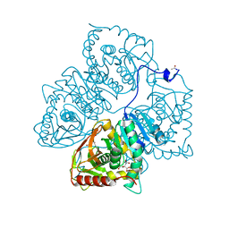
1EM1
 | | X-RAY CRYSTAL STRUCTURE FOR HUMAN MANGANESE SUPEROXIDE DISMUTASE, Q143A | | 分子名称: | MANGANESE (II) ION, MANGANESE SUPEROXIDE DISMUTASE, SULFATE ION | | 著者 | Leveque, V, Stroupe, M.E, Lepock, J.R, Cabelli, D.E, Tainer, J.A, Nick, H.S, Silverman, D.N. | | 登録日 | 2000-03-14 | | 公開日 | 2000-03-24 | | 最終更新日 | 2024-02-07 | | 実験手法 | X-RAY DIFFRACTION (2.13 Å) | | 主引用文献 | Multiple replacements of glutamine 143 in human manganese superoxide dismutase: effects on structure, stability, and catalysis.
Biochemistry, 39, 2000
|
|
5ADH
 | |
1ESL
 | |
1EXP
 | | BETA-1,4-GLYCANASE CEX-CD | | 分子名称: | BETA-1,4-D-GLYCANASE CEX-CD, beta-D-glucopyranose-(1-4)-2-deoxy-2-fluoro-alpha-D-glucopyranose | | 著者 | White, A, Tull, D, Johns, K.L, Withers, S.G, Rose, D.R. | | 登録日 | 1996-01-11 | | 公開日 | 1997-01-27 | | 最終更新日 | 2020-07-29 | | 実験手法 | X-RAY DIFFRACTION (1.8 Å) | | 主引用文献 | Crystallographic observation of a covalent catalytic intermediate in a beta-glycosidase.
Nat.Struct.Biol., 3, 1996
|
|
1E5G
 | | Solution structure of central CP module pair of a pox virus complement inhibitor | | 分子名称: | COMPLEMENT CONTROL PROTEIN C3 | | 著者 | Henderson, C.E, Bromek, K, Mullin, N.P, Smith, B.O, Uhrin, D, Barlow, P.N. | | 登録日 | 2000-07-25 | | 公開日 | 2000-08-31 | | 最終更新日 | 2013-07-03 | | 実験手法 | SOLUTION NMR | | 主引用文献 | Solution Structure and Dynamics of the Central Ccp Module Pair of a Poxvirus Complement Control Protein
J.Mol.Biol., 307, 2001
|
|


