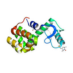
2XKS
 
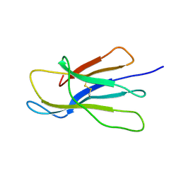 | | Prion-like conversion during amyloid formation at atomic resolution | | 分子名称: | BETA-2-MICROGLOBULIN | | 著者 | Eichner, T, Kalverda, A.P, Thompson, G.S, Radford, S.E, Homans, S.W. | | 登録日 | 2010-07-12 | | 公開日 | 2011-02-16 | | 最終更新日 | 2020-01-15 | | 実験手法 | SOLUTION NMR | | 主引用文献 | Conformational Conversion During Amyloid Formation at Atomic Resolution.
Mol.Cell, 41, 2011
|
|
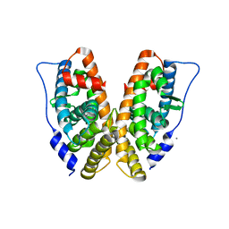
2PQK
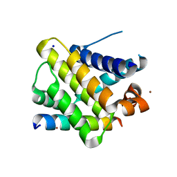 | | X-ray crystal structure of human Mcl-1 in complex with Bim BH3 | | 分子名称: | Bim BH3 peptide, Induced myeloid leukemia cell differentiation protein Mcl-1, SODIUM ION, ... | | 著者 | Bare, E, Grant, R.A, Keating, A.E. | | 登録日 | 2007-05-02 | | 公開日 | 2007-06-19 | | 最終更新日 | 2017-10-18 | | 実験手法 | X-RAY DIFFRACTION (2 Å) | | 主引用文献 | Mcl-1-Bim complexes accommodate surprising point mutations via minor structural changes.
Protein Sci., 19, 2010
|
|
2PXR
 | |
2PRH
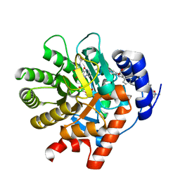 | | The structures of apo- and inhibitor bound human dihydroorotate dehydrogenase reveal conformational flexibility within the inhibitor binding site | | 分子名称: | 6-CHLORO-2-(2'-FLUOROBIPHENYL-4-YL)-3-METHYLQUINOLINE-4-CARBOXYLIC ACID, DECYLAMINE-N,N-DIMETHYL-N-OXIDE, Dihydroorotate dehydrogenase, ... | | 著者 | Walse, B, Dufe, V.T, Al-Karadaghi, S. | | 登録日 | 2007-05-04 | | 公開日 | 2008-05-20 | | 最終更新日 | 2023-08-30 | | 実験手法 | X-RAY DIFFRACTION (2.4 Å) | | 主引用文献 | The structures of human dihydroorotate dehydrogenase with and without inhibitor reveal conformational flexibility in the inhibitor and substrate binding sites
Biochemistry, 47, 2008
|
|
2PU1
 | | Crystal Structure of the T. brucei enolase complexed with Fluoro-phosphonoacetohydroxamate (FPAH) | | 分子名称: | 1,2-ETHANEDIOL, Enolase, ZINC ION, ... | | 著者 | Navarro, M.V.A.S, Rigden, D.J, Garratt, R.C, Dias, S.M.G. | | 登録日 | 2007-05-08 | | 公開日 | 2007-11-20 | | 最終更新日 | 2023-08-30 | | 実験手法 | X-RAY DIFFRACTION (1.8 Å) | | 主引用文献 | Structural flexibility in Trypanosoma brucei enolase revealed by X-ray crystallography and molecular dynamics.
Febs J., 274, 2007
|
|
2Q0M
 | | Tricarbonylmanganese(I)-lysozyme complex : a structurally characterized organometallic protein | | 分子名称: | CARBON MONOXIDE, CHLORIDE ION, Lysozyme C, ... | | 著者 | Razavet, M, Artero, V, Cavazza, C, Oudart, Y, Fontecilla-Camps, J.C, Fontecave, M. | | 登録日 | 2007-05-22 | | 公開日 | 2007-12-11 | | 最終更新日 | 2023-07-26 | | 実験手法 | X-RAY DIFFRACTION (1.7 Å) | | 主引用文献 | Tricarbonylmanganese(I)-lysozyme complex: a structurally characterized organometallic protein.
Chem.Commun.(Camb.), 2007
|
|
2H6V
 | |
2PS8
 | | Y295F Trichodiene Synthase: Complex With Mg and Pyrophosphate | | 分子名称: | 1,2-ETHANEDIOL, MAGNESIUM ION, PYROPHOSPHATE 2-, ... | | 著者 | Vedula, L.S, Cane, D.E, Christianson, D.W. | | 登録日 | 2007-05-04 | | 公開日 | 2007-12-18 | | 最終更新日 | 2023-08-30 | | 実験手法 | X-RAY DIFFRACTION (2.67 Å) | | 主引用文献 | Structural and mechanistic analysis of trichodiene synthase using site-directed mutagenesis: probing the catalytic function of tyrosine-295 and the asparagine-225/serine-229/glutamate-233-Mg2+B motif.
Arch.Biochem.Biophys., 469, 2008
|
|
2HFF
 | | Crystal structure of CB2 Fab | | 分子名称: | CB2 Fab, heavy chain, light chain | | 著者 | Hymowitz, S.G. | | 登録日 | 2006-06-23 | | 公開日 | 2006-11-07 | | 最終更新日 | 2023-08-30 | | 実験手法 | X-RAY DIFFRACTION (1.95 Å) | | 主引用文献 | Synthetic anti-BR3 antibodies that mimic BAFF binding and target both human and murine B cells.
Blood, 108, 2006
|
|
2Q59
 | |
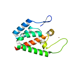
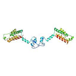
2PTL
 | | THREE-DIMENSIONAL SOLUTION STRUCTURE OF AN IMMUNOGLOBULIN LIGHT CHAIN-BINDING DOMAIN OF PROTEIN L. COMPARISON WITH THE IGG-BINDING DOMAINS OF PROTEIN G | | 分子名称: | PROTEIN L | | 著者 | Wikstroem, M, Drakenberg, T, Forsen, S, Sjoebring, U, Bjoerck, L. | | 登録日 | 1994-08-12 | | 公開日 | 1994-10-15 | | 最終更新日 | 2024-05-01 | | 実験手法 | SOLUTION NMR | | 主引用文献 | Three-dimensional solution structure of an immunoglobulin light chain-binding domain of protein L. Comparison with the IgG-binding domains of protein G.
Biochemistry, 33, 1994
|
|
2PVG
 | |
2PVO
 | |
2HFD
 | | NMR structure of protein Hydrogenase-1 operon protein hyaE from Escherichia coli: Northeast Structural Genomics Consortium Target ER415 | | 分子名称: | Hydrogenase-1 operon protein hyaE | | 著者 | Singarapu, K.K, Liu, G, Eletsky, A, Parish, D, Atreya, H.S, Xu, D, Janjua, H, Cunningham, K, Ma, L.C, Xiao, R, Liu, J, Baran, M, Swapna, G.V.T, Acton, T, Rost, B, Montelione, G.T, Szyperski, T, Northeast Structural Genomics Consortium (NESG) | | 登録日 | 2006-06-23 | | 公開日 | 2006-08-22 | | 最終更新日 | 2024-05-01 | | 実験手法 | SOLUTION NMR | | 主引用文献 | Protein chaperones Q8ZP25_SALTY from Salmonella typhimurium and HYAE_ECOLI from Escherichia coli exhibit thioredoxin-like structures despite lack of canonical thioredoxin active site sequence motif.
J.STRUCT.FUNCT.GENOM., 9, 2008
|
|
2Q9D
 | | Structure of spin-labeled T4 lysozyme mutant A41R1 | | 分子名称: | BETA-MERCAPTOETHANOL, Lysozyme, S-[(1-oxyl-2,2,5,5-tetramethyl-2,5-dihydro-1H-pyrrol-3-yl)methyl] methanesulfonothioate | | 著者 | Guo, Z, Cascio, D, Hideg, K, Hubbell, W.L. | | 登録日 | 2007-06-12 | | 公開日 | 2007-06-26 | | 最終更新日 | 2023-08-30 | | 実験手法 | EPR (1.4 Å), X-RAY DIFFRACTION | | 主引用文献 | Structural determinants of nitroxide motion in spin-labeled proteins: Solvent-exposed sites in helix B of T4 lysozyme.
Protein Sci., 17, 2008
|
|
2GP7
 | | Estrogen Related Receptor-gamma ligand binding domain | | 分子名称: | CALCIUM ION, Estrogen-related receptor gamma | | 著者 | Wang, L, Zuercher, W.J, Consler, T.G, Lambert, M.H, Miller, A.B, Osband-miller, L.A, McKee, D.D, Willson, T.M, Nolte, R.T. | | 登録日 | 2006-04-17 | | 公開日 | 2006-09-26 | | 最終更新日 | 2023-08-30 | | 実験手法 | X-RAY DIFFRACTION (2.45 Å) | | 主引用文献 | X-ray crystal structures of the estrogen-related receptor-gamma ligand binding domain in three functional states reveal the molecular basis of small molecule regulation.
J.Biol.Chem., 281, 2006
|
|
2GQ7
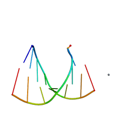 | | Crystal structure of an RNA racemate | | 分子名称: | CALCIUM ION, GLYCEROL, RNA (5'-R(*(0C)P*(0C)P*(0G)P*(0C)P*(0C)P*(0U)P*(0G)P*(0G))-3'), ... | | 著者 | Rypniewski, W, Vallazza, M, Perbandt, M, Klussmann, S, Betzel, C, Erdmann, V.A. | | 登録日 | 2006-04-20 | | 公開日 | 2006-06-27 | | 最終更新日 | 2024-04-03 | | 実験手法 | X-RAY DIFFRACTION (1.6 Å) | | 主引用文献 | The first crystal structure of an RNA racemate.
Acta Crystallogr.,Sect.D, 62, 2006
|
|
2F6N
 | |
2H2W
 | |
2NBW
 | |
2H0K
 | | Crystal Structure of a Mutant of Rat Annexin A5 | | 分子名称: | Annexin A5, CALCIUM ION | | 著者 | Granier, T, Langlois D'Estaintot, B, Gallois, B, Tessier, B, Brisson, A. | | 登録日 | 2006-05-15 | | 公開日 | 2007-06-19 | | 最終更新日 | 2023-08-30 | | 実験手法 | X-RAY DIFFRACTION (2.76 Å) | | 主引用文献 | Annexin-A5 assembled into two-dimensional arrays promotes cell membrane repair.
Nat Commun, 2, 2011
|
|
4COC
 | | HIV-1 capsid C-terminal domain mutant (Y169L) | | 分子名称: | CAPSID PROTEIN P24, SULFATE ION | | 著者 | Bharat, T.A.M, Castillo-Menendez, L.R, Hagen, W.J.H, Lux, V, Igonet, S, Schorb, M, Schur, F.K.M, Krausslich, H.-G, Briggs, J.A.G. | | 登録日 | 2014-01-28 | | 公開日 | 2014-06-04 | | 最終更新日 | 2023-12-20 | | 実験手法 | X-RAY DIFFRACTION (1.59 Å) | | 主引用文献 | Cryo-Electron Microscopy of Tubular Arrays of HIV-1 Gag Resolves Structures Essential for Immature Virus Assembly.
Proc.Natl.Acad.Sci.USA, 111, 2014
|
|
2H1E
 | |
2NAD
 | | HIGH RESOLUTION STRUCTURES OF HOLO AND APO FORMATE DEHYDROGENASE | | 分子名称: | AZIDE ION, NAD-DEPENDENT FORMATE DEHYDROGENASE, NICOTINAMIDE-ADENINE-DINUCLEOTIDE, ... | | 著者 | Lamzin, V.S, Dauter, Z, Popov, V.O, Harutyunyan, E.H, Wilson, K.S. | | 登録日 | 1994-07-06 | | 公開日 | 1995-01-26 | | 最終更新日 | 2024-02-21 | | 実験手法 | X-RAY DIFFRACTION (2.05 Å) | | 主引用文献 | High resolution structures of holo and apo formate dehydrogenase.
J.Mol.Biol., 236, 1994
|
|
2NO5
 | | Crystal Structure analysis of a Dehalogenase with intermediate complex | | 分子名称: | (2S)-2-CHLOROPROPANOIC ACID, (S)-2-haloacid dehalogenase IVA, CHLORIDE ION, ... | | 著者 | Schmidberger, J.W, Wilce, M.C.J. | | 登録日 | 2006-10-24 | | 公開日 | 2007-09-25 | | 最終更新日 | 2023-11-15 | | 実験手法 | X-RAY DIFFRACTION (2.6 Å) | | 主引用文献 | Crystal structures of the substrate free-enzyme, and reaction intermediate of the HAD superfamily member, haloacid dehalogenase DehIVa from Burkholderia cepacia MBA4
J.Mol.Biol., 368, 2007
|
|


