4JJ3
 
 | |
4JC3
 | |
4LCQ
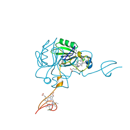
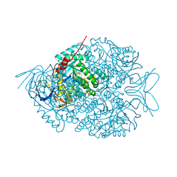
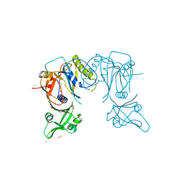
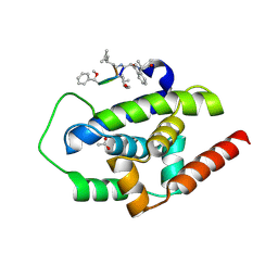
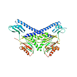
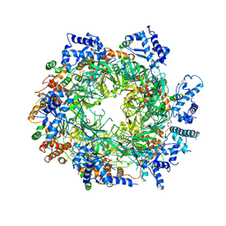
 | | The crystal structure of di-Zn dihydropyrimidinase in complex with NCBI | | 分子名称: | (2S)-3-(carbamoylamino)-2-methylpropanoic acid, ZINC ION, dihydropyrimidinase | | 著者 | Hsieh, Y.C, Chen, M.C, Hsu, C.C, Chan, S.I, Yang, Y.S, Chen, C.J. | | 登録日 | 2013-06-22 | | 公開日 | 2013-09-18 | | 最終更新日 | 2014-02-12 | | 実験手法 | X-RAY DIFFRACTION (1.81 Å) | | 主引用文献 | Crystal structures of vertebrate dihydropyrimidinase and complexes from Tetraodon nigroviridis with lysine carbamylation: metal and structural requirements for post-translational modification and function.
J.Biol.Chem., 288, 2013
|
|
4LCS
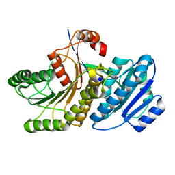
 | | The crystal structure of di-Zn dihydropyrimidinase in complex with hydantoin | | 分子名称: | Chromosome 8 SCAF14545, whole genome shotgun sequence, ZINC ION, ... | | 著者 | Hsieh, Y.C, Chen, M.C, Hsu, C.C, Chan, S.I, Yang, Y.S. | | 登録日 | 2013-06-23 | | 公開日 | 2013-09-18 | | 最終更新日 | 2014-02-12 | | 実験手法 | X-RAY DIFFRACTION (2.2 Å) | | 主引用文献 | Crystal structures of vertebrate dihydropyrimidinase and complexes from Tetraodon nigroviridis with lysine carbamylation: metal and structural requirements for post-translational modification and function.
J.Biol.Chem., 288, 2013
|
|
4LCR
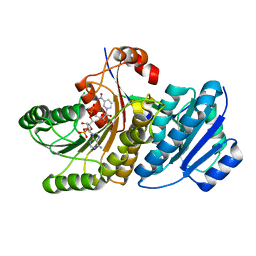
 | | The crystal structure of di-Zn dihydropyrimidinase in complex with NCBA | | 分子名称: | Chromosome 8 SCAF14545, whole genome shotgun sequence, N-(AMINOCARBONYL)-BETA-ALANINE, ... | | 著者 | Hsieh, Y.C, Chen, M.C, Hsu, C.C, Chan, S.I, Yang, Y.S, Chen, C.J. | | 登録日 | 2013-06-22 | | 公開日 | 2013-09-18 | | 最終更新日 | 2014-02-12 | | 実験手法 | X-RAY DIFFRACTION (2 Å) | | 主引用文献 | Crystal structures of vertebrate dihydropyrimidinase and complexes from Tetraodon nigroviridis with lysine carbamylation: metal and structural requirements for post-translational modification and function.
J.Biol.Chem., 288, 2013
|
|
4LDU
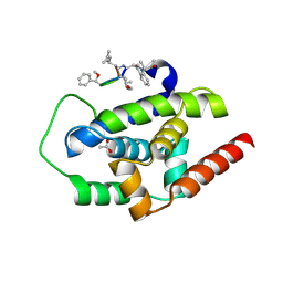
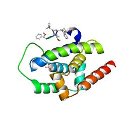
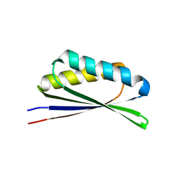
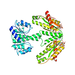
 | | Crystal structure of the DNA binding domain of Arabidopsis thaliana auxin response factor 5 | | 分子名称: | Auxin response factor 5, CHLORIDE ION | | 著者 | Boer, D.R, Freire-Rios, A, van den Berg, W.M.A, Weijers, D, Coll, M. | | 登録日 | 2013-06-25 | | 公開日 | 2014-02-12 | | 最終更新日 | 2024-02-28 | | 実験手法 | X-RAY DIFFRACTION (2.15 Å) | | 主引用文献 | Structural Basis for DNA Binding Specificity by the Auxin-Dependent ARF Transcription Factors.
Cell(Cambridge,Mass.), 156, 2014
|
|
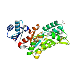
4KWI
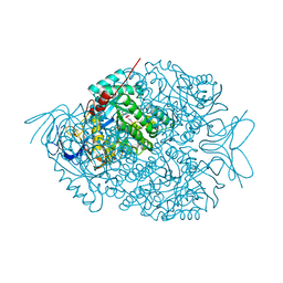
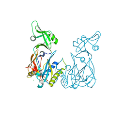
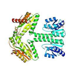
 | | The crystal structure of angucycline C-6 ketoreductase LanV with bound NADP and 11-deoxy-6-oxylandomycinone | | 分子名称: | 1,8-dihydroxy-3-methyltetraphene-6,7,12(5H)-trione, DI(HYDROXYETHYL)ETHER, NADP NICOTINAMIDE-ADENINE-DINUCLEOTIDE PHOSPHATE, ... | | 著者 | Paananen, P, Patrikainen, P, Mantsala, P, Niemi, J, Niiranen, L, Metsa-Ketela, M. | | 登録日 | 2013-05-24 | | 公開日 | 2013-07-31 | | 最終更新日 | 2023-09-20 | | 実験手法 | X-RAY DIFFRACTION (2 Å) | | 主引用文献 | Structural and functional analysis of angucycline C-6 ketoreductase LanV involved in landomycin biosynthesis.
Biochemistry, 52, 2013
|
|
4I1Q
 | |
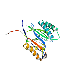
4LDV
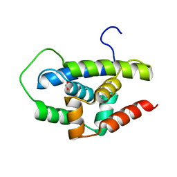
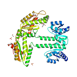
 | | Crystal structure of the DNA binding domain of A. thailana auxin response factor 1 | | 分子名称: | Auxin response factor 1, CHLORIDE ION, FORMIC ACID, ... | | 著者 | boer, D.R, Freire-Rios, A, van den Berg, W.M.A, Weijers, D, Coll, M. | | 登録日 | 2013-06-25 | | 公開日 | 2014-02-12 | | 最終更新日 | 2024-04-03 | | 実験手法 | X-RAY DIFFRACTION (1.45 Å) | | 主引用文献 | Structural Basis for DNA Binding Specificity by the Auxin-Dependent ARF Transcription Factors.
Cell(Cambridge,Mass.), 156, 2014
|
|
3WDE
 | |
3WDB
 | |
3WDD
 | |
3WDC
 | |
4J1S
 | |
4J1Q
 | |
6WI5
 | |
3E6S
 | |
8Q4S
 | |
5MQH
 | |
1ZY6
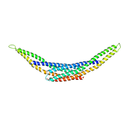
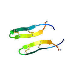 | | Membrane-bound dimer structure of Protegrin-1 (PG-1), a beta-Hairpin Antimicrobial Peptide in Lipid Bilayers from Rotational-Echo Double-Resonance Solid-State NMR | | 分子名称: | Protegrin 1 | | 著者 | Wu, X, Mani, R, Tang, M, Buffy, J.J, Waring, A.J, Sherman, M.A, Hong, M. | | 登録日 | 2005-06-09 | | 公開日 | 2006-06-13 | | 最終更新日 | 2022-03-02 | | 実験手法 | SOLID-STATE NMR | | 主引用文献 | Membrane-Bound Dimer Structure of a beta-Hairpin Antimicrobial Peptide from Rotational-Echo Double-Resonance Solid-State NMR.
Biochemistry, 45, 2006
|
|
5UCG
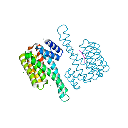
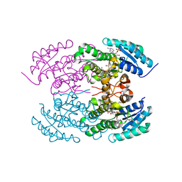
 | | Structure of the PP2C Phosphatase Domain and a Fragment of the Regulatory Domain of the Cell Fate Determinant SpoIIE from Bacillus Subtilis | | 分子名称: | Stage II sporulation protein E | | 著者 | Bradshaw, N, Levdikov, V, Zimanyi, C, Gaudet, R, Wilkinson, A, Losick, R. | | 登録日 | 2016-12-22 | | 公開日 | 2017-05-31 | | 最終更新日 | 2023-10-04 | | 実験手法 | X-RAY DIFFRACTION (3.906 Å) | | 主引用文献 | A widespread family of serine/threonine protein phosphatases shares a common regulatory switch with proteasomal proteases.
Elife, 6, 2017
|
|
6ZXM
 | | Diguanylate cyclase DgcR in complex with c-di-GMP | | 分子名称: | 9,9'-[(2R,3R,3aS,5S,7aR,9R,10R,10aS,12S,14aR)-3,5,10,12-tetrahydroxy-5,12-dioxidooctahydro-2H,7H-difuro[3,2-d:3',2'-j][1,3,7,9,2,8]tetraoxadiphosphacyclododecine-2,9-diyl]bis(2-amino-1,9-dihydro-6H-purin-6-one), MAGNESIUM ION, Putative GGDEF/response regulator receiver domain protein | | 著者 | Teixeira, R.D, Schirmer, T. | | 登録日 | 2020-07-29 | | 公開日 | 2021-03-31 | | 最終更新日 | 2024-01-31 | | 実験手法 | X-RAY DIFFRACTION (3.3 Å) | | 主引用文献 | Activation mechanism of a small prototypic Rec-GGDEF diguanylate cyclase.
Nat Commun, 12, 2021
|
|
6ZXC
 | |
6ZXB
 | |
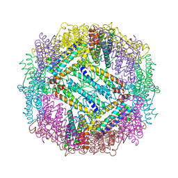
6EYC
 | | Re-refinement of the MCM2-7 double hexamer using ISOLDE | | 分子名称: | ADENOSINE-5'-DIPHOSPHATE, DNA replication licensing factor MCM2, DNA replication licensing factor MCM3, ... | | 著者 | Croll, T.I. | | 登録日 | 2017-11-11 | | 公開日 | 2018-06-20 | | 最終更新日 | 2024-05-08 | | 実験手法 | ELECTRON MICROSCOPY (3.8 Å) | | 主引用文献 | ISOLDE: a physically realistic environment for model building into low-resolution electron-density maps.
Acta Crystallogr D Struct Biol, 74, 2018
|
|


