1QYR
 
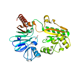
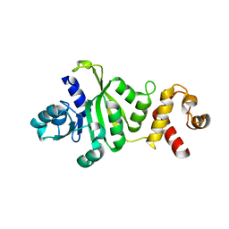 | | 2.1 Angstrom Crystal structure of KsgA: A Universally Conserved Adenosine Dimethyltransferase | | 分子名称: | High level Kasugamycin resistance protein | | 著者 | O'Farrell, H.C, Scarsdale, J.N, Wright, H.T, Rife, J.P. | | 登録日 | 2003-09-11 | | 公開日 | 2004-06-29 | | 最終更新日 | 2024-02-14 | | 実験手法 | X-RAY DIFFRACTION (2.1 Å) | | 主引用文献 | Crystal structure of KsgA, a universally conserved rRNA adenine dimethyltransferase in Escherichia coli
J.Mol.Biol., 339, 2004
|
|
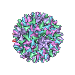
1QGT
 | |
1PXX
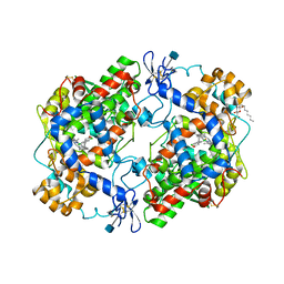 | | CRYSTAL STRUCTURE OF DICLOFENAC BOUND TO THE CYCLOOXYGENASE ACTIVE SITE OF COX-2 | | 分子名称: | 2-[2,6-DICHLOROPHENYL)AMINO]BENZENEACETIC ACID, 2-acetamido-2-deoxy-beta-D-glucopyranose, 2-acetamido-2-deoxy-beta-D-glucopyranose-(1-4)-2-acetamido-2-deoxy-beta-D-glucopyranose-(1-4)-2-acetamido-2-deoxy-beta-D-glucopyranose, ... | | 著者 | Kiefer, J.R, Rowlinson, S.W, Prusakiewicz, J.J, Pawlitz, J.L, Kozak, K.R, Kalgutkar, A.S, Stallings, W.C, Marnett, L.J, Kurumbail, R.G. | | 登録日 | 2003-07-07 | | 公開日 | 2003-09-09 | | 最終更新日 | 2020-07-29 | | 実験手法 | X-RAY DIFFRACTION (2.9 Å) | | 主引用文献 | A Novel Mechanism of Cyclooxygenase-2 Inhibition Involving Interactions with Ser-530 and Tyr-385.
J.Biol.Chem., 278, 2003
|
|
1PQS
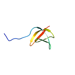
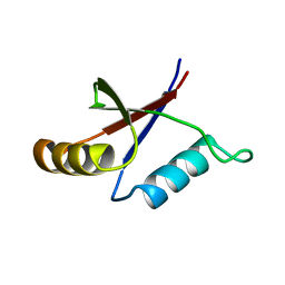 | | Solution structure of the C-terminal OPCA domain of yCdc24p | | 分子名称: | Cell division control protein 24 | | 著者 | Leitner, D, Wahl, M, Labudde, D, Diehl, A, Schmieder, P, Pires, J.R, Fossi, M, Leidert, M, Krause, G, Oschkinat, H. | | 登録日 | 2003-06-19 | | 公開日 | 2003-07-01 | | 最終更新日 | 2024-05-29 | | 実験手法 | SOLUTION NMR | | 主引用文献 | The solution structure of an N-terminally truncated version of the yeast CDC24p PB1 domain shows a different beta-sheet topology.
Febs Lett., 579, 2005
|
|
5P4C
 | |
5P1M
 | |
5P0P
 | |
5P38
 | |
5P3J
 | |
5Q64
 | | PanDDA analysis group deposition -- Crystal Structure of DCLRE1A after initial refinement with no ligand modelled (structure 141) | | 分子名称: | DCLRE1A, MALONATE ION, NICKEL (II) ION | | 著者 | Newman, J.A, Aitkenhead, H, Lee, S.Y, Kupinska, K, Burgess-Brown, N, Tallon, R, Krojer, T, von Delft, F, Arrowsmith, C.H, Edwards, A, Bountra, C, Gileadi, O. | | 登録日 | 2017-05-25 | | 公開日 | 2018-08-08 | | 最終更新日 | 2024-03-06 | | 実験手法 | X-RAY DIFFRACTION (1.42 Å) | | 主引用文献 | PanDDA analysis group deposition
To Be Published
|
|
1WG3
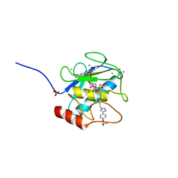
 | | Structural analysis of yeast nucleosome-assembly factor CIA1p | | 分子名称: | Anti-silencing protein 1 | | 著者 | Padmanabhan, B, Kataoka, K, Umehara, T, Adachi, N, Yokoyama, S, Horikoshi, M, RIKEN Structural Genomics/Proteomics Initiative (RSGI) | | 登録日 | 2004-05-27 | | 公開日 | 2005-06-14 | | 最終更新日 | 2023-10-25 | | 実験手法 | X-RAY DIFFRACTION (3 Å) | | 主引用文献 | Structural Similarity between Histone Chaperone Cia1p/Asf1p and DNA-Binding Protein NF-{kappa}B
J.Biochem.(Tokyo), 138, 2005
|
|
5Q6C
 | | PanDDA analysis group deposition -- Crystal Structure of DCLRE1A after initial refinement with no ligand modelled (structure 149) | | 分子名称: | DCLRE1A, MALONATE ION, NICKEL (II) ION | | 著者 | Newman, J.A, Aitkenhead, H, Lee, S.Y, Kupinska, K, Burgess-Brown, N, Tallon, R, Krojer, T, von Delft, F, Arrowsmith, C.H, Edwards, A, Bountra, C, Gileadi, O. | | 登録日 | 2017-05-25 | | 公開日 | 2018-08-08 | | 最終更新日 | 2024-03-06 | | 実験手法 | X-RAY DIFFRACTION (1.53 Å) | | 主引用文献 | PanDDA analysis group deposition
To Be Published
|
|
5Q8D
 | | PanDDA analysis group deposition -- Crystal Structure of DCLRE1A after initial refinement with no ligand modelled (structure 222) | | 分子名称: | DCLRE1A, MALONATE ION, NICKEL (II) ION | | 著者 | Newman, J.A, Aitkenhead, H, Lee, S.Y, Kupinska, K, Burgess-Brown, N, Tallon, R, Krojer, T, von Delft, F, Arrowsmith, C.H, Edwards, A, Bountra, C, Gileadi, O. | | 登録日 | 2017-05-25 | | 公開日 | 2018-08-08 | | 最終更新日 | 2024-03-06 | | 実験手法 | X-RAY DIFFRACTION (1.23 Å) | | 主引用文献 | PanDDA analysis group deposition
To Be Published
|
|
5Q8G
 | | PanDDA analysis group deposition -- Crystal Structure of DCLRE1A after initial refinement with no ligand modelled (structure 225) | | 分子名称: | DCLRE1A, MALONATE ION, NICKEL (II) ION | | 著者 | Newman, J.A, Aitkenhead, H, Lee, S.Y, Kupinska, K, Burgess-Brown, N, Tallon, R, Krojer, T, von Delft, F, Arrowsmith, C.H, Edwards, A, Bountra, C, Gileadi, O. | | 登録日 | 2017-05-25 | | 公開日 | 2018-08-08 | | 最終更新日 | 2024-03-06 | | 実験手法 | X-RAY DIFFRACTION (2 Å) | | 主引用文献 | PanDDA analysis group deposition
To Be Published
|
|
4JG4
 | |
4J8R
 | | Structure of an octapeptide repeat of the prion protein bound to the POM2 Fab antibody fragment | | 分子名称: | Heavy chain of POM2 Fab, Light chain of POM2 Fab, Major prion protein | | 著者 | Swayampakula, M, Baral, P.K, Kav, N.N.V, Aguzzi, A, James, M.N.G. | | 登録日 | 2013-02-14 | | 公開日 | 2013-05-22 | | 最終更新日 | 2023-09-20 | | 実験手法 | X-RAY DIFFRACTION (2.303 Å) | | 主引用文献 | The crystal structure of an octapeptide repeat of the Prion protein in complex with a Fab fragment of the POM2 antibody.
Protein Sci., 22, 2013
|
|
5P0N
 | |
5P80
 | |
5P78
 | |
5Q4P
 | | PanDDA analysis group deposition -- Crystal Structure of DCLRE1A after initial refinement with no ligand modelled (structure 90) | | 分子名称: | DCLRE1A, MALONATE ION, NICKEL (II) ION | | 著者 | Newman, J.A, Aitkenhead, H, Lee, S.Y, Kupinska, K, Burgess-Brown, N, Tallon, R, Krojer, T, von Delft, F, Arrowsmith, C.H, Edwards, A, Bountra, C, Gileadi, O. | | 登録日 | 2017-05-25 | | 公開日 | 2018-08-08 | | 最終更新日 | 2024-03-06 | | 実験手法 | X-RAY DIFFRACTION (1.4 Å) | | 主引用文献 | PanDDA analysis group deposition
To Be Published
|
|
1YVC
 | | Solution structure of the conserved protein from the gene locus MMP0076 of Methanococcus maripaludis. Northeast Structural Genomics target MrR5. | | 分子名称: | MrR5 | | 著者 | Northeast Structural Genomics Consortium (NESG), Rossi, P, Aramini, J.M, Xiao, R, Ho, C.K, Ma, L.C, Acton, T.B, Montelione, G.T. | | 登録日 | 2005-02-15 | | 公開日 | 2005-04-05 | | 最終更新日 | 2024-05-22 | | 実験手法 | SOLUTION NMR | | 主引用文献 |
To be Published
|
|
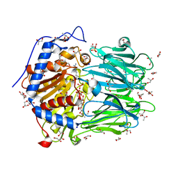
3O2X
 | | MMP-13 in complex with selective tetrazole core inhibitor | | 分子名称: | 4-(2-HYDROXYETHYL)-1-PIPERAZINE ETHANESULFONIC ACID, CALCIUM ION, Collagenase 3, ... | | 著者 | Kiefer, J.R, Barta, T.E, Becker, D.P, Mathis, K.J, Williams, J. | | 登録日 | 2010-07-22 | | 公開日 | 2011-08-10 | | 最終更新日 | 2024-02-21 | | 実験手法 | X-RAY DIFFRACTION (1.9 Å) | | 主引用文献 | MMP-13 in complex with selective tetrazole core inhibitor
To be Published
|
|
4AHC
 | |
4AIL
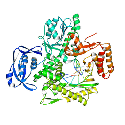 | |
2XE4
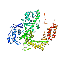
 | | Structure of Oligopeptidase B from Leishmania major | | 分子名称: | ANTIPAIN, CHLORIDE ION, GLYCEROL, ... | | 著者 | McLuskey, K, Paterson, N.G, Bland, N.D, Mottram, J.C, Isaacs, N.W. | | 登録日 | 2010-05-11 | | 公開日 | 2010-10-06 | | 最終更新日 | 2019-05-15 | | 実験手法 | X-RAY DIFFRACTION (1.65 Å) | | 主引用文献 | Crystal Structure of Leishmania Major Oligopeptidase B Gives Insight Into the Enzymatic Properties of a Trypanosomatid Virulence Factor.
J.Biol.Chem., 285, 2010
|
|


