1WZW
 
 | | Crystal Structure of UbcH8 | | 分子名称: | Ubiquitin-conjugating enzyme E2 L6 | | 著者 | Mizushima, T, Suzuki, M, Teshima, N, Yamane, T, Murata, S, Tanaka, K. | | 登録日 | 2005-03-10 | | 公開日 | 2005-03-22 | | 最終更新日 | 2024-03-13 | | 実験手法 | X-RAY DIFFRACTION (2.4 Å) | | 主引用文献 | Crystal Structure of UbcH8
To be Published
|
|
1WZX
 | | Crystal Structure of Family 30 Carbohydrate Binding Module. | | 分子名称: | COG3291: FOG: PKD repeat | | 著者 | Horiguchi, Y, Kono, M, Suzuki, A, Yamane, T, Arai, M, Sakka, K, Omiya, K. | | 登録日 | 2005-03-10 | | 公開日 | 2005-03-22 | | 最終更新日 | 2023-10-25 | | 実験手法 | X-RAY DIFFRACTION (3.52 Å) | | 主引用文献 | Crystal Structure of Family 30 Carbohydrate Binding Module
To be Published
|
|
1WZY
 | |
1WZZ
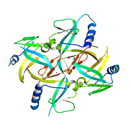
 | | Structure of endo-beta-1,4-glucanase CMCax from Acetobacter xylinum | | 分子名称: | Probable endoglucanase, SULFATE ION | | 著者 | Yasutake, Y, Kawano, S, Tajima, K, Yao, M, Satoh, Y, Munekata, M, Tanaka, I, Structural Genomics Consortium (SGC) | | 登録日 | 2005-03-10 | | 公開日 | 2006-03-14 | | 最終更新日 | 2011-07-13 | | 実験手法 | X-RAY DIFFRACTION (1.65 Å) | | 主引用文献 | Structural characterization of the Acetobacter xylinum endo-beta-1,4-glucanase CMCax required for cellulose biosynthesis.
Proteins, 64, 2006
|
|
1X01
 | |
1X02
 | | Solution structure of stereo array isotope labeled (SAIL) calmodulin | | 分子名称: | CALCIUM ION, calmodulin | | 著者 | Kainosho, M, Torizawa, T, Terauchi, T, Ono, A.M, Guntert, P. | | 登録日 | 2005-03-11 | | 公開日 | 2006-03-07 | | 最終更新日 | 2024-05-29 | | 実験手法 | SOLUTION NMR | | 主引用文献 | Optimal isotope labelling for NMR protein structure determinations.
Nature, 440, 2006
|
|
1X03
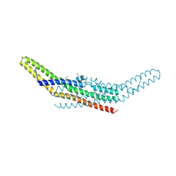 | | Crystal structure of endophilin BAR domain | | 分子名称: | SH3-containing GRB2-like protein 2 | | 著者 | Masuda, M, Takeda, S, Sone, M, Kamioka, Y, Mori, H, Mochizuki, N. | | 登録日 | 2005-03-14 | | 公開日 | 2006-05-02 | | 最終更新日 | 2024-10-09 | | 実験手法 | X-RAY DIFFRACTION (3.1 Å) | | 主引用文献 | Endophilin BAR domain drives membrane curvature by two newly identified structure-based mechanisms
Embo J., 25, 2006
|
|
1X04
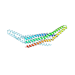 | | Crystal structure of endophilin BAR domain (mutant) | | 分子名称: | SH3-containing GRB2-like protein 2 | | 著者 | Masuda, M, Takeda, S, Sone, M, Kamioka, Y, Mori, H, Mochizuki, N. | | 登録日 | 2005-03-14 | | 公開日 | 2006-05-02 | | 最終更新日 | 2017-08-23 | | 実験手法 | X-RAY DIFFRACTION (2.9 Å) | | 主引用文献 | Endophilin BAR domain drives membrane curvature by two newly identified structure-based mechanisms
Embo J., 25, 2006
|
|
1X05
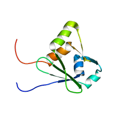
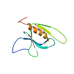 | | Solution structure of the C-terminal PH domain of human pleckstrin | | 分子名称: | Pleckstrin | | 著者 | Li, H, Tomizawa, T, Koshiba, S, Inoue, M, Kigawa, T, Yokoyama, S, RIKEN Structural Genomics/Proteomics Initiative (RSGI) | | 登録日 | 2005-03-15 | | 公開日 | 2005-09-15 | | 最終更新日 | 2024-05-29 | | 実験手法 | SOLUTION NMR | | 主引用文献 | Solution structure of the C-terminal PH domain of human pleckstrin
To be Published
|
|
1X06
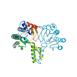 | | Crystal structure of undecaprenyl pyrophosphate synthase in complex with Mg, IPP and Fspp | | 分子名称: | MAGNESIUM ION, PHOSPHATE ION, S-[(2E,6E)-3,7,11-TRIMETHYLDODECA-2,6,10-TRIENYL] TRIHYDROGEN THIODIPHOSPHATE, ... | | 著者 | Guo, R.-T, Ko, T.-P, Chen, A.P.-C, Kuo, C.-J, Wang, A.H.-J, Liang, P.-H. | | 登録日 | 2005-03-15 | | 公開日 | 2005-03-22 | | 最終更新日 | 2023-10-25 | | 実験手法 | X-RAY DIFFRACTION (1.9 Å) | | 主引用文献 | Crystal structures of undecaprenyl pyrophosphate synthase in complex with magnesium, isopentenyl pyrophosphate, and farnesyl thiopyrophosphate: roles of the metal ion and conserved residues in catalysis.
J.Biol.Chem., 280, 2005
|
|
1X07
 | | Crystal structure of undecaprenyl pyrophosphate synthase in complex with Mg and IPP | | 分子名称: | 3-METHYLBUT-3-ENYL TRIHYDROGEN DIPHOSPHATE, MAGNESIUM ION, PHOSPHATE ION, ... | | 著者 | Guo, R.-T, Ko, T.-P, Chen, A.P.-C, Kuo, C.-J, Wang, A.H.-J, Liang, P.-H. | | 登録日 | 2005-03-15 | | 公開日 | 2005-03-22 | | 最終更新日 | 2023-10-25 | | 実験手法 | X-RAY DIFFRACTION (2.2 Å) | | 主引用文献 | Crystal structures of undecaprenyl pyrophosphate synthase in complex with magnesium, isopentenyl pyrophosphate, and farnesyl thiopyrophosphate: roles of the metal ion and conserved residues in catalysis.
J.Biol.Chem., 280, 2005
|
|
1X08
 | | Crystal structure of D26A mutant UPPs in complex with Mg, IPP and FsPP | | 分子名称: | 3-METHYLBUT-3-ENYL TRIHYDROGEN DIPHOSPHATE, S-[(2E,6E)-3,7,11-TRIMETHYLDODECA-2,6,10-TRIENYL] TRIHYDROGEN THIODIPHOSPHATE, Undecaprenyl pyrophosphate synthetase | | 著者 | Guo, R.-T, Ko, T.-P, Chen, A.P.-C, Kuo, C.-J, Wang, A.H.-J, Liang, P.-H. | | 登録日 | 2005-03-15 | | 公開日 | 2005-03-22 | | 最終更新日 | 2023-10-25 | | 実験手法 | X-RAY DIFFRACTION (1.9 Å) | | 主引用文献 | Crystal structures of undecaprenyl pyrophosphate synthase in complex with magnesium, isopentenyl pyrophosphate, and farnesyl thiopyrophosphate: roles of the metal ion and conserved residues in catalysis.
J.Biol.Chem., 280, 2005
|
|
1X09
 | | Crystal structure of the D26A mutant UPPs in complex with magnesium and isopentenyl pyrophosphate | | 分子名称: | 3-METHYLBUT-3-ENYL TRIHYDROGEN DIPHOSPHATE, MAGNESIUM ION, Undecaprenyl pyrophosphate synthetase | | 著者 | Guo, R.-T, Ko, T.-P, Chen, A.P.-C, Kuo, C.-J, Wang, A.H.-J, Liang, P.-H. | | 登録日 | 2005-03-15 | | 公開日 | 2005-03-22 | | 最終更新日 | 2023-10-25 | | 実験手法 | X-RAY DIFFRACTION (1.87 Å) | | 主引用文献 | Crystal structures of undecaprenyl pyrophosphate synthase in complex with magnesium, isopentenyl pyrophosphate, and farnesyl thiopyrophosphate: roles of the metal ion and conserved residues in catalysis.
J.Biol.Chem., 280, 2005
|
|
1X0A
 | |
1X0C
 | | Improved Crystal Structure of Isopullulanase from Aspergillus niger ATCC 9642 | | 分子名称: | 2-acetamido-2-deoxy-beta-D-glucopyranose, Isopullulanase | | 著者 | Mizuno, M, Tonozuka, T, Yamamura, A, Miyasaka, Y, Akeboshi, H, Kamitori, S, Nishikawa, A, Sakano, Y. | | 登録日 | 2005-03-17 | | 公開日 | 2006-06-13 | | 最終更新日 | 2020-07-29 | | 実験手法 | X-RAY DIFFRACTION (1.7 Å) | | 主引用文献 | Crystal Structure of Aspergillus niger Isopullulanase, a Member of Glycoside Hydrolase Family 49
J.Mol.Biol., 376, 2008
|
|
1X0F
 | | Complex structure of the C-terminal RNA-binding domain of hnRNP D(AUF1) with telomeric DNA | | 分子名称: | 5'-D(P*TP*AP*GP*G)-3', Heterogeneous nuclear ribonucleoprotein D0 | | 著者 | Enokizono, Y, Konishi, Y, Nagata, K, Ouhashi, K, Uesugi, S, Ishikawa, F, Katahira, M. | | 登録日 | 2005-03-22 | | 公開日 | 2005-04-05 | | 最終更新日 | 2024-05-29 | | 実験手法 | SOLUTION NMR | | 主引用文献 | Structure of hnRNP D complexed with single-stranded telomere DNA and unfolding of the quadruplex by heterogeneous nuclear ribonucleoprotein D.
J.Biol.Chem., 280, 2005
|
|
1X0G
 | | Crystal Structure of IscA with the [2Fe-2S] cluster | | 分子名称: | FE2/S2 (INORGANIC) CLUSTER, IscA, SODIUM ION | | 著者 | Morimoto, K, Yamashita, E, Kondou, Y, Lee, S.J, Tsukihara, T, Nakai, M. | | 登録日 | 2005-03-22 | | 公開日 | 2006-06-06 | | 最終更新日 | 2024-03-13 | | 実験手法 | X-RAY DIFFRACTION (2.5 Å) | | 主引用文献 | The Asymmetric IscA Homodimer with an Exposed [2Fe-2S] Cluster Suggests the Structural Basis of the Fe-S Cluster Biosynthetic Scaffold.
J.Mol.Biol., 360, 2006
|
|
1X0H
 | | Solution structure of the carboxyl-terminal RGC domain in human IQGAP1 | | 分子名称: | Ras GTPase-activating-like protein IQGAP1 | | 著者 | Saito, K, Koshiba, S, Inoue, M, Kigawa, T, Yokoyama, S, RIKEN Structural Genomics/Proteomics Initiative (RSGI) | | 登録日 | 2005-03-23 | | 公開日 | 2005-09-23 | | 最終更新日 | 2024-05-29 | | 実験手法 | SOLUTION NMR | | 主引用文献 | Solution structure of the carboxyl-terminal RGC domain in human IQGAP1
To be Published
|
|
1X0I
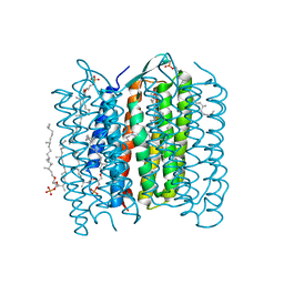
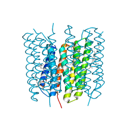 | | Crystal Structure of the Acid Blue Form of Bacteriorhodopsin | | 分子名称: | 2,3-DI-O-PHYTANLY-3-SN-GLYCERO-1-PHOSPHORYL-3'-SN-GLYCEROL-1'-PHOSPHATE, 2,3-DI-PHYTANYL-GLYCEROL, Bacteriorhodopsin, ... | | 著者 | Okumura, H, Murakami, M, Kouyama, T. | | 登録日 | 2005-03-23 | | 公開日 | 2005-08-02 | | 最終更新日 | 2023-10-25 | | 実験手法 | X-RAY DIFFRACTION (2.3 Å) | | 主引用文献 | Crystal Structures of Acid Blue and Alkaline Purple Forms of Bacteriorhodopsin
J.Mol.Biol., 351, 2005
|
|
1X0J
 | | Crystal structure analysis of the N-terminal bromodomain of human Brd2 | | 分子名称: | 2,3-DIHYDROXY-1,4-DITHIOBUTANE, 2-(N-MORPHOLINO)-ETHANESULFONIC ACID, Bromodomain-containing protein 2 | | 著者 | Nakamura, Y, Umehara, T, Shirouzu, M, Padmanabhan, B, Yokoyama, S, RIKEN Structural Genomics/Proteomics Initiative (RSGI) | | 登録日 | 2005-03-23 | | 公開日 | 2006-06-27 | | 最終更新日 | 2011-07-13 | | 実験手法 | X-RAY DIFFRACTION (1.8 Å) | | 主引用文献 | Structural basis for acetylated histone H4 recognition by the human Brd2 bromodomain
To be Published
|
|
1X0K
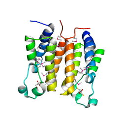
 | | Crystal Structure of Bacteriorhodopsin at pH 10 | | 分子名称: | 2,3-DI-O-PHYTANLY-3-SN-GLYCERO-1-PHOSPHORYL-3'-SN-GLYCEROL-1'-PHOSPHATE, 2,3-DI-PHYTANYL-GLYCEROL, Bacteriorhodopsin, ... | | 著者 | Okumura, H, Murakami, M, Kouyama, T. | | 登録日 | 2005-03-23 | | 公開日 | 2005-08-02 | | 最終更新日 | 2023-10-25 | | 実験手法 | X-RAY DIFFRACTION (2.6 Å) | | 主引用文献 | Crystal Structures of Acid Blue and Alkaline Purple Forms of Bacteriorhodopsin
J.Mol.Biol., 351, 2005
|
|
1X0L
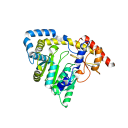
 | | Crystal structure of tetrameric homoisocitrate dehydrogenase from an extreme thermophile, Thermus thermophilus | | 分子名称: | Homoisocitrate dehydrogenase | | 著者 | Miyazaki, J, Asada, K, Fushinobu, S, Kuzuyama, T, Nishiyama, M. | | 登録日 | 2005-03-24 | | 公開日 | 2005-10-04 | | 最終更新日 | 2024-03-13 | | 実験手法 | X-RAY DIFFRACTION (1.85 Å) | | 主引用文献 | Crystal Structure of Tetrameric Homoisocitrate Dehydrogenase from an Extreme Thermophile, Thermus thermophilus: Involvement of Hydrophobic Dimer-Dimer Interaction in Extremely High Thermotolerance
J.Bacteriol., 187, 2005
|
|
1X0M
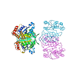
 | | a Human Kynurenine Aminotransferase II Homologue from Pyrococcus horikoshii OT3 | | 分子名称: | Aminotransferase II Homologue | | 著者 | Chon, H, Matsumura, H, Koga, Y, Takano, K, Kanaya, S. | | 登録日 | 2005-03-24 | | 公開日 | 2005-04-12 | | 最終更新日 | 2024-03-13 | | 実験手法 | X-RAY DIFFRACTION (2.2 Å) | | 主引用文献 | Crystal structure of a human kynurenine aminotransferase II homologue from Pyrococcus horikoshii OT3 at 2.20 A resolution
Proteins, 61, 2005
|
|
1X0N
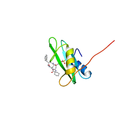 | | NMR structure of growth factor receptor binding protein SH2 domain complexed with the inhibitor | | 分子名称: | 4-[(10S,14S,18S)-18-(2-AMINO-2-OXOETHYL)-14-(1-NAPHTHYLMETHYL)-8,17,20-TRIOXO-7,16,19-TRIAZASPIRO[5.14]ICOS-11-EN-10-YL]BENZYLPHOSPHONIC ACID, Growth factor receptor-bound protein 2 | | 著者 | Ogura, K, Shiga, T, Yuzawa, S, Yokochi, M, Burke, T.R, Inagaki, F. | | 登録日 | 2005-03-24 | | 公開日 | 2005-04-19 | | 最終更新日 | 2024-05-29 | | 実験手法 | SOLUTION NMR | | 主引用文献 | NMR structure of growth factor receptor binding protein SH2 domain complexed with the inhibitor
To be Published
|
|
1X0O
 | | human ARNT C-terminal PAS domain | | 分子名称: | Aryl hydrocarbon receptor nuclear translocator | | 著者 | Card, P.B, Erbel, P.J, Gardner, K.H. | | 登録日 | 2005-03-25 | | 公開日 | 2005-10-25 | | 最終更新日 | 2024-05-29 | | 実験手法 | SOLUTION NMR | | 主引用文献 | Structural Basis of ARNT PAS-B Dimerization: Use of a Common Beta-sheet Interface for Hetero- and Homodimerization.
J.Mol.Biol., 353, 2005
|
|


