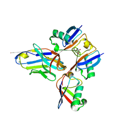
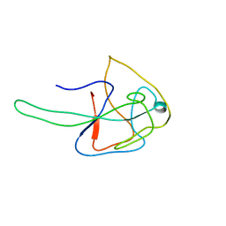
7NAF
 
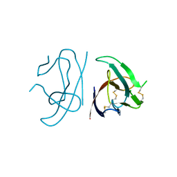
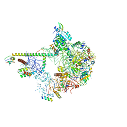 | | State E2 nucleolar 60S ribosomal biogenesis intermediate - Spb1-MTD local model | | 分子名称: | 25S rRNA, 25S rRNA (cytosine(2870)-C(5))-methyltransferase, 27S pre-rRNA (guanosine(2922)-2'-O)-methyltransferase, ... | | 著者 | Cruz, V.E, Sekulski, K, Peddada, N, Erzberger, J.P. | | 登録日 | 2021-06-21 | | 公開日 | 2022-11-09 | | 最終更新日 | 2024-06-05 | | 実験手法 | ELECTRON MICROSCOPY (3.13 Å) | | 主引用文献 | Sequence-specific remodeling of a topologically complex RNP substrate by Spb4.
Nat.Struct.Mol.Biol., 29, 2022
|
|
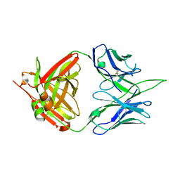
7NAN
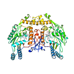
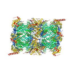 | | Human 20S proteasome core particle | | 分子名称: | Proteasome subunit alpha type-1, Proteasome subunit alpha type-2, Proteasome subunit alpha type-3, ... | | 著者 | Zhao, J, Makhija, S, Huang, B, Cheng, Y. | | 登録日 | 2021-06-22 | | 公開日 | 2022-11-02 | | 最終更新日 | 2025-05-14 | | 実験手法 | ELECTRON MICROSCOPY (2.8 Å) | | 主引用文献 | Structural insights into the human PA28-20S proteasome enabled by efficient tagging and purification of endogenous proteins.
Proc.Natl.Acad.Sci.USA, 119, 2022
|
|
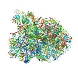
7NAD
 | | State E2 nucleolar 60S ribosomal biogenesis intermediate - Spb4 local refinement model | | 分子名称: | 25S rRNA, 5.8S rRNA, 60S ribosomal protein L17-A, ... | | 著者 | Cruz, V.E, Sekulski, K, Peddada, N, Erzberger, J.P. | | 登録日 | 2021-06-21 | | 公開日 | 2022-11-09 | | 最終更新日 | 2024-06-05 | | 実験手法 | ELECTRON MICROSCOPY (3.04 Å) | | 主引用文献 | Sequence-specific remodeling of a topologically complex RNP substrate by Spb4.
Nat.Struct.Mol.Biol., 29, 2022
|
|
7N6H
 | |
7MYX
 | |
7NBB
 | |
7MYT
 | |
7NAC
 | | State E2 nucleolar 60S ribosomal biogenesis intermediate - Composite model | | 分子名称: | 25S rRNA, 25S rRNA (cytosine(2870)-C(5))-methyltransferase, 27S pre-rRNA (guanosine(2922)-2'-O)-methyltransferase, ... | | 著者 | Cruz, V.E, Sekulski, K, Peddada, N, Erzberger, J.P. | | 登録日 | 2021-06-21 | | 公開日 | 2022-11-09 | | 最終更新日 | 2024-06-05 | | 実験手法 | ELECTRON MICROSCOPY (3.04 Å) | | 主引用文献 | Sequence-specific remodeling of a topologically complex RNP substrate by Spb4.
Nat.Struct.Mol.Biol., 29, 2022
|
|
7MX2
 | |
7MX9
 | |
7MLL
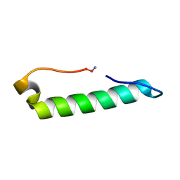 | |
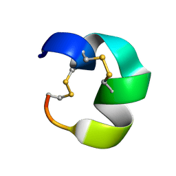
7MP7
 | | Rules for designing protein fold switches and their implications for the folding code | | 分子名称: | Sb3 | | 著者 | He, Y, Chen, Y, Ruan, B, Choi, J, Chen, Y, Motabar, D, Solomon, T, Simmerman, R, Kauffman, T, Gallagher, T, Bryan, P, Orban, J. | | 登録日 | 2021-05-04 | | 公開日 | 2022-05-18 | | 最終更新日 | 2024-05-15 | | 実験手法 | SOLUTION NMR | | 主引用文献 | Design and characterization of a protein fold switching network.
Nat Commun, 14, 2023
|
|
7N21
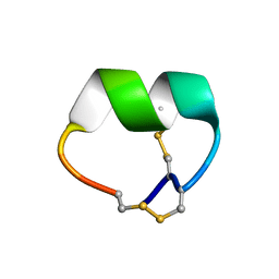
 | | NMR structure of AnIB-OH | | 分子名称: | Alpha-conotoxin AnIB | | 著者 | Conibear, A.C, Rosengren, K.J, Lee, H.S. | | 登録日 | 2021-05-28 | | 公開日 | 2021-11-17 | | 最終更新日 | 2024-11-06 | | 実験手法 | SOLUTION NMR | | 主引用文献 | Posttranslational modifications of alpha-conotoxins: sulfotyrosine and C-terminal amidation stabilise structures and increase acetylcholine receptor binding.
Rsc Med Chem, 12, 2021
|
|
7NE8
 | | Tick salivary protein BSAP1 | | 分子名称: | tick salivary protein BSAP1 | | 著者 | Denisov, S.S, Ippel, J.H, Castoldi, E, Hackeng, T.M, Dijkgraaf, I. | | 登録日 | 2021-02-03 | | 公開日 | 2021-06-16 | | 最終更新日 | 2024-10-16 | | 実験手法 | SOLUTION NMR | | 主引用文献 | Molecular basis of anticoagulant and anticomplement activity of the tick salivary protein Salp14 and its homologs.
J.Biol.Chem., 297, 2021
|
|
7MPA
 | |
7N22
 | |
7N20
 | | NMR structure of native AnIB | | 分子名称: | Alpha-conotoxin AnIB | | 著者 | Conibear, A.C, Rosengren, K.J, Lee, H.S. | | 登録日 | 2021-05-28 | | 公開日 | 2021-11-17 | | 最終更新日 | 2024-11-20 | | 実験手法 | SOLUTION NMR | | 主引用文献 | Posttranslational modifications of alpha-conotoxins: sulfotyrosine and C-terminal amidation stabilise structures and increase acetylcholine receptor binding.
Rsc Med Chem, 12, 2021
|
|
7N82
 | | NMR Solution structure of Se0862 | | 分子名称: | Biofilm-related protein | | 著者 | Zhang, N, LiWang, A.L. | | 登録日 | 2021-06-11 | | 公開日 | 2021-07-14 | | 最終更新日 | 2024-05-15 | | 実験手法 | SOLUTION NMR | | 主引用文献 | Assessment of prediction methods for protein structures determined by NMR in CASP14: Impact of AlphaFold2.
Proteins, 89, 2021
|
|
7N45
 | |
7N23
 | |
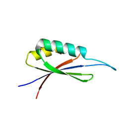
7MN1
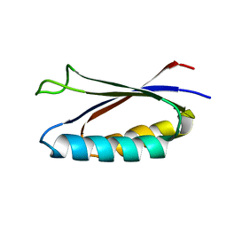 | | Rules for designing protein fold switches and their implications for the folding code | | 分子名称: | Sa1 | | 著者 | He, Y, Chen, Y, Ruan, B, Choi, J, Chen, Y, Motabar, D, Solomon, T, Simmerman, R, Kauffman, T, Gallagher, T, Bryan, P, Orban, J. | | 登録日 | 2021-04-30 | | 公開日 | 2022-05-18 | | 最終更新日 | 2024-05-15 | | 実験手法 | SOLUTION NMR | | 主引用文献 | Design and characterization of a protein fold switching network.
Nat Commun, 14, 2023
|
|
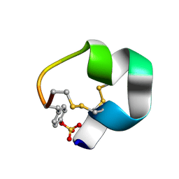
7MQ4
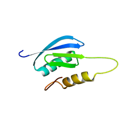 | | Rules for designing protein fold switches and their implications for the folding code | | 分子名称: | Sb1 | | 著者 | He, Y, Chen, Y, Ruan, B, Choi, J, Chen, Y, Motabar, D, Solomon, T, Simmerman, R, Kauffman, T, Gallagher, T, Bryan, P, Orban, J. | | 登録日 | 2021-05-05 | | 公開日 | 2022-05-18 | | 最終更新日 | 2024-05-15 | | 実験手法 | SOLUTION NMR | | 主引用文献 | Design and characterization of a protein fold switching network.
Nat Commun, 14, 2023
|
|
7N0T
 | |
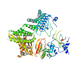
7MU6
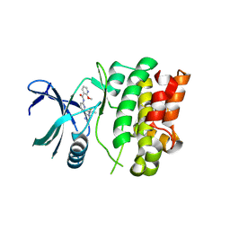 | | Ask1 bound to compound 28 | | 分子名称: | 2-methoxy-N-(6-{4-[(2S)-1,1,1-trifluoropropan-2-yl]-4H-1,2,4-triazol-3-yl}pyridin-2-yl)pyridine-3-carboxamide, Mitogen-activated protein kinase kinase kinase 5 | | 著者 | Chodaparambil, J.V, Marcotte, D.J. | | 登録日 | 2021-05-14 | | 公開日 | 2024-07-17 | | 実験手法 | X-RAY DIFFRACTION (2.165 Å) | | 主引用文献 | Discovery of Potent, Selective, and Brain-Penetrant Apoptosis Signal-Regulating Kinase 1 (ASK1) Inhibitors that Modulate Brain Inflammation In Vivo.
J.Med.Chem., 64, 2021
|
|
7NSE
 | | BOVINE ENDOTHELIAL NITRIC OXIDE SYNTHASE, H4B-FREE, ADMA COMPLEX | | 分子名称: | ACETATE ION, CACODYLIC ACID, GLYCEROL, ... | | 著者 | Raman, C.S, Li, H, Martasek, P, Masters, B.S.S, Poulos, T.L. | | 登録日 | 1999-01-13 | | 公開日 | 2002-05-29 | | 最終更新日 | 2023-12-27 | | 実験手法 | X-RAY DIFFRACTION (2.35 Å) | | 主引用文献 | Crystal Structures of the Heme Domain of Bovine Endothelial Nitric Oxide Synthase Complexed with Arginine Analogues
To be Published
|
|


