6ZZI
 
 | |
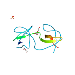
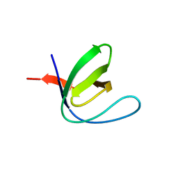
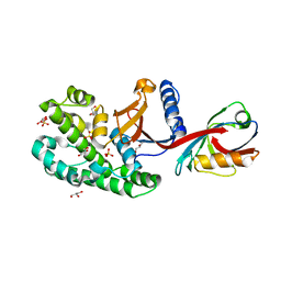
5MOL
 | | Human IgE-Fc crystal structure | | 分子名称: | 1,2-ETHANEDIOL, 2-(2-ETHOXYETHOXY)ETHANOL, 2-(2-METHOXYETHOXY)ETHANOL, ... | | 著者 | Dore, K.A, Davies, A.M, Drinkwater, N, Beavil, A.J, McDonnell, J.M, Sutton, B.J. | | 登録日 | 2016-12-14 | | 公開日 | 2018-01-10 | | 最終更新日 | 2024-01-17 | | 実験手法 | X-RAY DIFFRACTION (1.75 Å) | | 主引用文献 | Thermal sensitivity and flexibility of the C epsilon 3 domains in immunoglobulin E.
Biochim. Biophys. Acta, 1865, 2017
|
|
7A38
 | |
7A2X
 | |
7A30
 | |
7A34
 | |
7A2S
 | |
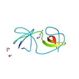
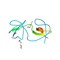
5MPO
 | | Crystal structure of human molybdopterin synthase complex | | 分子名称: | Molybdopterin synthase catalytic subunit, Molybdopterin synthase sulfur carrier subunit | | 著者 | Kopec, J, Bailey, H, Fitzpatrick, F, Strain-Damerell, C, Oberholzer, A.E, Williams, E, Burgess-Brown, N, von Delft, F, Arrowsmith, C, Edwards, A, Bountra, C, Yue, W.W. | | 登録日 | 2016-12-16 | | 公開日 | 2016-12-28 | | 最終更新日 | 2024-01-17 | | 実験手法 | X-RAY DIFFRACTION (2.43 Å) | | 主引用文献 | Crystal structure of human molybdopterin synthase complex
To Be Published
|
|
7A2K
 | |
7A3B
 | |
6SFI
 | | Crystal structure of p38 alpha in complex with compound 75 (MCP33) | | 分子名称: | Mitogen-activated protein kinase 14, ~{N}-[(2~{S})-1-azanyl-4-cyclohexyl-1-oxidanylidene-butan-2-yl]-2-[[[1-(2-methylphenyl)pyrazol-4-yl]carbonylamino]methyl]-1,3-thiazole-5-carboxamide | | 著者 | Chaikuad, A, Arrowsmith, C.H, Edwards, A.M, Bountra, C, Knapp, S, Structural Genomics Consortium (SGC) | | 登録日 | 2019-08-01 | | 公開日 | 2019-09-11 | | 最終更新日 | 2024-01-24 | | 実験手法 | X-RAY DIFFRACTION (1.6 Å) | | 主引用文献 | Fast Iterative Synthetic Approach toward Identification of Novel Highly Selective p38 MAP Kinase Inhibitors.
J.Med.Chem., 62, 2019
|
|
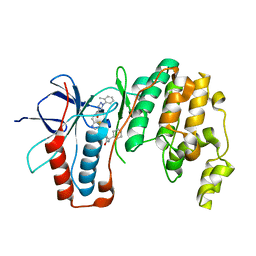
5MPR
 | | Single Amino Acid Variant of Human Mitochondrial Branched Chain Amino Acid Aminotransferase 2 | | 分子名称: | 1,2-ETHANEDIOL, Branched-chain-amino-acid aminotransferase, mitochondrial, ... | | 著者 | Hakansson, M, Walse, B, Nilsson, C, Anderson, L.C. | | 登録日 | 2016-12-18 | | 公開日 | 2017-07-19 | | 最終更新日 | 2019-10-16 | | 実験手法 | X-RAY DIFFRACTION (1.6 Å) | | 主引用文献 | Intact Protein Analysis at 21 Tesla and X-Ray Crystallography Define Structural Differences in Single Amino Acid Variants of Human Mitochondrial Branched-Chain Amino Acid Aminotransferase 2 (BCAT2).
J. Am. Soc. Mass Spectrom., 28, 2017
|
|
6SC9
 | | dAb3/HOIP-RBR-HOIPIN-8 | | 分子名称: | 2-[3-[2,6-bis(fluoranyl)-4-(1~{H}-pyrazol-4-yl)phenyl]-3-oxidanylidene-prop-1-enyl]-4-(1-methylpyrazol-4-yl)benzoic acid, CHLORIDE ION, E3 ubiquitin-protein ligase RNF31, ... | | 著者 | Tsai, Y.-C.I, Johansson, H, House, D, Rittinger, K. | | 登録日 | 2019-07-23 | | 公開日 | 2019-11-27 | | 最終更新日 | 2024-05-01 | | 実験手法 | X-RAY DIFFRACTION (2.47 Å) | | 主引用文献 | Single-Domain Antibodies as Crystallization Chaperones to Enable Structure-Based Inhibitor Development for RBR E3 Ubiquitin Ligases.
Cell Chem Biol, 27, 2020
|
|
7AAR
 | | sugar/H+ symporter STP10 in inward open conformation | | 分子名称: | CHLORIDE ION, Octyl Glucose Neopentyl Glycol, Sugar transport protein 10, ... | | 著者 | Bavnhoej, L, Paulsen, P.A, Pedersen, B.P. | | 登録日 | 2020-09-04 | | 公開日 | 2021-08-18 | | 最終更新日 | 2024-01-31 | | 実験手法 | X-RAY DIFFRACTION (2.64 Å) | | 主引用文献 | Molecular mechanism of sugar transport in plants unveiled by structures of glucose/H + symporter STP10.
Nat.Plants, 7, 2021
|
|
6S8X
 | |
7A2L
 | |
5M9F
 | |
7A2U
 | |
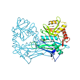
5MAQ
 | | Crystal Structure of Polyphosphate Kinase from Meiothermus ruber bound to ADP and PPi | | 分子名称: | ADENOSINE-5'-DIPHOSPHATE, MAGNESIUM ION, PYROPHOSPHATE, ... | | 著者 | Gerhardt, S, Einsle, O, Kemper, F, Schwarzer, N. | | 登録日 | 2016-11-04 | | 公開日 | 2017-12-20 | | 最終更新日 | 2024-01-17 | | 実験手法 | X-RAY DIFFRACTION (2.46 Å) | | 主引用文献 | Substrate recognition and mechanism revealed by ligand-bound polyphosphate kinase 2 structures.
Proc. Natl. Acad. Sci. U.S.A., 115, 2018
|
|
7A2O
 | |
6S96
 | |
6SAK
 | |
7A3D
 | |
6S9F
 | | Drosophila OTK, extracellular domains 3-5 | | 分子名称: | 2-acetamido-2-deoxy-beta-D-glucopyranose, SULFATE ION, Tyrosine-protein kinase-like otk | | 著者 | Rozbesky, D, Jones, E.Y. | | 登録日 | 2019-07-12 | | 公開日 | 2020-03-25 | | 最終更新日 | 2024-01-24 | | 実験手法 | X-RAY DIFFRACTION (1.969 Å) | | 主引用文献 | Drosophila OTK Is a Glycosaminoglycan-Binding Protein with High Conformational Flexibility.
Structure, 28, 2020
|
|
7A2F
 | | Crystal structure of T. brucei PDE-B1 catalytic domain with inhibitor NPD-656 | | 分子名称: | 1-cycloheptyl-3-{4-methoxy-3-[2-(4-methoxyphenyl)ethoxy]phenyl}-4,4-dimethyl-4,5-dihydro-1H-pyrazol-5-one, FORMIC ACID, GLYCEROL, ... | | 著者 | Singh, A.K, Blaazer, A.R, Zara, L, de Esch, I.J.P, Leurs, R, Brown, D.G. | | 登録日 | 2020-08-17 | | 公開日 | 2021-08-25 | | 最終更新日 | 2024-01-31 | | 実験手法 | X-RAY DIFFRACTION (2 Å) | | 主引用文献 | Crystal structure of T. brucei PDE-B1 catalytic domain with inhibitor NPD-656
To be published
|
|


