1WM1
 
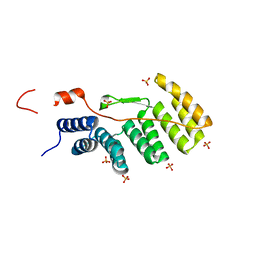
 | | Crystal Structure of Prolyl Aminopeptidase, Complex with Pro-TBODA | | 分子名称: | (5-TERT-BUTYL-1,3,4-OXADIAZOL-2-YL)[(2R)-PYRROLIDIN-2-YL]METHANONE, Proline iminopeptidase | | 著者 | Nakajima, Y, Inoue, T, Ito, K, Tozaka, T, Hatakeyama, S, Tanaka, N, Nakamura, K.T, Yoshimoto, T. | | 登録日 | 2004-07-01 | | 公開日 | 2004-07-20 | | 最終更新日 | 2024-03-13 | | 実験手法 | X-RAY DIFFRACTION (2.1 Å) | | 主引用文献 | Novel inhibitor for prolyl aminopeptidase from Serratia marcescens and studies on the mechanism of substrate recognition of the enzyme using the inhibitor
ARCH.BIOCHEM.BIOPHYS., 416, 2003
|
|
1WM2
 | |
1WM3
 | |
1WM4
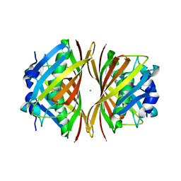
 | | Solution structure of mouse coactosin, an actin filament binding protein | | 分子名称: | Coactosin-like protein | | 著者 | Hellman, M, Paavilainen, V.O, Naumanen, P, Lappalainen, P, Annila, A, Permi, P. | | 登録日 | 2004-07-03 | | 公開日 | 2004-11-02 | | 最終更新日 | 2024-05-29 | | 実験手法 | SOLUTION NMR | | 主引用文献 | Solution structure of coactosin reveals structural homology to ADF/cofilin family proteins
Febs Lett., 576, 2004
|
|
1WM5
 | |
1WM6
 | |
1WM7
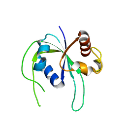
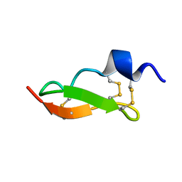 | | Solution Structure of BmP01 from the Venom of Scorpion Buthus martensii Karsch, 9 structures | | 分子名称: | Neurotoxin BmP01 | | 著者 | Wu, G, Li, Y, Wei, D, He, F, Jiang, S, Hu, G, Wu, H, Chen, X. | | 登録日 | 2004-07-05 | | 公開日 | 2004-07-27 | | 最終更新日 | 2022-03-02 | | 実験手法 | SOLUTION NMR | | 主引用文献 | Solution Structure of BmP01 from the Venom of Scorpion Buthus martensii Karsch
Biochem.Biophys.Res.Commun., 276, 2000
|
|
1WM8
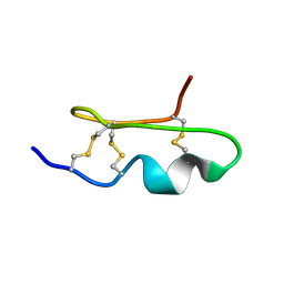 | | Solution Structure of BmP03 from the Venom of Scorpion Buthus martensii Karsch, 10 structures | | 分子名称: | Neurotoxin BmP03 | | 著者 | Wu, H, He, F, Li, Y, Wu, G, Cao, C, Chen, X. | | 登録日 | 2004-07-05 | | 公開日 | 2004-07-27 | | 最終更新日 | 2018-04-18 | | 実験手法 | SOLUTION NMR | | 主引用文献 | Three-Dimensional Structure of BmP03 from Venom of Scorpion Buthus martensii Karsch
Acta Chim.Sinica, 58, 2000
|
|
1WM9
 | |
1WMA
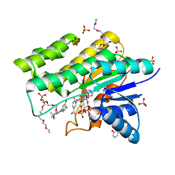 | | Crystal structure of human CBR1 in complex with Hydroxy-PP | | 分子名称: | 3,6,9,12,15,18,21,24-OCTAOXAHEXACOSAN-1-OL, 3,6,9,12,15,18-HEXAOXAICOSANE-1,20-DIOL, 3-(4-AMINO-1-TERT-BUTYL-1H-PYRAZOLO[3,4-D]PYRIMIDIN-3-YL)PHENOL, ... | | 著者 | Rauh, D, Bateman, R, Shokat, K.M. | | 登録日 | 2004-07-06 | | 公開日 | 2005-04-26 | | 最終更新日 | 2024-03-13 | | 実験手法 | X-RAY DIFFRACTION (1.24 Å) | | 主引用文献 | An unbiased cell morphology-based screen for new, biologically active small molecules
Plos Biol., 3, 2005
|
|
1WMB
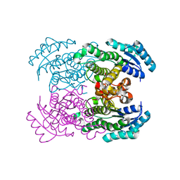 | | Crystal structure of NAD dependent D-3-hydroxybutylate dehydrogenase | | 分子名称: | CACODYLATE ION, D(-)-3-hydroxybutyrate dehydrogenase, MAGNESIUM ION | | 著者 | Ito, K, Nakajima, Y, Ichihara, E, Ogawa, K, Yoshimoto, T. | | 登録日 | 2004-07-06 | | 公開日 | 2005-09-06 | | 最終更新日 | 2024-03-13 | | 実験手法 | X-RAY DIFFRACTION (2 Å) | | 主引用文献 | d-3-Hydroxybutyrate Dehydrogenase from Pseudomonas fragi: Molecular Cloning of the Enzyme Gene and Crystal Structure of the Enzyme
J.Mol.Biol., 355, 2006
|
|
1WMD
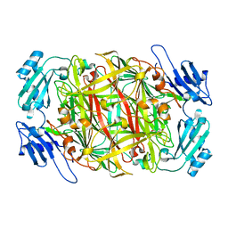
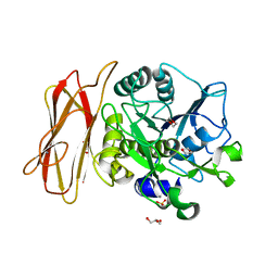 | | Crystal Structure of alkaline serine protease KP-43 from Bacillus sp. KSM-KP43 (1.30 angstrom, 100 K) | | 分子名称: | 1,4-DIETHYLENE DIOXIDE, CALCIUM ION, GLYCEROL, ... | | 著者 | Nonaka, T, Fujihashi, M, Kita, A, Saeki, K, Ito, S, Horikoshi, K, Miki, K. | | 登録日 | 2004-07-08 | | 公開日 | 2004-09-14 | | 最終更新日 | 2024-03-13 | | 実験手法 | X-RAY DIFFRACTION (1.3 Å) | | 主引用文献 | The Crystal Structure of an Oxidatively Stable Subtilisin-like Alkaline Serine Protease, KP-43, with a C-terminal {beta}-Barrel Domain
J.Biol.Chem., 279, 2004
|
|
1WME
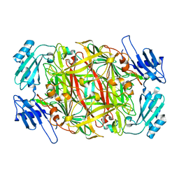
 | | Crystal Structure of alkaline serine protease KP-43 from Bacillus sp. KSM-KP43 (1.50 angstrom, 293 K) | | 分子名称: | CALCIUM ION, protease | | 著者 | Nonaka, T, Fujihashi, M, Kita, A, Saeki, K, Ito, S, Horikoshi, K, Miki, K. | | 登録日 | 2004-07-08 | | 公開日 | 2004-09-14 | | 最終更新日 | 2024-03-13 | | 実験手法 | X-RAY DIFFRACTION (1.5 Å) | | 主引用文献 | The Crystal Structure of an Oxidatively Stable Subtilisin-like Alkaline Serine Protease, KP-43, with a C-terminal {beta}-Barrel Domain
J.Biol.Chem., 279, 2004
|
|
1WMF
 | | Crystal Structure of alkaline serine protease KP-43 from Bacillus sp. KSM-KP43 (oxidized form, 1.73 angstrom) | | 分子名称: | 1,4-DIETHYLENE DIOXIDE, CALCIUM ION, GLYCEROL, ... | | 著者 | Nonaka, T, Fujihashi, M, Kita, A, Saeki, K, Ito, S, Horikoshi, K, Miki, K. | | 登録日 | 2004-07-08 | | 公開日 | 2004-09-14 | | 最終更新日 | 2011-07-13 | | 実験手法 | X-RAY DIFFRACTION (1.73 Å) | | 主引用文献 | The Crystal Structure of an Oxidatively Stable Subtilisin-like Alkaline Serine Protease, KP-43, with a C-terminal {beta}-Barrel Domain
J.Biol.Chem., 279, 2004
|
|
1WMG
 | | Crystal structure of the UNC5H2 death domain | | 分子名称: | SULFATE ION, SULFITE ION, netrin receptor Unc5h2 | | 著者 | Handa, N, Murayama, K, Shirouzu, M, Yokoyama, S, RIKEN Structural Genomics/Proteomics Initiative (RSGI) | | 登録日 | 2004-07-09 | | 公開日 | 2005-01-09 | | 最終更新日 | 2011-11-16 | | 実験手法 | X-RAY DIFFRACTION (2.1 Å) | | 主引用文献 | Structure of the UNC5H2 death domain
ACTA CRYSTALLOGR.,SECT.D, 62, 2006
|
|
1WMH
 | | Crystal structure of a PB1 domain complex of Protein kinase c iota and Par6 alpha | | 分子名称: | Partitioning defective-6 homolog alpha, Protein kinase C, iota type | | 著者 | Hirano, Y, Yoshinaga, S, Suzuki, N.N, Horiuchi, M, Kohjima, M, Takeya, R, Sumimoto, H, Inagaki, F. | | 登録日 | 2004-07-09 | | 公開日 | 2004-12-07 | | 最終更新日 | 2024-03-13 | | 実験手法 | X-RAY DIFFRACTION (1.5 Å) | | 主引用文献 | Structure of a Cell Polarity Regulator, a Complex between Atypical PKC and Par6 PB1 Domains
J.Biol.Chem., 280, 2005
|
|
1WMI
 | | Crystal structure of archaeal RelE-RelB complex from Pyrococcus horikoshii OT3 | | 分子名称: | hypothetical protein PHS013, hypothetical protein PHS014 | | 著者 | Takagi, H, Kakuta, Y, Kamachi, R, Yao, M, Tanaka, I, Kimura, M. | | 登録日 | 2004-07-09 | | 公開日 | 2005-03-15 | | 最終更新日 | 2024-03-13 | | 実験手法 | X-RAY DIFFRACTION (2.3 Å) | | 主引用文献 | Crystal structure of archaeal toxin-antitoxin RelE-RelB complex with implications for toxin activity and antitoxin effects
Nat.Struct.Mol.Biol., 12, 2005
|
|
1WMJ
 | |
1WMK
 | |
1WMM
 | |
1WMN
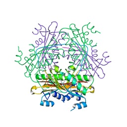
 | | Crystal structure of topaquinone-containing amine oxidase activated by cobalt ion | | 分子名称: | COBALT (II) ION, Phenylethylamine oxidase | | 著者 | Okajima, T, Kishishita, S, Chiu, Y.C, Murakawa, T, Kim, M, Yamaguchi, H, Hirota, S, Kuroda, S, Tanizawa, K. | | 登録日 | 2004-07-13 | | 公開日 | 2005-08-02 | | 最終更新日 | 2011-07-13 | | 実験手法 | X-RAY DIFFRACTION (1.8 Å) | | 主引用文献 | Reinvestigation of metal ion specificity for quinone cofactor biogenesis in bacterial copper amine oxidase
Biochemistry, 44, 2005
|
|
1WMO
 | | Crystal structure of topaquinone-containing amine oxidase activated by nickel ion | | 分子名称: | NICKEL (II) ION, Phenylethylamine oxidase | | 著者 | Okajima, T, Kishishita, S, Chiu, Y.C, Murakawa, T, Kim, M, Yamaguchi, H, Hirota, S, Kuroda, S, Tanizawa, K. | | 登録日 | 2004-07-13 | | 公開日 | 2005-08-02 | | 最終更新日 | 2011-07-13 | | 実験手法 | X-RAY DIFFRACTION (1.8 Å) | | 主引用文献 | Reinvestigation of metal ion specificity for quinone cofactor biogenesis in bacterial copper amine oxidase
Biochemistry, 44, 2005
|
|
1WMP
 | | Crystal structure of amine oxidase complexed with cobalt ion | | 分子名称: | COBALT (II) ION, Phenylethylamine oxidase | | 著者 | Okajima, T, Kishishita, S, Chiu, Y.C, Murakawa, T, Kim, M, Yamaguchi, H, Hirota, S, Kuroda, S, Tanizawa, K. | | 登録日 | 2004-07-13 | | 公開日 | 2005-08-02 | | 最終更新日 | 2011-07-13 | | 実験手法 | X-RAY DIFFRACTION (2 Å) | | 主引用文献 | Reinvestigation of metal ion specificity for quinone cofactor biogenesis in bacterial copper amine oxidase
Biochemistry, 44, 2005
|
|
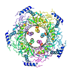
1WMQ
 | | Structure of the HutP antitermination complex bound to a single stranded region of hut mRNA | | 分子名称: | 5'-R(P*UP*UP*UP*AP*GP*UP*U)-3', HISTIDINE, Hut operon positive regulatory protein, ... | | 著者 | Kumarevel, T.S, Mizuno, H, Kumar, P.K.R. | | 登録日 | 2004-07-14 | | 公開日 | 2005-03-15 | | 最終更新日 | 2023-10-25 | | 実験手法 | X-RAY DIFFRACTION (1.6 Å) | | 主引用文献 | Structural basis of HutP-mediated anti-termination and roles of the Mg2+ ion and L-histidine ligand.
Nature, 434, 2005
|
|
1WMR
 | | Crystal Structure of Isopullulanase from Aspergillus niger ATCC 9642 | | 分子名称: | 2-acetamido-2-deoxy-beta-D-glucopyranose, Isopullulanase | | 著者 | Mizuno, M, Tonozuka, T, Miyasaka, Y, Akeboshi, H, Kamitori, S, Nishikawa, A, Sakano, Y. | | 登録日 | 2004-07-15 | | 公開日 | 2005-07-19 | | 最終更新日 | 2023-10-25 | | 実験手法 | X-RAY DIFFRACTION (2.4 Å) | | 主引用文献 | Crystal Structure of Aspergillus niger Isopullulanase, a Member of Glycoside Hydrolase Family 49
J.Mol.Biol., 376, 2008
|
|


