Loading
PDBj
メニューPDBj@FacebookPDBj@TwitterPDBj@YouTubewwPDB FoundationwwPDB
RCSB PDBPDBeBMRBAdv. SearchSearch help
PDB: 2 件

6BXI
DownloadVisualize
BU of 6bxi by Molmil
X-ray crystal structure of NDR1 kinase domain
分子名称: MAGNESIUM ION, PHOSPHOAMINOPHOSPHONIC ACID-ADENYLATE ESTER, Serine/threonine-protein kinase 38
著者Xiong, S, Sicheri, F.
登録日2017-12-18
公開日2018-08-29
最終更新日2024-03-13
実験手法X-RAY DIFFRACTION (2.2 Å)
主引用文献Structural Basis for Auto-Inhibition of the NDR1 Kinase Domain by an Atypically Long Activation Segment.
Structure, 26, 2018
1PSB
DownloadVisualize
BU of 1psb by Molmil
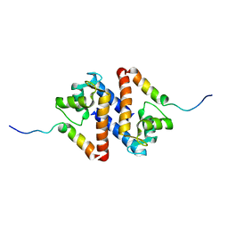
Solution structure of calcium loaded S100B complexed to a peptide from N-Terminal regulatory domain of NDR kinase.
分子名称: Ndr Ser/Thr kinase-like protein, S-100 protein, beta chain
著者Bhattacharya, S, Large, E, Heizmann, C.W, Hemmings, B, Chazin, W.J.
登録日2003-06-21
公開日2003-12-16
最終更新日2024-05-22
実験手法SOLUTION NMR
主引用文献Structure of the Ca(2+)/S100B/NDR Kinase Peptide Complex: Insights into S100 Target Specificity and Activation of the Kinase.
Biochemistry, 42, 2003

222415

件を2024-07-10に公開中

PDB statisticsPDBj update infoContact PDBjnumon