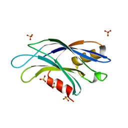
2YOA
 
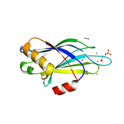 | | Synaptotagmin-1 C2B domain with phosphoserine | | 分子名称: | CALCIUM ION, PHOSPHOSERINE, SYNAPTOTAGMIN-1, ... | | 著者 | Honigmann, A, van den Bogaart, G, Iraheta, E, Risselada, H.J, Milovanovic, D, Mueller, V, Muellar, S, Diederichsen, U, Fasshauer, D, Grubmuller, H, Hell, S.W, Eggeling, C, Kuhnel, K, Jahn, R. | | 登録日 | 2012-10-22 | | 公開日 | 2013-03-20 | | 最終更新日 | 2023-12-20 | | 実験手法 | X-RAY DIFFRACTION (1.5 Å) | | 主引用文献 | Phosphatidylinositol 4,5-Bisphosphate Clusters Act as Molecular Beacons for Vesicle Recruitment
Nat.Struct.Mol.Biol., 20, 2013
|
|
9BKY
 | |
4V29
 | |
9BKW
 | |
2Z0U
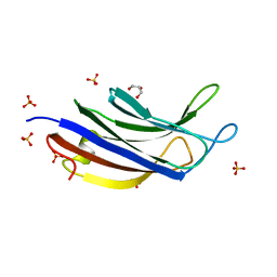
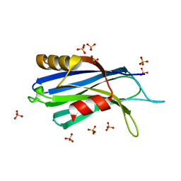
 | | Crystal structure of C2 domain of KIBRA protein | | 分子名称: | WW domain-containing protein 1 | | 著者 | Murayama, K, Kato-Murayama, M, Terada, T, Shirouzu, M, Yokoyama, S, RIKEN Structural Genomics/Proteomics Initiative (RSGI) | | 登録日 | 2007-05-07 | | 公開日 | 2008-05-13 | | 最終更新日 | 2011-07-13 | | 実験手法 | X-RAY DIFFRACTION (2.2 Å) | | 主引用文献 | Crystal structure of C2 domain of KIBRA protein
To be Published
|
|
5W4S
 | |
5LO8
 | |
6TZ3
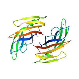
 | | Crystal Structure of Human Synaptotagmin 1 C2B without Ca2+ | | 分子名称: | SULFATE ION, Synaptotagmin-1 | | 著者 | Dominguez, M.J, Karmakar, S, Meyer, A.G, Sutton, R.B. | | 登録日 | 2019-08-09 | | 公開日 | 2020-05-13 | | 最終更新日 | 2023-10-11 | | 実験手法 | X-RAY DIFFRACTION (1.17 Å) | | 主引用文献 | Molecular Basis for Synaptotagmin-1-Associated Neurodevelopmental Disorder.
Neuron, 107, 2020
|
|
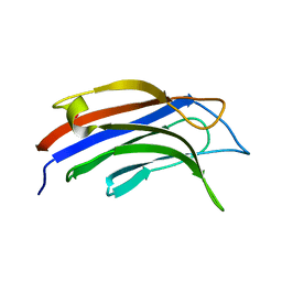
6U41
 | | 1.7 angstrom structure of a pathogenic human Syt 1 C2B (D304G) | | 分子名称: | SULFATE ION, Synaptotagmin-1 | | 著者 | Dominguez, M.J, Bradberry, M.M, Chapman, E.R, Sutton, R.B. | | 登録日 | 2019-08-22 | | 公開日 | 2020-05-13 | | 最終更新日 | 2023-10-11 | | 実験手法 | X-RAY DIFFRACTION (1.7 Å) | | 主引用文献 | Molecular Basis for Synaptotagmin-1-Associated Neurodevelopmental Disorder.
Neuron, 107, 2020
|
|
5LOB
 | |
5LOW
 | |
6U4U
 | | 1.3 A structure of a pathogenic human Syt 1 C2B (I368T) | | 分子名称: | SULFATE ION, Synaptotagmin-1 | | 著者 | Dominguez, M.J, Bradberry, M.M, Chapman, E.R, Sutton, R.B. | | 登録日 | 2019-08-26 | | 公開日 | 2020-05-13 | | 最終更新日 | 2023-10-11 | | 実験手法 | X-RAY DIFFRACTION (1.3 Å) | | 主引用文献 | Molecular Basis for Synaptotagmin-1-Associated Neurodevelopmental Disorder.
Neuron, 107, 2020
|
|
7JOF
 | | Calcium-bound C2A Domain from Human Dysferlin | | 分子名称: | CALCIUM ION, Isoform 6 of Dysferlin | | 著者 | Tadayon, R, Wang, Y, Santamaria, L, Mercier, P, Forristal, C, Shaw, G.S. | | 登録日 | 2020-08-06 | | 公開日 | 2021-06-16 | | 最終更新日 | 2023-10-18 | | 実験手法 | X-RAY DIFFRACTION (2 Å) | | 主引用文献 | Calcium binds and rigidifies the dysferlin C2A domain in a tightly coupled manner.
Biochem.J., 478, 2021
|
|
4Y1S
 | | Structural basis for Ca2+-mediated interaction of the perforin C2 domain with lipid membranes | | 分子名称: | CALCIUM ION, Perforin-1 | | 著者 | Conroy, P.J, Yagi, H, Whisstock, J.C, Norton, R.S. | | 登録日 | 2015-02-09 | | 公開日 | 2015-09-02 | | 最終更新日 | 2023-09-27 | | 実験手法 | X-RAY DIFFRACTION (1.611 Å) | | 主引用文献 | Structural Basis for Ca2+-mediated Interaction of the Perforin C2 Domain with Lipid Membranes.
J.Biol.Chem., 290, 2015
|
|
7K6B
 | |
7KRB
 | |
4Y1T
 | | Structural basis for Ca2+-mediated interaction of the perforin C2 domain with lipid membranes | | 分子名称: | CALCIUM ION, Perforin-1 | | 著者 | Conroy, P.J, Yagi, H, Whisstock, J.C, Norton, R.S. | | 登録日 | 2015-02-09 | | 公開日 | 2015-09-02 | | 最終更新日 | 2023-09-27 | | 実験手法 | X-RAY DIFFRACTION (2.666 Å) | | 主引用文献 | Structural Basis for Ca2+-mediated Interaction of the Perforin C2 Domain with Lipid Membranes.
J.Biol.Chem., 290, 2015
|
|
3W56
 | |
6NYT
 | | Munc13-1 C2B-domain, calcium bound | | 分子名称: | CALCIUM ION, CHLORIDE ION, GLYCEROL, ... | | 著者 | Tomchick, D.R, Rizo, J, Machius, M, Lu, J. | | 登録日 | 2019-02-12 | | 公開日 | 2019-02-20 | | 最終更新日 | 2023-10-11 | | 実験手法 | X-RAY DIFFRACTION (1.369 Å) | | 主引用文献 | Munc13 C2B domain is an activity-dependent Ca2+ regulator of synaptic exocytosis.
Nat. Struct. Mol. Biol., 17, 2010
|
|
3W57
 | | Structure of a C2 domain | | 分子名称: | C2 domain protein, CALCIUM ION | | 著者 | Traore, D.A.K, Whisstock, J.C. | | 登録日 | 2013-01-24 | | 公開日 | 2013-10-23 | | 最終更新日 | 2023-11-08 | | 実験手法 | X-RAY DIFFRACTION (1.662 Å) | | 主引用文献 | Defining the interaction of perforin with calcium and the phospholipid membrane.
Biochem.J., 456, 2013
|
|
6NYC
 | | Munc13-1 C2B-domain, calcium free | | 分子名称: | 2-[3-(2-HYDROXY-1,1-DIHYDROXYMETHYL-ETHYLAMINO)-PROPYLAMINO]-2-HYDROXYMETHYL-PROPANE-1,3-DIOL, CHLORIDE ION, Munc13-1 | | 著者 | Tomchick, D.R, Rizo, J, Machius, M, Lu, J. | | 登録日 | 2019-02-11 | | 公開日 | 2019-02-20 | | 最終更新日 | 2023-10-11 | | 実験手法 | X-RAY DIFFRACTION (1.893 Å) | | 主引用文献 | Munc13 C2B domain is an activity-dependent Ca2+ regulator of synaptic exocytosis.
Nat. Struct. Mol. Biol., 17, 2010
|
|
3TWY
 | | RAT PKC C2 DOMAIN BOUND TO PB | | 分子名称: | LEAD (II) ION, Protein kinase C alpha type, SULFATE ION | | 著者 | Li, P. | | 登録日 | 2011-09-22 | | 公開日 | 2011-11-02 | | 最終更新日 | 2024-02-28 | | 実験手法 | X-RAY DIFFRACTION (1.5 Å) | | 主引用文献 | Pb2+ as Modulator of Protein-Membrane Interactions.
J.Am.Chem.Soc., 133, 2011
|
|
7A1R
 | |
7ATP
 | |
7AS6
 | | 2.0 angstrom structure of plant Extended Synaptotagmin 1, C2A domain | | 分子名称: | 1,2-ETHANEDIOL, CADMIUM ION, CHLORIDE ION, ... | | 著者 | Benavente, J.L, Albert, A. | | 登録日 | 2020-10-27 | | 公開日 | 2021-08-18 | | 最終更新日 | 2024-01-31 | | 実験手法 | X-RAY DIFFRACTION (2 Å) | | 主引用文献 | The structure and flexibility analysis of the Arabidopsis synaptotagmin 1 reveal the basis of its regulation at membrane contact sites.
Life Sci Alliance, 4, 2021
|
|


