Loading
PDBj
メニューPDBj@FacebookPDBj@TwitterPDBj@YouTubewwPDB FoundationwwPDB
RCSB PDBPDBeBMRBAdv. SearchSearch help
PDB: 523 件

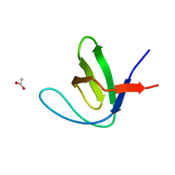
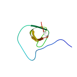
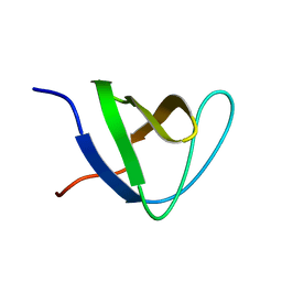
7A2T
DownloadVisualize
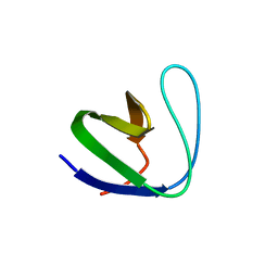
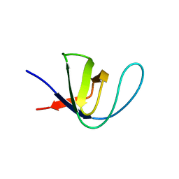
BU of 7a2t by Molmil
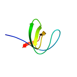
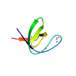
Crystal structure of the Fyn SH3 domain L112V-S114N-S115T-E121L-R123H mutant at pH 4.0
分子名称: ACETATE ION, Tyrosine-protein kinase Fyn
著者Camara-Artigas, A, Plaza-Garrido, M, Salinas-Garcia, M.C.
登録日2020-08-18
公開日2021-08-25
最終更新日2024-01-31
実験手法X-RAY DIFFRACTION (1.22 Å)
主引用文献Crystal structure of the Fyn SH3 domain L112V-S114N-S115T-E121L-R123H mutant at pH 4.0
To be published
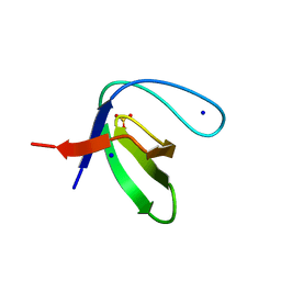
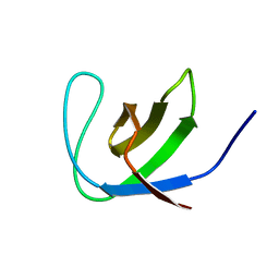
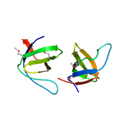
7A35
DownloadVisualize
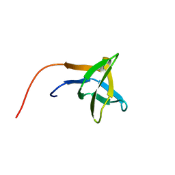
BU of 7a35 by Molmil
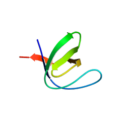
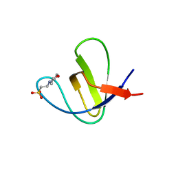
Crystal structure of the c-Src SH3 domain mutant V111L-N113S-T114S at pH 7.0
分子名称: Proto-oncogene tyrosine-protein kinase Src, SULFATE ION
著者Camara-Artigas, A, Plaza-Garrido, M, Salinas-Garcia, M.C.
登録日2020-08-18
公開日2021-08-25
最終更新日2024-01-31
実験手法X-RAY DIFFRACTION (1.314 Å)
主引用文献Crystal structure of the c-Src SH3 domain mutant V111L-N113S-T114S at pH 7.0
To be published
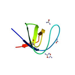
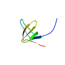
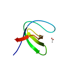
7A2U
DownloadVisualize
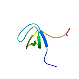
BU of 7a2u by Molmil
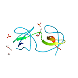
Crystal structure of the Fyn SH3 domain L112V-S114N-S115T at pH 4.5
分子名称: ACETATE ION, Tyrosine-protein kinase Fyn
著者Camara-Artigas, A, Plaza-Garrido, M, Salinas-Garcia, M.C.
登録日2020-08-18
公開日2021-08-25
最終更新日2024-01-31
実験手法X-RAY DIFFRACTION (1.7 Å)
主引用文献Crystal structure of the Fyn SH3 domain L112V-S114N-S115T at pH 4.5
To be published
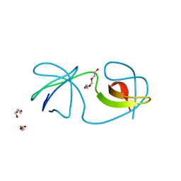
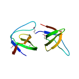
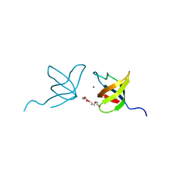
7A34
DownloadVisualize
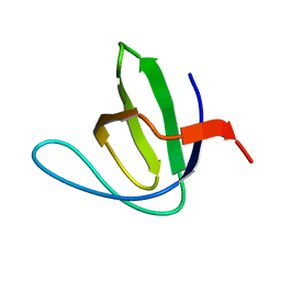
BU of 7a34 by Molmil
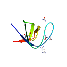
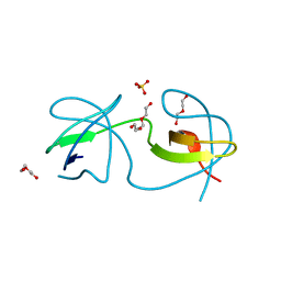
Intertwined dimer of the c-Src SH3 domain mutant V111L-N113S-T114S
分子名称: DI(HYDROXYETHYL)ETHER, Proto-oncogene tyrosine-protein kinase Src, TRIETHYLENE GLYCOL
著者Camara-Artigas, A, Plaza-Garrido, M, Salinas-Garcia, M.C.
登録日2020-08-18
公開日2021-08-25
最終更新日2024-01-31
実験手法X-RAY DIFFRACTION (1.85 Å)
主引用文献Intertwined dimer of the c-Src SH3 domain mutant V111L-N113S-T114S
To be published
7A2M
DownloadVisualize
BU of 7a2m by Molmil
Crystal structure of the Fyn SH3 domain A95S-D99T mutant in C21 space group
分子名称: FORMIC ACID, SODIUM ION, SULFATE ION, ...
著者Camara-Artigas, A, Plaza-Garrido, M, Salinas-Garcia, M.C.
登録日2020-08-18
公開日2021-08-25
最終更新日2024-01-31
実験手法X-RAY DIFFRACTION (1.5 Å)
主引用文献Crystal structure of the Fyn SH3 domain A95S-D99T mutant in C21 space group
To be published
7A37
DownloadVisualize
BU of 7a37 by Molmil
Crystal structure of the c-Src SH3 domain mutant V111L-N113S-T114S in 3 M urea
分子名称: Proto-oncogene tyrosine-protein kinase Src, UREA
著者Camara-Artigas, A, Plaza-Garrido, M, Salinas-Garcia, M.C.
登録日2020-08-18
公開日2021-08-25
最終更新日2024-01-31
実験手法X-RAY DIFFRACTION (1.52 Å)
主引用文献Crystal structure of the c-Src SH3 domain mutant V111L-N113S-T114S in 3 M urea
To be published
7A2S
DownloadVisualize
BU of 7a2s by Molmil
Crystal structure of the Fyn SH3 domain L112V-S114N-S115T-E121L-R123H mutant at pH 5.0
分子名称: Tyrosine-protein kinase Fyn
著者Camara-Artigas, A, Plaza-Garrido, M, Salinas-Garcia, M.C.
登録日2020-08-18
公開日2021-08-25
最終更新日2024-01-31
実験手法X-RAY DIFFRACTION (1.02 Å)
主引用文献Crystal structure of the Fyn SH3 domain L112V-S114N-S115T-E121L-R123H mutant at pH 5.0
To Be Published
7A2K
DownloadVisualize
BU of 7a2k by Molmil
Crystal structure of the Fyn SH3 domain in space group P1 at pH 7.5
分子名称: SODIUM ION, Tyrosine-protein kinase Fyn
著者Camara-Artigas, A, Plaza-Garrido, M, Salinas-Garcia, M.C.
登録日2020-08-18
公開日2021-08-25
最終更新日2024-01-31
実験手法X-RAY DIFFRACTION (1.5 Å)
主引用文献Crystal structure of the Fyn SH3 domain in space group P1 at pH 7.5
To be published
7A3A
DownloadVisualize
BU of 7a3a by Molmil
Intertwined dimer of the c-Src SH3 domain mutant V111L-N113S-T114S-Q128E at pH 6.0
分子名称: 1,2-ETHANEDIOL, DI(HYDROXYETHYL)ETHER, Proto-oncogene tyrosine-protein kinase Src, ...
著者Camara-Artigas, A, Plaza-Garrido, M, Salinas-Garcia, M.C.
登録日2020-08-18
公開日2021-08-25
最終更新日2024-01-31
実験手法X-RAY DIFFRACTION (1.8 Å)
主引用文献Intertwined dimer of the c-Src SH3 domain mutant V111L-N113S-T114S-Q128E at pH 6.0
To be published
7A36
DownloadVisualize
BU of 7a36 by Molmil
Crystal structure of the c-Src SH3 domain mutant V111L-N113S-T114S in 7 M urea
分子名称: Proto-oncogene tyrosine-protein kinase Src, SODIUM ION, UREA
著者Camara-Artigas, A, Plaza-Garrido, M, Salinas-Garcia, M.C.
登録日2020-08-18
公開日2021-08-25
最終更新日2024-01-31
実験手法X-RAY DIFFRACTION (1.5 Å)
主引用文献Crystal structure of the c-Src SH3 domain mutant V111L-N113S-T114S in 7 M urea
To be published
7A2N
DownloadVisualize
BU of 7a2n by Molmil
Crystal structure of the Fyn SH3 domain A95S-D99T mutant in space group P21
分子名称: FORMIC ACID, SODIUM ION, Tyrosine-protein kinase Fyn
著者Camara-Artigas, A, Plaza-Garrido, M, Salinas-Garcia, M.C.
登録日2020-08-18
公開日2021-08-25
最終更新日2024-01-31
実験手法X-RAY DIFFRACTION (1.4 Å)
主引用文献Crystal structure of the Fyn SH3 domain A95S-D99T mutant in space group P21
To be published
7A3E
DownloadVisualize
BU of 7a3e by Molmil
Intertwined dimer of the c-Src SH3 domain mutant T126S
分子名称: DI(HYDROXYETHYL)ETHER, Proto-oncogene tyrosine-protein kinase Src, SULFATE ION, ...
著者Camara-Artigas, A, Plaza-Garrido, M, Salinas-Garcia, M.C.
登録日2020-08-18
公開日2021-08-25
最終更新日2024-01-31
実験手法X-RAY DIFFRACTION (1.52 Å)
主引用文献Intertwined dimer of the c-Src SH3 domain mutant T126S
To be published
7A2W
DownloadVisualize
BU of 7a2w by Molmil
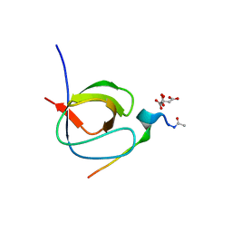
Crystal structure of the Fyn SH3 domain L112V-S114N-S115T-E121L-R123H mutant in complex with VSL12 at pH 3.0
分子名称: CITRIC ACID, Tyrosine-protein kinase Fyn, VSL12
著者Camara-Artigas, A, Plaza-Garrido, M, Salinas-Garcia, M.C.
登録日2020-08-18
公開日2021-08-25
最終更新日2024-01-31
実験手法X-RAY DIFFRACTION (0.99 Å)
主引用文献Crystal structure of the Fyn SH3 domain L112V-S114N-S115T-E121L-R123H mutant in complex with VSL12 at pH 3.0
To be published
7A38
DownloadVisualize
BU of 7a38 by Molmil
Crystal structure of the c-Src SH3 domain mutant V111L-N113S-T114S-Q128E at pH 6.0
分子名称: 2-(N-MORPHOLINO)-ETHANESULFONIC ACID, Proto-oncogene tyrosine-protein kinase Src, SULFATE ION
著者Camara-Artigas, A, Plaza-Garrido, M, Salinas-Garcia, M.C.
登録日2020-08-18
公開日2021-08-25
最終更新日2024-01-31
実験手法X-RAY DIFFRACTION (1.62 Å)
主引用文献Crystal structure of the c-Src SH3 domain mutant V111L-N113S-T114S-Q128E at pH 6.0
To be published
2GNC
DownloadVisualize
BU of 2gnc by Molmil
Crystal structure of srGAP1 SH3 domain in the slit-robo signaling pathway
分子名称: SLIT-ROBO Rho GTPase-activating protein 1
著者Li, X, Liu, Y, Gao, F, Bartlam, M, Wu, J.Y, Rao, Z.
登録日2006-04-10
公開日2006-07-18
最終更新日2023-10-25
実験手法X-RAY DIFFRACTION (1.8 Å)
主引用文献Structural Basis of Robo Proline-rich Motif Recognition by the srGAP1 Src Homology 3 Domain in the Slit-Robo Signaling Pathway
J.Biol.Chem., 281, 2006
2HSP
DownloadVisualize
BU of 2hsp by Molmil
SOLUTION STRUCTURE OF THE SH3 DOMAIN OF PHOSPHOLIPASE CGAMMA
分子名称: PHOSPHOLIPASE C-GAMMA (SH3 DOMAIN)
著者Kohda, D, Hatanaka, H, Odaka, M, Inagaki, F.
登録日1994-06-13
公開日1994-08-31
最終更新日2024-05-01
実験手法SOLUTION NMR
主引用文献Solution structure of the SH3 domain of phospholipase C-gamma.
Cell(Cambridge,Mass.), 72, 1993
2JW4
DownloadVisualize
BU of 2jw4 by Molmil
NMR solution structure of the N-terminal SH3 domain of human Nckalpha
分子名称: Cytoplasmic protein NCK1
著者Santiveri, C.M, Borroto, A, Simon, L, Rico, M, Ortiz, A.R, Alarcon, B, Jimenez, M.
登録日2007-10-05
公開日2008-08-26
最終更新日2024-05-29
実験手法SOLUTION NMR
主引用文献Interaction between the N-terminal SH3 domain of Nckalpha and CD3epsilon-derived peptides: Non-canonical and canonical recognition motifs
BIOCHEM.BIOPHYS.ACTA PROTEINS & PROTEOMICS, 1794, 2009
2K3B
DownloadVisualize
BU of 2k3b by Molmil
Seeing the Invisible: Structures of Excited Protein States by Relaxation Dispersion NMR
分子名称: Actin-binding protein
著者Vallurupalli, P, Hansen, F.D, Kay, L.E.
登録日2008-04-30
公開日2008-07-29
最終更新日2024-05-29
実験手法SOLUTION NMR
主引用文献Structures of invisible, excited protein states by relaxation dispersion NMR spectroscopy
Proc.Natl.Acad.Sci.Usa, 105, 2008
2JXB
DownloadVisualize
BU of 2jxb by Molmil
Structure of CD3epsilon-Nck2 first SH3 domain complex
分子名称: T-cell surface glycoprotein CD3 epsilon chain, Cytoplasmic protein NCK2
著者Takeuchi, K, Yang, H, Ng, E, Park, S, Sun, Z.J, Reinherz, E.L, Wagner, G.
登録日2007-11-09
公開日2008-09-23
最終更新日2024-05-29
実験手法SOLUTION NMR
主引用文献Structural and functional evidence that Nck interaction with CD3epsilon regulates T-cell receptor activity.
J.Mol.Biol., 380, 2008
2JTE
DownloadVisualize
BU of 2jte by Molmil
Third SH3 domain of CD2AP
分子名称: CD2-associated protein
著者van Nuland, N.A.J, Ortega, R.J, Romero Romero, M, Ab, E, Ora, A, Lopez, M.O, Azuaga, A.I.
登録日2007-07-28
公開日2007-12-11
最終更新日2024-05-29
実験手法SOLUTION NMR
主引用文献The high resolution NMR structure of the third SH3 domain of CD2AP.
J.Biomol.Nmr, 39, 2007
2J06
DownloadVisualize
BU of 2j06 by Molmil
Crystal structure of the RasGAP SH3 domain at 1.8 Angstrom resolution
分子名称: RAS GTPASE-ACTIVATING PROTEIN 1
著者Ross, B, Gajhede, M, Kristensen, O.
登録日2006-08-01
公開日2007-01-02
最終更新日2011-07-13
実験手法X-RAY DIFFRACTION (1.8 Å)
主引用文献High Resolution Crystal Structures of the P120 Rasgap SH3 Domain.
Biochem.Biophys.Res.Commun., 353, 2007
2JS2
DownloadVisualize
BU of 2js2 by Molmil
Solution structure of first SH3 domain of adaptor Nck
分子名称: Cytoplasmic protein NCK1
著者Hake, M.J, Choowongkomon, K, Carlin, C.R, Sonnichsen, F.D.
登録日2007-06-29
公開日2008-02-26
最終更新日2024-05-29
実験手法SOLUTION NMR
主引用文献Specificity determinants of a novel Nck interaction with the juxtamembrane domain of the epidermal growth factor receptor.
Biochemistry, 47, 2008
2JS0
DownloadVisualize
BU of 2js0 by Molmil
Solution structure of second SH3 domain of adaptor Nck
分子名称: Cytoplasmic protein NCK1
著者Hake, M.J, Choowongkomon, K, Carlin, C.R, Sonnichsen, F.D.
登録日2007-06-29
公開日2008-02-26
最終更新日2024-05-29
実験手法SOLUTION NMR
主引用文献Specificity determinants of a novel Nck interaction with the juxtamembrane domain of the epidermal growth factor receptor.
Biochemistry, 47, 2008
2J05
DownloadVisualize
BU of 2j05 by Molmil
Crystal structure of the RasGAP SH3 domain at 1.5 Angstrom resolution
分子名称: RAS GTPASE-ACTIVATING PROTEIN 1
著者Ross, B, Gajhede, M, Kristensen, O.
登録日2006-08-01
公開日2007-01-02
最終更新日2011-07-13
実験手法X-RAY DIFFRACTION (1.5 Å)
主引用文献High Resolution Crystal Structures of the P120 Rasgap SH3 Domain.
Biochem.Biophys.Res.Commun., 353, 2007
2IIM
DownloadVisualize
BU of 2iim by Molmil
SH3 Domain of Human Lck
分子名称: CALCIUM ION, Proto-oncogene tyrosine-protein kinase LCK, TETRAETHYLENE GLYCOL, ...
著者Romir, J, Egerer-Sieber, C, Muller, Y.A.
登録日2006-09-28
公開日2006-11-07
最終更新日2023-08-30
実験手法X-RAY DIFFRACTION (1 Å)
主引用文献Crystal structure analysis and solution studies of human Lck-SH3; zinc-induced homodimerization competes with the binding of proline-rich motifs.
J.Mol.Biol., 365, 2007

221051

件を2024-06-12に公開中

PDB statisticsPDBj update infoContact PDBjnumon