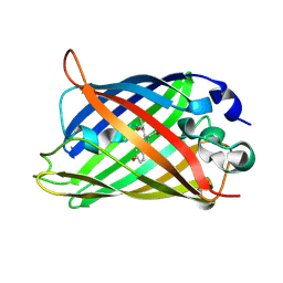
4B5Y
 
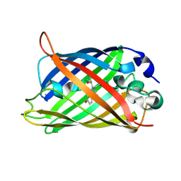
 | | X-ray structure of the cyan fluorescent protein mTurquoise-GL (K206A mutant) in space group C222(1) | | 分子名称: | GREEN FLUORESCENT PROTEIN | | 著者 | von Stetten, D, Lelimousin, M, Oost, K, Noirclerc-Savoye, M, Gadella, T.W.J, Goedhart, J, Royant, A. | | 登録日 | 2012-08-08 | | 公開日 | 2013-08-28 | | 最終更新日 | 2023-12-20 | | 実験手法 | X-RAY DIFFRACTION (1.45 Å) | | 主引用文献 | Influence of the H148G Mutation on Fluorescence Properties of Cyan Fluorescent Proteins
To be Published
|
|
1Q4A
 | |
5DTX
 | |
2O24
 | |
3EVP
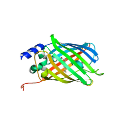
 | | crystal structure of circular-permutated EGFP | | 分子名称: | Green fluorescent protein,Green fluorescent protein | | 著者 | Wang, Q, Shui, B, Kotlikoff, M.I, Sondermann, H. | | 登録日 | 2008-10-13 | | 公開日 | 2008-12-09 | | 最終更新日 | 2023-12-27 | | 実験手法 | X-RAY DIFFRACTION (1.453 Å) | | 主引用文献 | Structural Basis for Calcium Sensing by GCaMP2.
Structure, 16, 2008
|
|
8A7V
 | |
6QQ8
 | |
3DQF
 | |
3DQL
 | |
3DQ4
 | |
2YE0
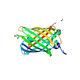
 | | X-ray structure of the cyan fluorescent protein mTurquoise (K206A mutant) | | 分子名称: | GREEN FLUORESCENT PROTEIN | | 著者 | von Stetten, D, Goedhart, J, Noirclerc-Savoye, M, Lelimousin, M, Joosen, L, Hink, M.A, van Weeren, L, Gadella, T.W.J, Royant, A. | | 登録日 | 2011-03-25 | | 公開日 | 2012-03-21 | | 最終更新日 | 2023-12-20 | | 実験手法 | X-RAY DIFFRACTION (1.47 Å) | | 主引用文献 | Structure-Guided Evolution of Cyan Fluorescent Proteins Towards a Quantum Yield of 93%
Nat.Commun, 3, 2012
|
|
2H6V
 | |
6OFM
 | | Crystal structure of green fluorescent protein (GFP); S65T, Y66(3-CH3Y); ih circular permutant (50-51) | | 分子名称: | Green fluorescent protein (GFP); S65T, Y66(3-CH3Y); ih circular permutant (50-51) | | 著者 | Lin, C.-Y, Romei, M.G, Mathews, I.I, Boxer, S.G. | | 登録日 | 2019-03-31 | | 公開日 | 2019-07-10 | | 最終更新日 | 2023-11-15 | | 実験手法 | X-RAY DIFFRACTION (1.48 Å) | | 主引用文献 | Unified Model for Photophysical and Electro-Optical Properties of Green Fluorescent Proteins.
J.Am.Chem.Soc., 141, 2019
|
|
1Q4B
 | |
3CBE
 | |
3DQC
 | |
3SVE
 | |
3SS0
 | |
4XGY
 | | GFP based antibody (fluorobody) | | 分子名称: | 3,6,9,12,15,18,21,24-OCTAOXAHEXACOSAN-1-OL, Green fluorescent protein, mAb LCDR3 | | 著者 | Shi, N, Chen, Y.G, Wang, S.H. | | 登録日 | 2015-01-03 | | 公開日 | 2015-07-01 | | 最終更新日 | 2023-11-15 | | 実験手法 | X-RAY DIFFRACTION (1.494 Å) | | 主引用文献 | The structure of a GFP-based antibody (fluorobody) to TLH, a toxin from Vibrio parahaemolyticus.
Acta Crystallogr.,Sect.F, 71, 2015
|
|
6UN4
 | | Crystal structure of rsEGFP2, Y67(3-ClY), Y107(3-ClY) | | 分子名称: | Green fluorescent protein, SULFATE ION | | 著者 | Lin, C.-Y, Romei, M.G, Boxer, S.G, Chang, J. | | 登録日 | 2019-10-10 | | 公開日 | 2020-07-15 | | 最終更新日 | 2023-11-15 | | 実験手法 | X-RAY DIFFRACTION (1.499 Å) | | 主引用文献 | Structural and spectroscopic characterization of photoactive yellow protein and photoswitchable fluorescent protein constructs containing heavy atoms.
J Photochem Photobiol A Chem, 401, 2020
|
|
1S6Z
 | | Enhanced Green Fluorescent Protein Containing the Y66L Substitution | | 分子名称: | CHLORIDE ION, green fluorescent protein | | 著者 | Rosenow, M.A, Huffman, H.A, Phail, M.E, Wachter, R.M. | | 登録日 | 2004-01-28 | | 公開日 | 2004-05-04 | | 最終更新日 | 2024-07-10 | | 実験手法 | X-RAY DIFFRACTION (1.5 Å) | | 主引用文献 | The Crystal Structure of the Y66L Variant of Green Fluorescent Protein Supports a Cyclization-Oxidation-Dehydration Mechanism for Chromophore Maturation(,).
Biochemistry, 43, 2004
|
|
5DTY
 | |
2DUF
 | |
3W1D
 | |
1JBZ
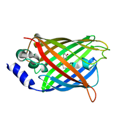
 | | CRYSTAL STRUCTURE ANALYSIS OF A DUAL-WAVELENGTH EMISSION GREEN FLUORESCENT PROTEIN VARIANT AT HIGH PH | | 分子名称: | 1,2-ETHANEDIOL, GREEN FLUORESCENT PROTEIN, MAGNESIUM ION | | 著者 | Hanson, G.T, McAnaney, T.B, Park, E.S, Rendell, M.E.P, Yarbrough, D.K, Chu, S, Xi, L, Boxer, S.G, Montrose, M.H, Remington, S.J. | | 登録日 | 2001-06-07 | | 公開日 | 2003-01-07 | | 最終更新日 | 2023-11-15 | | 実験手法 | X-RAY DIFFRACTION (1.5 Å) | | 主引用文献 | Green Fluorescent Protein Variants as Ratiometric Dual Emission pH Sensors. 1. Structural Characterization and Preliminary Application.
Biochemistry, 41, 2002
|
|


