1RI0
 
 | |
1RI1
 | | Structure and mechanism of mRNA cap (guanine N-7) methyltransferase | | 分子名称: | 7-METHYL-GUANOSINE-5'-TRIPHOSPHATE-5'-GUANOSINE, S-ADENOSYL-L-HOMOCYSTEINE, mRNA CAPPING ENZYME | | 著者 | Fabrega, C, Hausmann, S, Shen, V, Shuman, S, Lima, C.D. | | 登録日 | 2003-11-16 | | 公開日 | 2004-02-03 | | 最終更新日 | 2024-02-14 | | 実験手法 | X-RAY DIFFRACTION (2.5 Å) | | 主引用文献 | Structure and mechanism of mRNA cap (Guanine-n7) methyltransferase
Mol.Cell, 13, 2004
|
|
1RI2
 | | Structure and mechanism of mRNA cap (guanine N-7) methyltransferase | | 分子名称: | 7-METHYL-GUANOSINE-5'-TRIPHOSPHATE-5'-GUANOSINE, mRNA CAPPING ENZYME | | 著者 | Fabrega, C, Hausmann, S, Shen, V, Shuman, S, Lima, C.D. | | 登録日 | 2003-11-16 | | 公開日 | 2004-02-03 | | 最終更新日 | 2024-02-14 | | 実験手法 | X-RAY DIFFRACTION (2.7 Å) | | 主引用文献 | Structure and mechanism of mRNA cap (Guanine-n7) methyltransferase
Mol.Cell, 13, 2004
|
|
1RI3
 | | Structure and mechanism of mRNA cap (guanine N-7) methyltransferase | | 分子名称: | S-ADENOSYL-L-HOMOCYSTEINE, mRNA CAPPING ENZYME | | 著者 | Fabrega, C, Hausmann, S, Shen, V, Shuman, S, Lima, C.D. | | 登録日 | 2003-11-16 | | 公開日 | 2004-02-03 | | 最終更新日 | 2024-02-14 | | 実験手法 | X-RAY DIFFRACTION (2.5 Å) | | 主引用文献 | Structure and mechanism of mRNA cap (Guanine-n7) methyltransferase
Mol.Cell, 13, 2004
|
|
1RI4
 | | Structure and mechanism of mRNA cap (guanine N-7) methyltransferase | | 分子名称: | S-ADENOSYLMETHIONINE, mRNA CAPPING ENZYME | | 著者 | Fabrega, C, Hausmann, S, Shen, V, Shuman, S, Lima, C.D. | | 登録日 | 2003-11-16 | | 公開日 | 2004-02-03 | | 最終更新日 | 2024-02-14 | | 実験手法 | X-RAY DIFFRACTION (2.4 Å) | | 主引用文献 | Structure and mechanism of mRNA cap (Guanine-n7) methyltransferase
Mol.Cell, 13, 2004
|
|
1RI5
 | | Structure and mechanism of mRNA cap (guanine N-7) methyltransferase | | 分子名称: | mRNA CAPPING ENZYME | | 著者 | Fabrega, C, Hausmann, S, Shen, V, Shuman, S, Lima, C.D, Burley, S.K, New York SGX Research Center for Structural Genomics (NYSGXRC) | | 登録日 | 2003-11-16 | | 公開日 | 2004-02-03 | | 最終更新日 | 2024-02-14 | | 実験手法 | X-RAY DIFFRACTION (2.1 Å) | | 主引用文献 | Structure and mechanism of mRNA cap (Guanine-n7) methyltransferase
Mol.Cell, 13, 2004
|
|
1RI6
 | | Structure of a putative isomerase from E. coli | | 分子名称: | putative isomerase ybhE | | 著者 | Lima, C.D, Kniewel, R, Solorzano, V, Wu, J, Burley, S.K, New York SGX Research Center for Structural Genomics (NYSGXRC) | | 登録日 | 2003-11-16 | | 公開日 | 2003-11-25 | | 最終更新日 | 2024-02-14 | | 実験手法 | X-RAY DIFFRACTION (2 Å) | | 主引用文献 | Structure of a putative 7-bladed propeller isomerase
To be Published
|
|
1RI7
 | |
1RI8
 | | Crystal Structure of the Camelid Single Domain Antibody 1D2L19 in complex with Hen Egg White Lysozyme | | 分子名称: | GLYCEROL, Lysozyme C, camelid ANTIBODY HEAVY CHAIN | | 著者 | De Genst, E, Silence, K, Ghahroudi, M.A, Decanniere, K, Loris, R, Kinne, J, Wyns, L, Muyldermans, S. | | 登録日 | 2003-11-17 | | 公開日 | 2005-02-01 | | 最終更新日 | 2024-11-20 | | 実験手法 | X-RAY DIFFRACTION (1.85 Å) | | 主引用文献 | Strong in vivo maturation compensates for structurally restricted H3 loops in antibody repertoires.
J.Biol.Chem., 280, 2005
|
|
1RI9
 | | Structure of a helically extended SH3 domain of the T cell adapter protein ADAP | | 分子名称: | FYN-binding protein | | 著者 | Heuer, K, Kofler, M, Langdon, G, Thiemke, K, Freund, C. | | 登録日 | 2003-11-17 | | 公開日 | 2004-04-20 | | 最終更新日 | 2024-05-08 | | 実験手法 | SOLUTION NMR | | 主引用文献 | Structure of a Helically Extended SH3 Domain of the T Cell Adapter Protein ADAP.
STRUCTURE, 12, 2004
|
|
1RIB
 | |
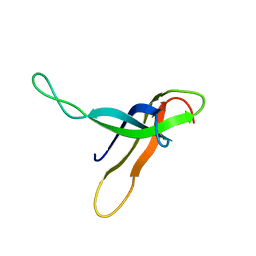
1RIE
 | | STRUCTURE OF A WATER SOLUBLE FRAGMENT OF THE RIESKE IRON-SULFUR PROTEIN OF THE BOVINE HEART MITOCHONDRIAL CYTOCHROME BC1-COMPLEX | | 分子名称: | FE2/S2 (INORGANIC) CLUSTER, RIESKE IRON-SULFUR PROTEIN | | 著者 | Iwata, S, Saynovits, M, Link, T.A, Michel, H. | | 登録日 | 1996-02-23 | | 公開日 | 1996-12-07 | | 最終更新日 | 2024-10-23 | | 実験手法 | X-RAY DIFFRACTION (1.5 Å) | | 主引用文献 | Structure of a water soluble fragment of the 'Rieske' iron-sulfur protein of the bovine heart mitochondrial cytochrome bc1 complex determined by MAD phasing at 1.5 A resolution.
Structure, 4, 1996
|
|
1RIF
 | |
1RIH
 | | Crystal Structure of Fab 14F7, a unique anti-tumor antibody specific for N-glycolyl GM3 | | 分子名称: | heavy chain of antibody 14F7, light chain of antibody 14F7 | | 著者 | Krengel, U, Olsson, L.-L, Martinez, C, Talavera, A, Rojas, G, Mier, E, Angstrom, J, Moreno, E. | | 登録日 | 2003-11-17 | | 公開日 | 2004-01-13 | | 最終更新日 | 2024-10-30 | | 実験手法 | X-RAY DIFFRACTION (2.5 Å) | | 主引用文献 | Structure and Molecular Interactions of a Unique Antitumor Antibody Specific for N-Glycolyl GM3.
J.Biol.Chem., 279, 2004
|
|
1RII
 | | Crystal structure of phosphoglycerate mutase from M. Tuberculosis | | 分子名称: | 2,3-bisphosphoglycerate-dependent phosphoglycerate mutase, GLYCEROL | | 著者 | Mueller, P, Sawaya, M.R, Chan, S, Wu, Y, Pashkova, I, Perry, J, Eisenberg, D, TB Structural Genomics Consortium (TBSGC) | | 登録日 | 2003-11-17 | | 公開日 | 2004-10-05 | | 最終更新日 | 2023-08-23 | | 実験手法 | X-RAY DIFFRACTION (1.7 Å) | | 主引用文献 | The 1.70 angstroms X-ray crystal structure of Mycobacterium tuberculosis phosphoglycerate mutase.
Acta Crystallogr.,Sect.D, 61, 2005
|
|
1RIJ
 | | E6-bind Trp-cage (E6apn1) | | 分子名称: | E6apn1 peptide | | 著者 | Liu, Y, Liu, Z, Androphy, E, Chen, J, Baleja, J.D. | | 登録日 | 2003-11-17 | | 公開日 | 2004-08-03 | | 最終更新日 | 2024-05-22 | | 実験手法 | SOLUTION NMR | | 主引用文献 | Design and characterization of helical peptides that inhibit the E6 protein of papillomavirus.
Biochemistry, 43, 2004
|
|
1RIK
 | | E6-binding zinc finger (E6apc1) | | 分子名称: | E6apc1 peptide | | 著者 | Liu, Y, Liu, Z, Androphy, E, Chen, J, Baleja, J.D. | | 登録日 | 2003-11-17 | | 公開日 | 2004-08-03 | | 最終更新日 | 2024-05-22 | | 実験手法 | SOLUTION NMR | | 主引用文献 | Design and characterization of helical peptides that inhibit the E6 protein of papillomavirus.
Biochemistry, 43, 2004
|
|
1RIL
 | | CRYSTAL STRUCTURE OF RIBONUCLEASE H FROM THERMUS THERMOPHILUS HB8 REFINED AT 2.8 ANGSTROMS RESOLUTION | | 分子名称: | RIBONUCLEASE H | | 著者 | Ishikawa, K, Okumura, M, Katayanagi, K, Kimura, S, Kanaya, S, Nakamura, H, Morikawa, K. | | 登録日 | 1993-01-14 | | 公開日 | 1993-10-31 | | 最終更新日 | 2024-02-14 | | 実験手法 | X-RAY DIFFRACTION (2.8 Å) | | 主引用文献 | Crystal structure of ribonuclease H from Thermus thermophilus HB8 refined at 2.8 A resolution.
J.Mol.Biol., 230, 1993
|
|
1RIM
 | | E6-binding zinc finger (E6apc2) | | 分子名称: | E6apc2 peptide | | 著者 | Liu, Y, Liu, Z, Androphy, E, Chen, J, Baleja, J.D. | | 登録日 | 2003-11-17 | | 公開日 | 2004-08-03 | | 最終更新日 | 2024-05-22 | | 実験手法 | SOLUTION NMR | | 主引用文献 | Design and characterization of helical peptides that inhibit the E6 protein of papillomavirus.
Biochemistry, 43, 2004
|
|
1RIN
 | | X-RAY CRYSTAL STRUCTURE OF A PEA LECTIN-TRIMANNOSIDE COMPLEX AT 2.6 ANGSTROMS RESOLUTION | | 分子名称: | CALCIUM ION, MANGANESE (II) ION, PEA LECTIN, ... | | 著者 | Rini, J.M, Hardman, K.D, Einspahr, H, Suddath, F.L, Carver, J.P. | | 登録日 | 1993-01-27 | | 公開日 | 1993-10-31 | | 最終更新日 | 2024-02-14 | | 実験手法 | X-RAY DIFFRACTION (2.6 Å) | | 主引用文献 | X-ray crystal structure of a pea lectin-trimannoside complex at 2.6 A resolution.
J.Biol.Chem., 268, 1993
|
|
1RIO
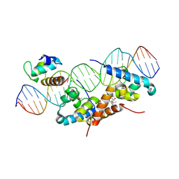 | | Structure of bacteriophage lambda cI-NTD in complex with sigma-region4 of Thermus aquaticus bound to DNA | | 分子名称: | (4S)-2-METHYL-2,4-PENTANEDIOL, 27-MER, CALCIUM ION, ... | | 著者 | Jain, D, Nickels, B.E, Sun, L, Hochschild, A, Darst, S.A. | | 登録日 | 2003-11-17 | | 公開日 | 2004-01-27 | | 最終更新日 | 2024-11-20 | | 実験手法 | X-RAY DIFFRACTION (2.3 Å) | | 主引用文献 | Structure of a ternary transcription activation complex.
Mol.Cell, 13, 2004
|
|
1RIP
 | |
1RIQ
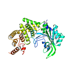 | | The crystal structure of the catalytic fragment of the alanyl-tRNA synthetase | | 分子名称: | Alanyl-tRNA synthetase | | 著者 | Swairjo, M.A, Otero, F.J, Yang, X.-L, Lovato, M.A, Skene, R.J, McRee, D.E, Ribas de Pouplana, L, Schimmel, P. | | 登録日 | 2003-11-17 | | 公開日 | 2004-04-06 | | 最終更新日 | 2024-10-30 | | 実験手法 | X-RAY DIFFRACTION (2.14 Å) | | 主引用文献 | Alanyl-tRNA Synthetase Crystal Structure and Design for Acceptor-Stem Recognition
Mol.Cell, 13, 2004
|
|
1RIR
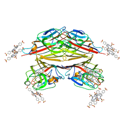 | | Crystal structure of meso-tetrasulphonatophenylporphyrin in complex with Peanut lectin. | | 分子名称: | 5,10,15,20-TETRAKIS(4-SULPFONATOPHENYL)-21H,23H-PORPHINE, CALCIUM ION, Galactose-binding lectin, ... | | 著者 | Goel, M, Kaur, K.J, Maiya, B.G, Swamy, M.J, Salunke, D.M. | | 登録日 | 2003-11-17 | | 公開日 | 2004-12-28 | | 最終更新日 | 2023-08-23 | | 実験手法 | X-RAY DIFFRACTION (2.9 Å) | | 主引用文献 | Crystal structures of the PNA-porphyrin complex in the presence and absence of lactose: mapping the conformational changes on lactose binding, interacting surfaces, and supramolecular aggregations.
Biochemistry, 44, 2005
|
|
1RIS
 | | CRYSTAL STRUCTURE OF THE RIBOSOMAL PROTEIN S6 FROM THERMUS THERMOPHILUS | | 分子名称: | RIBOSOMAL PROTEIN S6 | | 著者 | Lindahl, M, Svensson, L.A, Liljas, A, Sedelnikova, S.E, Eliseikina, I.A, Fomenkova, N.P, Nevskaya, N, Nikonov, S.V, Garber, M.B, Muranova, T.A, Rykonova, A.I, Amons, R. | | 登録日 | 1994-05-31 | | 公開日 | 1994-09-30 | | 最終更新日 | 2024-02-14 | | 実験手法 | X-RAY DIFFRACTION (2 Å) | | 主引用文献 | Crystal structure of the ribosomal protein S6 from Thermus thermophilus.
EMBO J., 13, 1994
|
|


