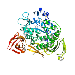
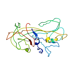
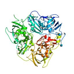
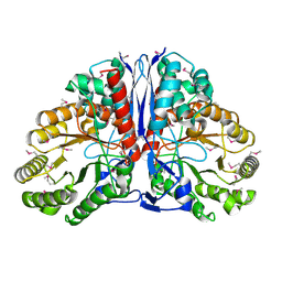
1KCE
 
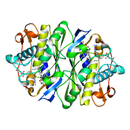 | | E. COLI THYMIDYLATE SYNTHASE MUTANT E58Q IN COMPLEX WITH CB3717 AND 2'-DEOXYURIDINE 5'-MONOPHOSPHATE (DUMP) | | 分子名称: | 10-PROPARGYL-5,8-DIDEAZAFOLIC ACID, 2'-DEOXYURIDINE 5'-MONOPHOSPHATE, THYMIDYLATE SYNTHASE | | 著者 | Sage, C.R, Rutenber, E.E, Stout, T.J, Stroud, R.M. | | 登録日 | 1996-10-22 | | 公開日 | 1997-04-21 | | 最終更新日 | 2024-06-05 | | 実験手法 | X-RAY DIFFRACTION (2 Å) | | 主引用文献 | An essential role for water in an enzyme reaction mechanism: the crystal structure of the thymidylate synthase mutant E58Q.
Biochemistry, 35, 1996
|
|
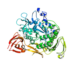
1KCF
 | | Crystal Structure of the Yeast Mitochondrial Holliday Junction Resolvase, Ydc2 | | 分子名称: | HYPOTHETICAL 30.2 KD PROTEIN C25G10.02 IN CHROMOSOME I, SULFATE ION | | 著者 | Ceschini, S, Keeley, A, McAlister, M.S.B, Oram, M, Phelan, J, Pearl, L.H, Tsaneva, I.R, Barrett, T.E. | | 登録日 | 2001-11-08 | | 公開日 | 2001-11-28 | | 最終更新日 | 2024-02-07 | | 実験手法 | X-RAY DIFFRACTION (2.3 Å) | | 主引用文献 | Crystal structure of the fission yeast mitochondrial Holliday junction resolvase Ydc2.
EMBO J., 20, 2001
|
|
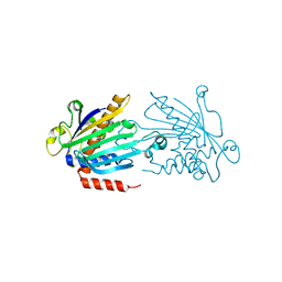
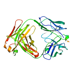
1KCG
 | | NKG2D in complex with ULBP3 | | 分子名称: | GLUTATHIONE, NKG2-D type II integral membrane protein, ULBP3 protein | | 著者 | Radaev, S, Sun, P. | | 登録日 | 2001-11-08 | | 公開日 | 2002-01-09 | | 最終更新日 | 2011-07-13 | | 実験手法 | X-RAY DIFFRACTION (2.6 Å) | | 主引用文献 | Conformational plasticity revealed by the cocrystal structure of NKG2D and its class I MHC-like ligand ULBP3.
Immunity, 15, 2001
|
|
1KCI
 | | Crystal Structure of 9-amino-N-[2-(4-morpholinyl)ethyl]-4-acridinecarboxamide Bound to d(CGTACG)2 | | 分子名称: | 5'-D(*CP*GP*TP*AP*CP*G)-3', 9-AMINO-N-[2-(4-MORPHOLINYL)ETHYL]-4-ACRIDINECARBOXAMIDE | | 著者 | Adams, A, Guss, J.M, Denny, W.A, Wakelin, L.P.G. | | 登録日 | 2001-11-08 | | 公開日 | 2002-02-01 | | 最終更新日 | 2024-04-03 | | 実験手法 | X-RAY DIFFRACTION (1.8 Å) | | 主引用文献 | Crystal structure of 9-amino-N-[2-(4-morpholinyl)ethyl]-4-acridinecarboxamide bound to d(CGTACG)2: implications for structure-activity relationships of acridinecarboxamide topoisomerase poisons.
Nucleic Acids Res., 30, 2002
|
|
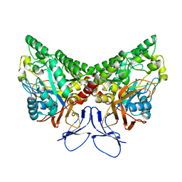
1KCK
 | | Bacillus circulans strain 251 Cyclodextrin glycosyl transferase mutant N193G | | 分子名称: | 1-AMINO-2,3-DIHYDROXY-5-HYDROXYMETHYL CYCLOHEX-5-ENE, CALCIUM ION, CYCLODEXTRIN GLYCOSYLTRANSFERASE, ... | | 著者 | Rozeboom, H.J, Uitdehaag, J.C.M, Dijkstra, B.W. | | 登録日 | 2001-11-09 | | 公開日 | 2002-01-16 | | 最終更新日 | 2023-08-16 | | 実験手法 | X-RAY DIFFRACTION (2.43 Å) | | 主引用文献 | The remote substrate binding subsite -6 in cyclodextrin-glycosyltransferase controls the transferase activity of the enzyme via an induced-fit mechanism.
J.Biol.Chem., 277, 2002
|
|
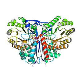
1KCL
 | | Bacillus ciruclans strain 251 Cyclodextrin glycosyl transferase mutant G179L | | 分子名称: | (4S)-2-METHYL-2,4-PENTANEDIOL, CALCIUM ION, Cyclodextrin glycosyltransferase, ... | | 著者 | Rozeboom, H.J, Uitdehaag, J.C.M, Dijkstra, B.W. | | 登録日 | 2001-11-09 | | 公開日 | 2002-01-16 | | 最終更新日 | 2023-08-16 | | 実験手法 | X-RAY DIFFRACTION (1.94 Å) | | 主引用文献 | The remote substrate binding subsite -6 in cyclodextrin-glycosyltransferase controls the transferase activity of the enzyme via an induced-fit mechanism.
J.Biol.Chem., 277, 2002
|
|
1KCM
 | | Crystal Structure of Mouse PITP Alpha Void of Bound Phospholipid at 2.0 Angstroms Resolution | | 分子名称: | Phosphatidylinositol Transfer Protein alpha | | 著者 | Schouten, A, Agianian, B, Westerman, J, Kroon, J, Wirtz, K.W.A, Gros, P. | | 登録日 | 2001-11-09 | | 公開日 | 2002-05-08 | | 最終更新日 | 2023-08-16 | | 実験手法 | X-RAY DIFFRACTION (2 Å) | | 主引用文献 | Structure of apo-phosphatidylinositol transfer protein alpha provides insight into membrane association.
EMBO J., 21, 2002
|
|
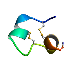
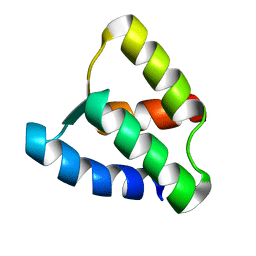
1KCN
 | | Structure of e109 Zeta Peptide, an Antagonist of the High-Affinity IgE Receptor | | 分子名称: | e109 zeta peptide | | 著者 | Nakamura, G.R, Reynolds, M.E, Chen, Y.M, Starovasnik, M.A, Lowman, H.B. | | 登録日 | 2001-11-09 | | 公開日 | 2002-03-06 | | 最終更新日 | 2022-02-23 | | 実験手法 | SOLUTION NMR | | 主引用文献 | Stable "zeta" peptides that act as potent antagonists of the high-affinity IgE receptor.
Proc.Natl.Acad.Sci.USA, 99, 2002
|
|
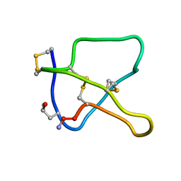
1KCO
 | | Structure of e131 Zeta Peptide, a Potent Antagonist of the High-Affinity IgE Receptor | | 分子名称: | e131 Zeta Peptide | | 著者 | Nakamura, G.R, Reynolds, M.E, Chen, Y.M, Starovasnik, M.A, Lowman, H.B. | | 登録日 | 2001-11-09 | | 公開日 | 2002-03-06 | | 最終更新日 | 2022-02-23 | | 実験手法 | SOLUTION NMR | | 主引用文献 | Stable "zeta" peptides that act as potent antagonists of the high-affinity IgE receptor.
Proc.Natl.Acad.Sci.USA, 99, 2002
|
|
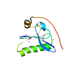
1KCP
 | | 3D STRUCTURE OF K-CONOTOXIN PVIIA, A NOVEL POTASSIUM CHANNEL-BLOCKING TOXIN FROM CONE SNAILS, NMR, 22 STRUCTURES | | 分子名称: | KAPPA-CONOTOXIN PVIIA | | 著者 | Savarin, P, Guenneugues, M, Gilquin, B, Lamthanh, H, Gasparini, S, Zinn-Justin, S, Menez, A. | | 登録日 | 1998-01-27 | | 公開日 | 1998-10-14 | | 最終更新日 | 2017-11-29 | | 実験手法 | SOLUTION NMR | | 主引用文献 | Three-dimensional structure of kappa-conotoxin PVIIA, a novel potassium channel-blocking toxin from cone snails.
Biochemistry, 37, 1998
|
|
1KCQ
 | | Human Gelsolin Domain 2 with a Cd2+ bound | | 分子名称: | CADMIUM ION, GELSOLIN | | 著者 | Kazmirski, S.L, Isaacson, R.L, An, C, Buckle, A, Johnson, C.M, Daggett, V, Fersht, A.R. | | 登録日 | 2001-11-09 | | 公開日 | 2002-01-04 | | 最終更新日 | 2023-08-16 | | 実験手法 | X-RAY DIFFRACTION (1.65 Å) | | 主引用文献 | Loss of a metal-binding site in gelsolin leads to familial amyloidosis-Finnish type.
Nat.Struct.Biol., 9, 2002
|
|
1KCR
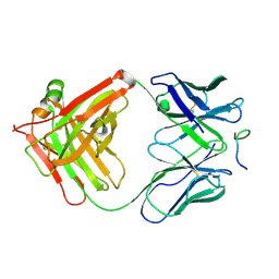 | | CRYSTAL STRUCTURE OF ANTIBODY PC283 IN COMPLEX WITH PS1 PEPTIDE | | 分子名称: | PC283 IMMUNOGLOBULIN, PS1 peptide | | 著者 | Nair, D.T, Singh, K, Sahu, N, Rao, K.V.S, Salunke, D.M. | | 登録日 | 2001-11-11 | | 公開日 | 2002-05-11 | | 最終更新日 | 2011-07-13 | | 実験手法 | X-RAY DIFFRACTION (2.9 Å) | | 主引用文献 | Crystal structure of an antibody bound to an immunodominant peptide epitope: novel features in peptide-antibody recognition.
J.Immunol., 165, 2000
|
|
1KCS
 | | CRYSTAL STRUCTURE OF ANTIBODY PC282 IN COMPLEX WITH PS1 PEPTIDE | | 分子名称: | PC282 IMMUNOGLOBULIN, PS1 peptide | | 著者 | Nair, D.T, Singh, K, Siddiqui, Z, Nayak, B.P, Rao, K.V.S, Salunke, D.M. | | 登録日 | 2001-11-11 | | 公開日 | 2002-05-11 | | 最終更新日 | 2011-07-13 | | 実験手法 | X-RAY DIFFRACTION (2.5 Å) | | 主引用文献 | Epitope recognition by diverse antibodies suggests conformational convergence in an antibody response.
J.Immunol., 168, 2002
|
|
1KCT
 | | ALPHA1-ANTITRYPSIN | | 分子名称: | ALPHA1-ANTITRYPSIN | | 著者 | Song, H.K, Suh, S.W. | | 登録日 | 1996-08-06 | | 公開日 | 1997-01-11 | | 最終更新日 | 2024-02-07 | | 実験手法 | X-RAY DIFFRACTION (3.46 Å) | | 主引用文献 | Crystal structure of an uncleaved alpha 1-antitrypsin reveals the conformation of its inhibitory reactive loop.
FEBS Lett., 377, 1995
|
|
1KCU
 | | CRYSTAL STRUCTURE OF ANTIBODY PC287 | | 分子名称: | PC287 IMMUNOGLOBULIN | | 著者 | Nair, D.T, Singh, K, Siddiqui, Z, Nayak, B.P, Rao, K.V.S, Salunke, D.M. | | 登録日 | 2001-11-11 | | 公開日 | 2002-05-11 | | 最終更新日 | 2011-07-13 | | 実験手法 | X-RAY DIFFRACTION (2.2 Å) | | 主引用文献 | Epitope recognition by diverse antibodies suggests conformational convergence in an antibody response.
J.Immunol., 168, 2002
|
|
1KCV
 | | Crystal structure of antibody pc282 | | 分子名称: | PC282 IMMUNOGLOBULIN | | 著者 | Nair, D.T, Singh, K, Siddiqui, Z, Nayak, B.P, Rao, K.V, Salunke, D.M. | | 登録日 | 2001-11-11 | | 公開日 | 2002-05-11 | | 最終更新日 | 2011-07-13 | | 実験手法 | X-RAY DIFFRACTION (1.8 Å) | | 主引用文献 | Epitope recognition by diverse antibodies suggests conformational convergence in an antibody response.
J.Immunol., 168, 2002
|
|
1KCW
 | | X-RAY CRYSTAL STRUCTURE OF HUMAN CERULOPLASMIN AT 3.0 ANGSTROMS | | 分子名称: | 2-acetamido-2-deoxy-beta-D-glucopyranose, CERULOPLASMIN, COPPER (II) ION, ... | | 著者 | Card, G.L, Zaitsev, V.N, Lindley, P.F. | | 登録日 | 1996-09-25 | | 公開日 | 1997-02-12 | | 最終更新日 | 2020-07-29 | | 実験手法 | X-RAY DIFFRACTION (3 Å) | | 主引用文献 | The X-ray structure of human serum ceruloplasmin at 3.1 angstrom: Nature of the copper centres.
J.Biol.Inorg.Chem., 1, 1996
|
|
1KCX
 | | X-ray structure of NYSGRC target T-45 | | 分子名称: | DIHYDROPYRIMIDINASE RELATED PROTEIN-1 | | 著者 | Deo, R.C, Schmidt, E.F, Strittmatter, S.M, Burley, S.K, New York SGX Research Center for Structural Genomics (NYSGXRC) | | 登録日 | 2001-11-11 | | 公開日 | 2003-08-05 | | 最終更新日 | 2024-02-07 | | 実験手法 | X-RAY DIFFRACTION (2.12 Å) | | 主引用文献 | Structural bases for CRMP function in plexin-dependent semaphorin3A signaling
Embo J., 23, 2004
|
|
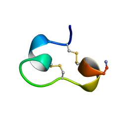
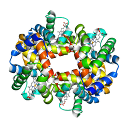
1KCY
 | | NMR solution structure of apo calbindin D9k (F36G + P43M mutant) | | 分子名称: | calbindin D9k | | 著者 | Nelson, M.R, Thulin, E, Fagan, P.A, Forsen, S, Chazin, W.J. | | 登録日 | 2001-11-12 | | 公開日 | 2001-11-21 | | 最終更新日 | 2024-05-22 | | 実験手法 | SOLUTION NMR | | 主引用文献 | The EF-hand domain: a globally cooperative structural unit.
Protein Sci., 11, 2002
|
|
1KCZ
 | | Crystal Structure of beta-methylaspartase from Clostridium tetanomorphum. Mg-complex. | | 分子名称: | 1,2-ETHANEDIOL, MAGNESIUM ION, beta-methylaspartase | | 著者 | Asuncion, M, Blankenfeldt, W, Barlow, J.N, Gani, D, Naismith, J.H. | | 登録日 | 2001-11-12 | | 公開日 | 2001-12-19 | | 最終更新日 | 2011-07-13 | | 実験手法 | X-RAY DIFFRACTION (1.9 Å) | | 主引用文献 | The structure of 3-methylaspartase from Clostridium tetanomorphum functions via the common enolase chemical step.
J.Biol.Chem., 277, 2002
|
|
1KD0
 | | Crystal Structure of beta-methylaspartase from Clostridium tetanomorphum. Apo-structure. | | 分子名称: | 1,2-ETHANEDIOL, beta-methylaspartase | | 著者 | Asuncion, M, Blankenfeldt, W, Barlow, J.N, Gani, D, Naismith, J.H. | | 登録日 | 2001-11-12 | | 公開日 | 2001-12-19 | | 最終更新日 | 2011-07-13 | | 実験手法 | X-RAY DIFFRACTION (1.9 Å) | | 主引用文献 | The structure of 3-methylaspartase from Clostridium tetanomorphum functions via the common enolase chemical step.
J.Biol.Chem., 277, 2002
|
|
1KD1
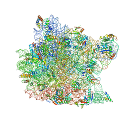 | | Co-crystal Structure of Spiramycin bound to the 50S Ribosomal Subunit of Haloarcula marismortui | | 分子名称: | 23S RRNA, 5S RRNA, CADMIUM ION, ... | | 著者 | Hansen, J.L, Ippolito, J.A, Ban, N, Nissen, P, Moore, P.B, Steitz, T.A. | | 登録日 | 2001-11-12 | | 公開日 | 2002-07-19 | | 最終更新日 | 2023-08-16 | | 実験手法 | X-RAY DIFFRACTION (3 Å) | | 主引用文献 | The structures of four macrolide antibiotics bound to the large ribosomal subunit.
Mol.Cell, 10, 2002
|
|
1KD2
 | |
1KD3
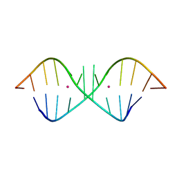 | | The Crystal Structure of r(GGUCACAGCCC)2, Thallium form | | 分子名称: | 5'-R(*GP*GP*UP*CP*AP*CP*AP*GP*CP*CP*C)-3', THALLIUM (I) ION | | 著者 | Kacer, V, Scaringe, S.A, Scarsdale, J.N, Rife, J.P. | | 登録日 | 2001-11-12 | | 公開日 | 2003-03-04 | | 最終更新日 | 2024-02-07 | | 実験手法 | X-RAY DIFFRACTION (1.8 Å) | | 主引用文献 | Crystal structures of r(GGUCACAGCCC)2.
Acta Crystallogr.,Sect.D, 59, 2003
|
|
1KD4
 | | The Crystal Structure of r(GGUCACAGCCC)2, Barium form | | 分子名称: | 5'-R(*GP*GP*UP*CP*AP*CP*AP*GP*CP*CP*C)-3', BARIUM ION | | 著者 | Kacer, V, Scaringe, S.A, Scarsdale, J.N, Rife, J.P. | | 登録日 | 2001-11-12 | | 公開日 | 2003-03-04 | | 最終更新日 | 2024-02-07 | | 実験手法 | X-RAY DIFFRACTION (1.85 Å) | | 主引用文献 | Crystal structures of r(GGUCACAGCCC)2.
Acta Crystallogr.,Sect.D, 59, 2003
|
|


