Loading
PDBj
MenuPDBj@FacebookPDBj@TwitterPDBj@YouTubewwPDB FoundationwwPDB
RCSB PDBPDBeBMRBAdv. SearchSearch help

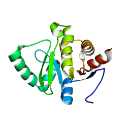
5N03
DownloadVisualize
BU of 5n03 by Molmil
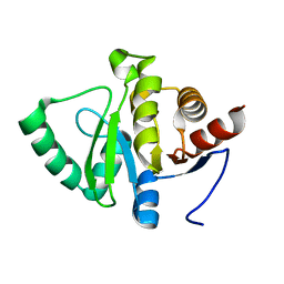
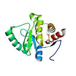
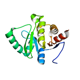
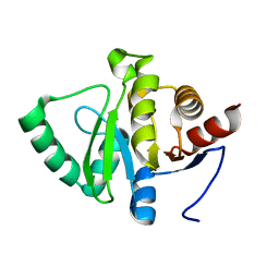
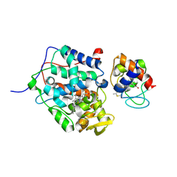
Crystal structure of the decarboxylase AibA/AibB C56V variant
Descriptor: ACETATE ION, Glutaconate CoA-transferase family, subunit A, ...
Authors:Bock, T, Luxenburger, E, Hoffmann, J, Schuetza, V, Feiler, C, Mueller, R, Blankenfeldt, W.
Deposit date:2017-02-02
Release date:2017-05-31
Last modified:2024-01-17
Method:X-RAY DIFFRACTION (2.1 Å)
Cite:AibA/AibB Induces an Intramolecular Decarboxylation in Isovalerate Biosynthesis by Myxococcus xanthus.
Angew. Chem. Int. Ed. Engl., 56, 2017
2JKW
DownloadVisualize
BU of 2jkw by Molmil
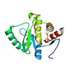
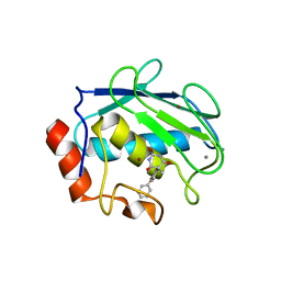
Pseudoazurin M16F
Descriptor: COPPER (II) ION, PSEUDOAZURIN
Authors:Yanagisawa, S, Crowley, P.B, Firbank, S.J, Lawler, A.T, Hunter, D.M, McFarlane, W, Li, C, Kohzuma, T, Banfield, M.J, Dennison, C.
Deposit date:2008-09-01
Release date:2008-11-04
Last modified:2023-12-13
Method:X-RAY DIFFRACTION (1.6 Å)
Cite:Pi-Interaction Tuning of the Active Site Properties of Metalloproteins.
J.Am.Chem.Soc., 130, 2008
6GT2
DownloadVisualize
BU of 6gt2 by Molmil
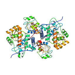
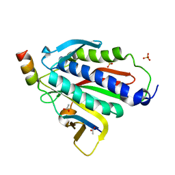
Reduced copper nitrite reductase from Achromobacter cycloclastes determined by serial femtosecond rotation crystallography
Descriptor: COPPER (II) ION, Copper-containing nitrite reductase, MALONATE ION
Authors:Halsted, T.P, Yamashita, K, Gopalasingam, C.C, Shenoy, R.T, Hirata, K, Ago, H, Ueno, G, Eady, R.R, Antonyuk, S.V, Yamamoto, M, Hasnain, S.S.
Deposit date:2018-06-15
Release date:2019-07-03
Last modified:2024-05-15
Method:X-RAY DIFFRACTION (1.6 Å)
Cite:Catalytically important damage-free structures of a copper nitrite reductase obtained by femtosecond X-ray laser and room-temperature neutron crystallography.
Iucrj, 6, 2019
2WGS
DownloadVisualize
BU of 2wgs by Molmil
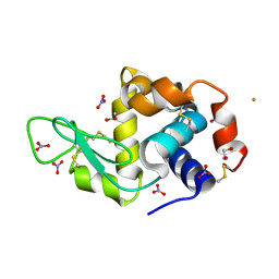
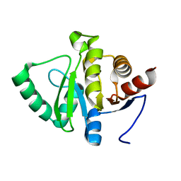
Crystal structure of Mycobacterium Tuberculosis Glutamine Synthetase in complex with a purine analogue inhibitor.
Descriptor: 1-(3,4-dichlorobenzyl)-3,7-dimethyl-8-morpholin-4-yl-3,7-dihydro-1H-purine-2,6-dione, CHLORIDE ION, GLUTAMINE SYNTHETASE 1
Authors:Nilsson, M.T, Krajewski, W.W, Jones, T.A, Mowbray, S.L.
Deposit date:2009-04-27
Release date:2009-09-01
Last modified:2023-12-13
Method:X-RAY DIFFRACTION (2.55 Å)
Cite:Structural Basis for the Inhibition of Mycobacterium Tuberculosis Glutamine Synthetase by Novel ATP-Competitive Inhibitors.
J.Mol.Biol., 393, 2009
2OAX
DownloadVisualize
BU of 2oax by Molmil
Crystal structure of the S810L mutant mineralocorticoid receptor associated with SC9420
Descriptor: Mineralocorticoid receptor, SPIRONOLACTONE
Authors:Huyet, J, Pinon, G.M, Fay, M.R, Rafestin-Oblin, M.E, Fagart, J.
Deposit date:2006-12-18
Release date:2007-08-14
Last modified:2023-08-30
Method:X-RAY DIFFRACTION (2.29 Å)
Cite:Structural basis of spirolactone recognition by the mineralocorticoid receptor
Mol.Pharmacol., 72, 2007
1XJA
DownloadVisualize
BU of 1xja by Molmil
Apo form of the Y31V mutant dimerization domain fragment of Escherichia coli regulatory protein AraC
Descriptor: 1,2-ETHANEDIOL, Arabinose operon regulatory protein
Authors:Weldon, J.E, Larkin, C, Schleif, R.F.
Deposit date:2004-09-23
Release date:2004-10-12
Last modified:2023-08-23
Method:X-RAY DIFFRACTION (2.4 Å)
Cite:Structure and properties of a truely apo form of AraC dimerization domain.
Proteins, 66, 2007
6H1T
DownloadVisualize
BU of 6h1t by Molmil
Structure of the BM3 heme domain in complex with clotrimazole
Descriptor: (4S)-2-METHYL-2,4-PENTANEDIOL, 1,2-ETHANEDIOL, 1-[(2-CHLOROPHENYL)(DIPHENYL)METHYL]-1H-IMIDAZOLE, ...
Authors:Jeffreys, L.N, Munro, A.W.M, Leys, D.
Deposit date:2018-07-12
Release date:2019-02-20
Last modified:2024-01-17
Method:X-RAY DIFFRACTION (2.08 Å)
Cite:Novel insights into P450 BM3 interactions with FDA-approved antifungal azole drugs.
Sci Rep, 9, 2019
4HBM
DownloadVisualize
BU of 4hbm by Molmil
Ordering of the N Terminus of Human MDM2 by Small Molecule Inhibitors
Descriptor: E3 ubiquitin-protein ligase Mdm2, {(3R,5R,6S)-5-(3-chlorophenyl)-6-(4-chlorophenyl)-1-[(2S)-1-hydroxybutan-2-yl]-2-oxopiperidin-3-yl}acetic acid
Authors:Huang, X.
Deposit date:2012-09-28
Release date:2012-10-17
Last modified:2024-02-28
Method:X-RAY DIFFRACTION (1.9 Å)
Cite:Ordering of the N-terminus of human MDM2 by small molecule inhibitors.
J.Am.Chem.Soc., 134, 2012
2C9T
DownloadVisualize
BU of 2c9t by Molmil
Crystal Structure Of Acetylcholine Binding Protein (AChBP) From Aplysia Californica In Complex With alpha-Conotoxin ImI
Descriptor: ALPHA-CONOTOXIN IMI, SOLUBLE ACETYLCHOLINE RECEPTOR
Authors:Ulens, C, Hogg, R.C, Celie, P.H, Bertrand, D, Tsetlin, V, Smit, A.B, Sixma, T.K.
Deposit date:2005-12-14
Release date:2006-02-13
Last modified:2023-12-13
Method:X-RAY DIFFRACTION (2.25 Å)
Cite:Structural Determinants of Selective {Alpha}-Conotoxin Binding to a Nicotinic Acetylcholine Receptor Homolog Achbp.
Proc.Natl.Acad.Sci.USA, 103, 2006
4QT8
DownloadVisualize
BU of 4qt8 by Molmil
Crystal Structure of RON Sema-PSI-IPT1 extracellular domains in complex with MSP beta-chain
Descriptor: 2-acetamido-2-deoxy-beta-D-glucopyranose-(1-4)-2-acetamido-2-deoxy-beta-D-glucopyranose, Hepatocyte growth factor-like protein, Macrophage-stimulating protein receptor, ...
Authors:Herzberg, O, Chao, K.L.
Deposit date:2014-07-07
Release date:2014-09-17
Last modified:2023-09-20
Method:X-RAY DIFFRACTION (3 Å)
Cite:Structural basis for the binding specificity of human Recepteur d'Origine Nantais (RON) receptor tyrosine kinase to macrophage-stimulating protein.
J.Biol.Chem., 289, 2014
6H7V
DownloadVisualize
BU of 6h7v by Molmil
Crystal structure of BauA, the Ferric preacinetobactin receptor from Acinetobacter baumannii
Descriptor: (HYDROXYETHYLOXY)TRI(ETHYLOXY)OCTANE, 1,2-ETHANEDIOL, Ligand-gated channel protein
Authors:Moynie, L, Naismith, J.H.
Deposit date:2018-07-31
Release date:2018-10-10
Last modified:2019-01-16
Method:X-RAY DIFFRACTION (2.54 Å)
Cite:Preacinetobactin not acinetobactin is essential for iron uptake by the BauA transporter of the pathogenAcinetobacter baumannii.
Elife, 7, 2018
4DLP
DownloadVisualize
BU of 4dlp by Molmil
Crystal structure of methionyl-tRNA synthetase MetRS from Brucella melitensis bound to selenomethionine
Descriptor: Aminoacyl-tRNA synthetase, class I:Aminoacyl-tRNA synthetase, class Ia:Methionyl-tRNA synthetase, ...
Authors:Seattle Structural Genomics Center for Infectious Disease (SSGCID)
Deposit date:2012-02-06
Release date:2012-02-15
Last modified:2023-12-06
Method:X-RAY DIFFRACTION (2.65 Å)
Cite:Brucella melitensis Methionyl-tRNA-Synthetase (MetRS), a Potential Drug Target for Brucellosis.
Plos One, 11, 2016
2ABE
DownloadVisualize
BU of 2abe by Molmil
Carbonic anhydrase activators: X-ray crystal structure of the adduct of human isozyme II with L-histidine as a platform for the design of stronger activators
Descriptor: Carbonic anhydrase II, HISTIDINE, MERCURY (II) ION, ...
Authors:Temperini, C, Scozzafava, A, Puccetti, L, Supuran, C.T.
Deposit date:2005-07-15
Release date:2005-10-25
Last modified:2024-02-14
Method:X-RAY DIFFRACTION (2 Å)
Cite:Carbonic anhydrase activators: X-ray crystal structure of the adduct of human isozyme II with l-histidine as a platform for the design of stronger activators.
Bioorg.Med.Chem.Lett., 15, 2005
4CPP
DownloadVisualize
BU of 4cpp by Molmil
CRYSTAL STRUCTURES OF CYTOCHROME P450-CAM COMPLEXED WITH CAMPHANE, THIOCAMPHOR, AND ADAMANTANE: FACTORS CONTROLLING P450 SUBSTRATE HYDROXYLATION
Descriptor: ADAMANTANE, CYTOCHROME P450-CAM, PROTOPORPHYRIN IX CONTAINING FE
Authors:Raag, R, Poulos, T.L.
Deposit date:1990-05-18
Release date:1991-07-15
Last modified:2024-02-28
Method:X-RAY DIFFRACTION (2.11 Å)
Cite:Crystal structures of cytochrome P-450CAM complexed with camphane, thiocamphor, and adamantane: factors controlling P-450 substrate hydroxylation.
Biochemistry, 30, 1991
4LG6
DownloadVisualize
BU of 4lg6 by Molmil
Crystal structure of ANKRA2-CCDC8 complex
Descriptor: Ankyrin repeat family A protein 2, Coiled-coil domain-containing protein 8, UNKNOWN ATOM OR ION
Authors:Xu, C, Bian, C, Tempel, W, Mackenzie, F, Bountra, C, Arrowsmith, C.H, Edwards, A.M, Min, J, Structural Genomics Consortium (SGC)
Deposit date:2013-06-27
Release date:2013-09-25
Last modified:2023-09-20
Method:X-RAY DIFFRACTION (1.8 Å)
Cite:Ankyrin Repeats of ANKRA2 Recognize a PxLPxL Motif on the 3M Syndrome Protein CCDC8.
Structure, 23, 2015
2POT
DownloadVisualize
BU of 2pot by Molmil
tRNA guanine transglycosylase (TGT) E235Q mutant in complex with guanine
Descriptor: GLYCEROL, GUANINE, Queuine tRNA-ribosyltransferase, ...
Authors:Tidten, N, Heine, A, Reuter, K, Klebe, G.
Deposit date:2007-04-27
Release date:2007-10-30
Last modified:2023-08-30
Method:X-RAY DIFFRACTION (1.8 Å)
Cite:Glutamate versus Glutamine Exchange Swaps Substrate Selectivity in tRNA-Guanine Transglycosylase: Insight into the Regulation of Substrate Selectivity by Kinetic and Crystallographic Studies.
J.Mol.Biol., 374, 2007
2VNG
DownloadVisualize
BU of 2vng by Molmil
Family 51 carbohydrate binding module from a family 98 glycoside hydrolase produced by Clostridium perfringens in complex with blood group A-trisaccharide ligand.
Descriptor: CALCIUM ION, CPE0329, alpha-L-fucopyranose-(1-2)-[2-acetamido-2-deoxy-alpha-D-galactopyranose-(1-3)]beta-D-galactopyranose
Authors:Gregg, K.J, Finn, R, Abbott, D.W, Boraston, A.B.
Deposit date:2008-02-04
Release date:2008-02-19
Last modified:2024-05-08
Method:X-RAY DIFFRACTION (1.4 Å)
Cite:Divergent Modes of Glycan Recognition by a New Family of Carbohydrate-Binding Modules
J.Biol.Chem., 283, 2008
8UQE
DownloadVisualize
BU of 8uqe by Molmil
Crystal structure of RNF168 (RING)-UbcH5c fused to H2A-H2B via a 26-residue linker (RING not modeled in density)
Descriptor: E3 ubiquitin-protein ligase RNF168,Ubiquitin-conjugating enzyme E2 D3,Histone H2B type 2-E,Histone H2A type 1-B/E
Authors:Hu, Q, Botuyan, M.V, Mer, G.
Deposit date:2023-10-23
Release date:2024-01-17
Last modified:2024-03-20
Method:X-RAY DIFFRACTION (3.562 Å)
Cite:Mechanisms of RNF168 nucleosome recognition and ubiquitylation.
Mol.Cell, 84, 2024
8UQB
DownloadVisualize
BU of 8uqb by Molmil
Crystal structure of RNF168 (RING)-UbcH5c fused to H2A-H2B via a 20-residue linker (crystallization condition 1)
Descriptor: CHLORIDE ION, E3 ubiquitin-protein ligase RNF168,Ubiquitin-conjugating enzyme E2 D3,Histone H2B type 2-E,Histone H2A type 1-B/E, ZINC ION
Authors:Hu, Q, Botuyan, M.V, Mer, G.
Deposit date:2023-10-23
Release date:2024-01-17
Last modified:2024-03-20
Method:X-RAY DIFFRACTION (2.484 Å)
Cite:Mechanisms of RNF168 nucleosome recognition and ubiquitylation.
Mol.Cell, 84, 2024
8UPF
DownloadVisualize
BU of 8upf by Molmil
Cryo-EM structure of the human nucleosome core particle in complex with RNF168-UbcH5c
Descriptor: DNA (147-MER), E3 ubiquitin-protein ligase RNF168, Histone H2A type 1-B/E, ...
Authors:Hu, Q, Botuyan, M.V, Zhao, D, Cui, G, Mer, G.
Deposit date:2023-10-22
Release date:2024-01-17
Last modified:2024-03-20
Method:ELECTRON MICROSCOPY (3.2 Å)
Cite:Mechanisms of RNF168 nucleosome recognition and ubiquitylation.
Mol.Cell, 84, 2024
8UQA
DownloadVisualize
BU of 8uqa by Molmil
Crystal structure of RNF168 (RING)-UbcH5c fused to H2A-H2B via a 12-residue linker
Descriptor: CHLORIDE ION, E3 ubiquitin-protein ligase RNF168,Ubiquitin-conjugating enzyme E2 D3,Histone H2B type 2-E,Histone H2A type 1-B/E, SODIUM ION, ...
Authors:Hu, Q, Botuyan, M.V, Mer, G.
Deposit date:2023-10-23
Release date:2024-01-17
Last modified:2024-03-20
Method:X-RAY DIFFRACTION (2.049 Å)
Cite:Mechanisms of RNF168 nucleosome recognition and ubiquitylation.
Mol.Cell, 84, 2024
8UQ8
DownloadVisualize
BU of 8uq8 by Molmil
Crystal structure of RNF168 (RING)-UbcH5c fused to H2A-H2B via a 2-residue linker
Descriptor: CHLORIDE ION, E3 ubiquitin-protein ligase RNF168,Ubiquitin-conjugating enzyme E2 D3,Histone H2B type 2-E,Histone H2A type 1-B/E, GLYCEROL, ...
Authors:Hu, Q, Botuyan, M.V, Mer, G.
Deposit date:2023-10-23
Release date:2024-01-17
Last modified:2024-03-20
Method:X-RAY DIFFRACTION (2.34 Å)
Cite:Mechanisms of RNF168 nucleosome recognition and ubiquitylation.
Mol.Cell, 84, 2024
8UQ9
DownloadVisualize
BU of 8uq9 by Molmil
Crystal structure of RNF168 (RING)-UbcH5c fused to H2A-H2B via a 4-residue linker
Descriptor: CHLORIDE ION, E3 ubiquitin-protein ligase RNF168,Ubiquitin-conjugating enzyme E2 D3,Histone H2B type 2-E,Histone H2A type 1-B/E, GLYCEROL, ...
Authors:Hu, Q, Botuyan, M.V, Mer, G.
Deposit date:2023-10-23
Release date:2024-01-17
Last modified:2024-03-20
Method:X-RAY DIFFRACTION (2.3 Å)
Cite:Mechanisms of RNF168 nucleosome recognition and ubiquitylation.
Mol.Cell, 84, 2024
8UQD
DownloadVisualize
BU of 8uqd by Molmil
Crystal structure of RNF168 (RING)-UbcH5c fused to H2A-H2B via a 20-residue linker (condition 2. RING not modeled in density)
Descriptor: E3 ubiquitin-protein ligase RNF168,Ubiquitin-conjugating enzyme E2 D3,Histone H2B type 2-E,Histone H2A type 1-B/E
Authors:Hu, Q, Botuyan, M.V, Mer, G.
Deposit date:2023-10-23
Release date:2024-01-17
Last modified:2024-03-20
Method:X-RAY DIFFRACTION (3.893 Å)
Cite:Mechanisms of RNF168 nucleosome recognition and ubiquitylation.
Mol.Cell, 84, 2024
8UQC
DownloadVisualize
BU of 8uqc by Molmil
Crystal structure of RNF168 (RING)-UbcH5c fused to H2A-H2B via a 20-residue linker (crystallization condition 2)
Descriptor: E3 ubiquitin-protein ligase RNF168,Ubiquitin-conjugating enzyme E2 D3,Histone H2B type 2-E,Histone H2A type 1-B/E, ZINC ION
Authors:Hu, Q, Botuyan, M.V, Mer, G.
Deposit date:2023-10-23
Release date:2024-01-17
Last modified:2024-03-20
Method:X-RAY DIFFRACTION (2.61 Å)
Cite:Mechanisms of RNF168 nucleosome recognition and ubiquitylation.
Mol.Cell, 84, 2024

226262

PDB entries from 2024-10-16

PDB statisticsPDBj update infoContact PDBjnumon