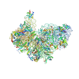
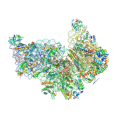
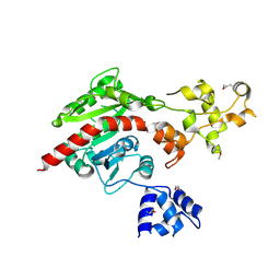
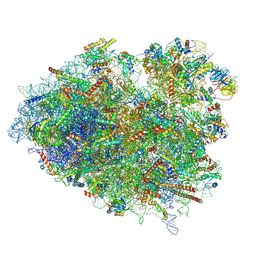
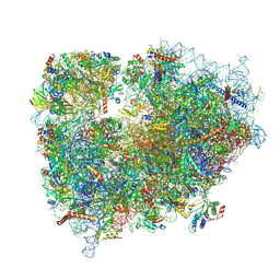
9FO0
 
 | | PF30S ribosomal subunit - control | | Descriptor: | 16S rRNA, Large ribosomal subunit protein eL8, RNA-binding protein, ... | | Authors: | Hassan, A.H, Demo, G. | | Deposit date: | 2024-06-11 | | Release date: | 2025-01-15 | | Last modified: | 2025-01-22 | | Method: | ELECTRON MICROSCOPY (3.4 Å) | | Cite: | Novel archaeal ribosome dimerization factor facilitating unique 30S-30S dimerization.
Nucleic Acids Res., 53, 2025
|
|
9FSF
 | | Cryo-EM structure of Saccharolobus solfataricus 30S initiation complex bound to Ss-MAP leaderless mRNA with h44 in up position | | Descriptor: | ADENOSINE-5'-TRIPHOSPHATE, LSU ribosomal protein S30E (Rps30E), Large ribosomal subunit protein eL8, ... | | Authors: | Bourgeois, G, Coureux, P.D, Mechulam, Y, Schmitt, E. | | Deposit date: | 2024-06-21 | | Release date: | 2025-01-15 | | Method: | ELECTRON MICROSCOPY (2.8 Å) | | Cite: | Structures of Saccharolobus solfataricus initiation complexes with leaderless mRNAs highlight archaeal features and eukaryotic proximity.
Nat Commun, 16, 2025
|
|
9FY0
 | | Cryo-EM structure of Saccharolobus solfataricus 30S initiation complex bound to Ss-aIF2beta leaderless mRNA | | Descriptor: | GUANOSINE-5'-TRIPHOSPHATE, LSU ribosomal protein S26E (Rps26E), LSU ribosomal protein S30E (Rps30E), ... | | Authors: | Bourgeois, G, Coureux, P.D, Mechulam, Y, Schmitt, E. | | Deposit date: | 2024-07-02 | | Release date: | 2025-01-15 | | Method: | ELECTRON MICROSCOPY (2.9 Å) | | Cite: | Structures of Saccharolobus solfataricus initiation complexes with leaderless mRNAs highlight archaeal features and eukaryotic proximity.
Nat Commun, 16, 2025
|
|
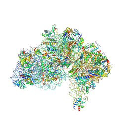
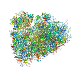
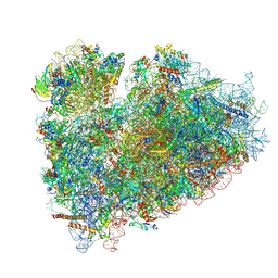
9FNY
 | | PF30S-PF30S dimer mediated by aRDF from P. furiosus (Structure I) | | Descriptor: | 16S rRNA, Archaeal Ribosome Dimerizing Factor (aRDF), Large ribosomal subunit protein eL8, ... | | Authors: | Hassan, A.H, Demo, G. | | Deposit date: | 2024-06-11 | | Release date: | 2025-01-15 | | Last modified: | 2025-01-22 | | Method: | ELECTRON MICROSCOPY (3.2 Å) | | Cite: | Novel archaeal ribosome dimerization factor facilitating unique 30S-30S dimerization.
Nucleic Acids Res., 53, 2025
|
|
9FRL
 | | Cryo-EM structure of Saccharolobus solfataricus 30S initiation complex bound to SD mRNA with h44 in up position | | Descriptor: | LSU ribosomal protein S30E (Rps30E), Large ribosomal subunit protein eL8, MAGNESIUM ION, ... | | Authors: | Bourgeois, G, Coureux, P.D, Mechulam, Y, Schmitt, E. | | Deposit date: | 2024-06-19 | | Release date: | 2025-01-15 | | Method: | ELECTRON MICROSCOPY (2.97 Å) | | Cite: | Structures of Saccharolobus solfataricus initiation complexes with leaderless mRNAs highlight archaeal features and eukaryotic proximity.
Nat Commun, 16, 2025
|
|
9FS6
 | | Cryo-EM structure of Saccharolobus solfataricus 30S initiation complex bound to Ss-aIF2beta leaderless mRNA with h44 in up position | | Descriptor: | GUANOSINE-5'-TRIPHOSPHATE, LSU ribosomal protein S26E (Rps26E), LSU ribosomal protein S30E (Rps30E), ... | | Authors: | Bourgeois, G, Coureux, P.D, Mechulam, Y, Schmitt, E. | | Deposit date: | 2024-06-20 | | Release date: | 2025-01-15 | | Method: | ELECTRON MICROSCOPY (2.9 Å) | | Cite: | Structures of Saccharolobus solfataricus initiation complexes with leaderless mRNAs highlight archaeal features and eukaryotic proximity.
Nat Commun, 16, 2025
|
|
9FS8
 | | Cryo-EM structure of Saccharolobus solfataricus 30S initiation complex bound to Ss-aEF1A-like mRNA | | Descriptor: | LSU ribosomal protein S26E (Rps26E), LSU ribosomal protein S30E (Rps30E), Large ribosomal subunit protein eL8, ... | | Authors: | Bourgeois, G, Coureux, P.D, Mechulam, Y, Schmitt, E. | | Deposit date: | 2024-06-20 | | Release date: | 2025-01-15 | | Method: | ELECTRON MICROSCOPY (3.7 Å) | | Cite: | Structures of Saccharolobus solfataricus initiation complexes with leaderless mRNAs highlight archaeal features and eukaryotic proximity.
Nat Commun, 16, 2025
|
|
9FNZ
 | |
1GP1
 | |
1HMY
 | |
1JS4
 | | ENDO/EXOCELLULASE:CELLOBIOSE FROM THERMOMONOSPORA | | Descriptor: | CALCIUM ION, ENDO/EXOCELLULASE E4, beta-D-glucopyranose, ... | | Authors: | Sakon, J, Wilson, D.B, Karplus, P.A. | | Deposit date: | 1997-05-30 | | Release date: | 1997-09-17 | | Last modified: | 2024-11-13 | | Method: | X-RAY DIFFRACTION (2 Å) | | Cite: | Structure and mechanism of endo/exocellulase E4 from Thermomonospora fusca.
Nat.Struct.Biol., 4, 1997
|
|
3L0P
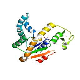 | | Crystal structures of Iron containing Adenylate kinase from Desulfovibrio gigas | | Descriptor: | Adenylate kinase, FE (III) ION, GLYCEROL | | Authors: | Mukhopadhyay, A, Trincao, J, Romao, M.J. | | Deposit date: | 2009-12-10 | | Release date: | 2010-12-15 | | Last modified: | 2024-03-20 | | Method: | X-RAY DIFFRACTION (3 Å) | | Cite: | Crystal structure of the zinc-, cobalt-, and iron-containing adenylate kinase from Desulfovibrio gigas: a novel metal-containing adenylate kinase from Gram-negative bacteria
J.Biol.Inorg.Chem., 16, 2011
|
|
3KGV
 | |
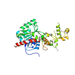
3L0S
 | | Crystal structures of Zinc, Cobalt and Iron containing Adenylate kinase from Gram-negative bacteria Desulfovibrio gigas | | Descriptor: | Adenylate kinase, COBALT (II) ION, D(-)-TARTARIC ACID | | Authors: | Mukhopadhyay, A, Trincao, J, Romao, M.J. | | Deposit date: | 2009-12-10 | | Release date: | 2010-12-15 | | Last modified: | 2024-03-20 | | Method: | X-RAY DIFFRACTION (2 Å) | | Cite: | Crystal structure of the zinc-, cobalt-, and iron-containing adenylate kinase from Desulfovibrio gigas: a novel metal-containing adenylate kinase from Gram-negative bacteria
J.Biol.Inorg.Chem., 16, 2011
|
|
3LX6
 | |
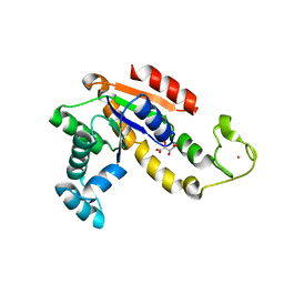
3ME5
 | | Crystal structure of putative dna cytosine methylase from shigella flexneri 2a str. 2457T | | Descriptor: | Cytosine-specific methyltransferase | | Authors: | Ramagopal, U.A, Malashkevich, V.N, Toro, R, Sauder, J.M, Burley, S.K, Almo, S.C, New York SGX Research Center for Structural Genomics (NYSGXRC) | | Deposit date: | 2010-03-31 | | Release date: | 2010-04-21 | | Last modified: | 2024-11-06 | | Method: | X-RAY DIFFRACTION (1.75 Å) | | Cite: | Crystal structure of putative dna cytosine methylase from shigella flexneri 2a str. 2457T
To be Published
|
|
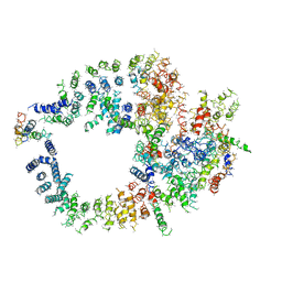
3M9S
 | | Crystal structure of respiratory complex I from Thermus thermophilus | | Descriptor: | FE2/S2 (INORGANIC) CLUSTER, FLAVIN MONONUCLEOTIDE, IRON/SULFUR CLUSTER, ... | | Authors: | Efremov, R.G, Baradaran, R, Sazanov, L.A. | | Deposit date: | 2010-03-22 | | Release date: | 2010-05-26 | | Last modified: | 2024-11-27 | | Method: | X-RAY DIFFRACTION (4.5 Å) | | Cite: | The architecture of respiratory complex I
Nature, 465, 2010
|
|
8UTI
 | | Eukaryotic 80S ribosome with Reh1 and A/P site tRNA | | Descriptor: | 18S rRNA, 25S rRNA, 4-{(2R)-2-[(1S,3S,5S)-3,5-dimethyl-2-oxocyclohexyl]-2-hydroxyethyl}piperidine-2,6-dione, ... | | Authors: | Yelland, J.N, Taylor, D.W, Johnson, A.W. | | Deposit date: | 2023-10-31 | | Release date: | 2024-11-06 | | Last modified: | 2025-02-19 | | Method: | ELECTRON MICROSCOPY (3.13 Å) | | Cite: | The assembly factor Reh1 is released from the ribosome during its initial round of translation.
Nat Commun, 16, 2025
|
|
8UT0
 | | Eukaryotic 80S ribosome with Reh1, eIF5A and A/P site tRNA | | Descriptor: | 18S rRNA, 25S rRNA, 4-{(2R)-2-[(1S,3S,5S)-3,5-dimethyl-2-oxocyclohexyl]-2-hydroxyethyl}piperidine-2,6-dione, ... | | Authors: | Yelland, J.N, Taylor, D.W, Johnson, A.W. | | Deposit date: | 2023-10-30 | | Release date: | 2024-11-06 | | Last modified: | 2025-02-19 | | Method: | ELECTRON MICROSCOPY (3.22 Å) | | Cite: | The assembly factor Reh1 is released from the ribosome during its initial round of translation.
Nat Commun, 16, 2025
|
|
8VFT
 | |
4X8L
 | | Crystal structure of E. coli Adenylate kinase P177A mutant in complex with inhibitor Ap5a | | Descriptor: | Adenylate kinase, BIS(ADENOSINE)-5'-PENTAPHOSPHATE, MAGNESIUM ION, ... | | Authors: | Sauer-Eriksson, A.E, Kovermann, M, Aden, J, Grundstrom, C, Wolf-Watz, M, Sauer, U.H. | | Deposit date: | 2014-12-10 | | Release date: | 2015-07-22 | | Last modified: | 2024-01-10 | | Method: | X-RAY DIFFRACTION (1.7 Å) | | Cite: | Structural basis for catalytically restrictive dynamics of a high-energy enzyme state.
Nat Commun, 6, 2015
|
|
8UIY
 | | In situ human P-Z state 80S ribosome | | Descriptor: | 18S rRNA, 28S rRNA, 40S ribosomal protein S10, ... | | Authors: | Wei, Z, Yong, X. | | Deposit date: | 2023-10-10 | | Release date: | 2025-04-09 | | Method: | ELECTRON MICROSCOPY (3.44 Å) | | Cite: | In situ human P-Z state 80S ribosome
To Be Published
|
|
8UIK
 | |
8UJB
 | |
8UJJ
 | | In situ HHT and CHX treated A-P state 80S ribosome | | Descriptor: | (3beta)-O~3~-[(2R)-2,6-dihydroxy-2-(2-methoxy-2-oxoethyl)-6-methylheptanoyl]cephalotaxine, 18S rRNA, 28S rRNA, ... | | Authors: | Wei, Z, Yong, X. | | Deposit date: | 2023-10-11 | | Release date: | 2025-04-16 | | Method: | ELECTRON MICROSCOPY (3.56 Å) | | Cite: | In situ HHT and CHX treated human A-P state 80S ribosome
To Be Published
|
|


