Loading
PDBj
MenuPDBj@FacebookPDBj@TwitterPDBj@YouTubewwPDB FoundationwwPDB
RCSB PDBPDBeBMRBAdv. SearchSearch help

4I60
DownloadVisualize
BU of 4i60 by Molmil
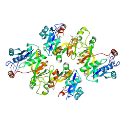
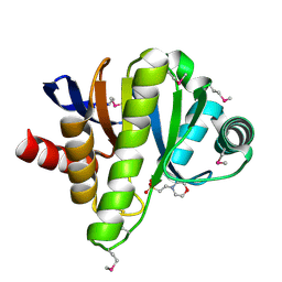
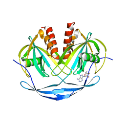
Crystal structure of avidin - biotinylruthenocene complex
Descriptor: 2-acetamido-2-deoxy-beta-D-glucopyranose-(1-4)-2-acetamido-2-deoxy-beta-D-glucopyranose, Avidin, [(1,2,3,4,5-eta)-cyclopentadienyl][(1,2,3,4,5-eta)-{5-[(3aS,4S,6aR)-2-oxohexahydro-1H-thieno[3,4-d]imidazol-4-yl]pentanoyl}cyclopentadienyl]ruthenium
Authors:Strzelczyk, P, Bujacz, A, Bujacz, G.
Deposit date:2012-11-29
Release date:2013-08-14
Last modified:2023-11-08
Method:X-RAY DIFFRACTION (2.5 Å)
Cite:Structural investigation of the interactions of biotinylruthenocene with avidin.
Chem.Biol.Interact, 204, 2013
6RGW
DownloadVisualize
BU of 6rgw by Molmil
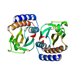
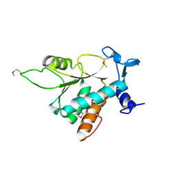
Photorhabdus asymbiotica lectin PHL in complex with O-methylated PGL-1-derived disaccharide
Descriptor: 1,2-ETHANEDIOL, 3,6-O-dimethyl-D-glucose, 4-(2-azanylethoxy)phenol, ...
Authors:Houser, J, Fujdiarova, E, Jancarikova, G, Wimmerova, M.
Deposit date:2019-04-17
Release date:2020-07-01
Last modified:2024-01-24
Method:X-RAY DIFFRACTION (1.75 Å)
Cite:Heptabladed beta-propeller lectins PLL2 and PHL from Photorhabdus spp. recognize O-methylated sugars and influence the host immune system.
Febs J., 288, 2021
6S0W
DownloadVisualize
BU of 6s0w by Molmil
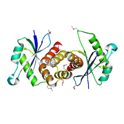
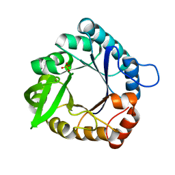
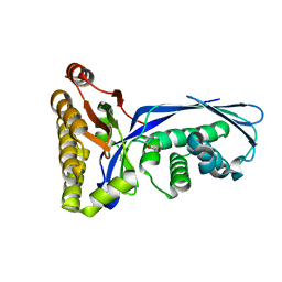
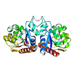
The crystal structure of kanamycin B dioxygenase (KanJ) from Streptomyces kanamyceticus in complex with nickel and kanamycin B sulfate
Descriptor: (1R,2S,3S,4R,6S)-4,6-DIAMINO-3-[(3-AMINO-3-DEOXY-ALPHA-D-GLUCOPYRANOSYL)OXY]-2-HYDROXYCYCLOHEXYL 2,6-DIAMINO-2,6-DIDEOXY-ALPHA-D-GLUCOPYRANOSIDE, DI(HYDROXYETHYL)ETHER, Kanamycin B dioxygenase, ...
Authors:Mrugala, B, Niedzialkowska, E, Minor, W, Borowski, T.
Deposit date:2019-06-18
Release date:2020-07-08
Last modified:2024-01-24
Method:X-RAY DIFFRACTION (2.36 Å)
Cite:A study on the structure, mechanism, and biochemistry of kanamycin B dioxygenase (KanJ)-an enzyme with a broad range of substrates.
Febs J., 288, 2021
6TQP
DownloadVisualize
BU of 6tqp by Molmil
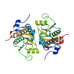
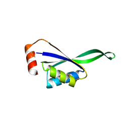
Structural insight into tanapoxvirus mediated inhibition of apoptosis
Descriptor: 16L protein, Bcl-2-binding component 3, isoforms 1/2, ...
Authors:Suraweera, C.D, Hinds, M.G, Kvansakul, M.
Deposit date:2019-12-17
Release date:2020-06-03
Last modified:2024-05-01
Method:X-RAY DIFFRACTION (1.84940946 Å)
Cite:Structural insight into tanapoxvirus-mediated inhibition of apoptosis.
Febs J., 287, 2020
6TRR
DownloadVisualize
BU of 6trr by Molmil
Structural insight into tanapoxvirus mediated inhibition of apoptosis
Descriptor: 16L protein, Apoptosis regulator BAX, SODIUM ION
Authors:Suraweera, C.D, Hinds, M.G, Kvansakul, M.
Deposit date:2019-12-19
Release date:2020-06-03
Last modified:2024-01-24
Method:X-RAY DIFFRACTION (2.12007356 Å)
Cite:Structural insight into tanapoxvirus-mediated inhibition of apoptosis.
Febs J., 287, 2020
6RG2
DownloadVisualize
BU of 6rg2 by Molmil
Photorhabdus laumondii lectin PLL2 in complex with 3-O-methyl-D-glucose
Descriptor: 3-O-methyl-alpha-D-glucopyranose, 3-O-methyl-beta-D-glucopyranose, ACETATE ION, ...
Authors:Houser, J, Fujdiarova, E, Wimmerova, M.
Deposit date:2019-04-16
Release date:2020-07-01
Last modified:2024-05-15
Method:X-RAY DIFFRACTION (1.12 Å)
Cite:Heptabladed beta-propeller lectins PLL2 and PHL from Photorhabdus spp. recognize O-methylated sugars and influence the host immune system.
Febs J., 288, 2021
6RGG
DownloadVisualize
BU of 6rgg by Molmil
Photorhabdus laumondii lectin PLL2 in complex with O-methylated PGL-1-derived disaccharide
Descriptor: 1,2-ETHANEDIOL, 3,6-O-dimethyl-D-glucose, 6-deoxy-2,3-di-O-methyl-alpha-L-mannopyranose, ...
Authors:Houser, J, Fujdiarova, E, Wimmerova, M.
Deposit date:2019-04-16
Release date:2020-07-01
Last modified:2024-05-15
Method:X-RAY DIFFRACTION (2.2 Å)
Cite:Heptabladed beta-propeller lectins PLL2 and PHL from Photorhabdus spp. recognize O-methylated sugars and influence the host immune system.
Febs J., 288, 2021
4UM4
DownloadVisualize
BU of 4um4 by Molmil
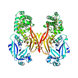
STRUCTURE OF INORGANIC PYROPHOSPHATASE FROM ESCHERICHIA COLI IN COMPLEX WITH SULFATE
Descriptor: INORGANIC PYROPHOSPHATASE, SULFATE ION
Authors:Campos-Acevedo, A.A, Rudino-Pinera, E.
Deposit date:2014-05-15
Release date:2014-11-26
Last modified:2024-01-10
Method:X-RAY DIFFRACTION (2.65 Å)
Cite:First attempts to crystallize a non-homogeneous sample of thioredoxin from Litopenaeus vannamei: What to do when you have diffraction data of a protein that is not the target?
Biochem Biophys Rep, 8, 2016
6S0U
DownloadVisualize
BU of 6s0u by Molmil
The crystal structure of kanamycin B dioxygenase (KanJ) from Streptomyces kanamyceticus in complex with nickel and 2-oxoglutarate
Descriptor: 2-OXOGLUTARIC ACID, CHLORIDE ION, DI(HYDROXYETHYL)ETHER, ...
Authors:Mrugala, B, Porebski, P.J, Niedzialkowska, E, Minor, W, Borowski, T.
Deposit date:2019-06-18
Release date:2020-07-08
Last modified:2024-01-24
Method:X-RAY DIFFRACTION (2.15 Å)
Cite:A study on the structure, mechanism, and biochemistry of kanamycin B dioxygenase (KanJ)-an enzyme with a broad range of substrates.
Febs J., 288, 2021
5T0N
DownloadVisualize
BU of 5t0n by Molmil
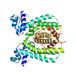
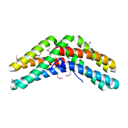
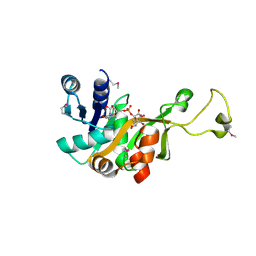
Pseudo-apo structure of Sestrin2 at 3.0 angstrom resolution
Descriptor: LEUCINE, Sestrin-2
Authors:Saxton, R.A, Knockenhauer, K.E, Schwartz, T.U.
Deposit date:2016-08-16
Release date:2016-10-05
Last modified:2023-10-04
Method:X-RAY DIFFRACTION (3.004 Å)
Cite:The apo-structure of the leucine sensor Sestrin2 is still elusive.
Sci.Signal., 9, 2016
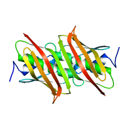
6T76
DownloadVisualize
BU of 6t76 by Molmil
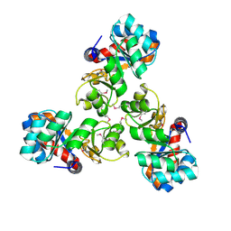
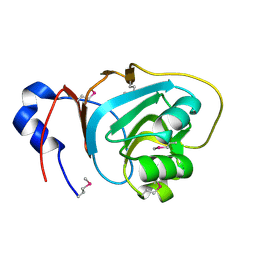
PII-like protein CutA from Nostoc sp. PCC 7120 in apo form
Descriptor: Periplasmic divalent cation tolerance protein, SULFATE ION
Authors:Selim, K.A, Albrecht, R, Forchhammer, K, Hartmann, M.D.
Deposit date:2019-10-21
Release date:2020-07-22
Last modified:2024-01-24
Method:X-RAY DIFFRACTION (1.9 Å)
Cite:Functional and structural characterization of PII-like protein CutA does not support involvement in heavy metal tolerance and hints at a small-molecule carrying/signaling role.
Febs J., 288, 2021
6T7E
DownloadVisualize
BU of 6t7e by Molmil
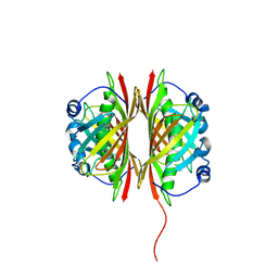
PII-like protein CutA from Nostoc sp. PCC7120 in complex with MES
Descriptor: 2-(N-MORPHOLINO)-ETHANESULFONIC ACID, Periplasmic divalent cation tolerance protein
Authors:Selim, K.A, Albrecht, R, Forchhammer, K, Hartmann, M.D.
Deposit date:2019-10-21
Release date:2020-07-22
Last modified:2024-01-24
Method:X-RAY DIFFRACTION (2.45 Å)
Cite:Functional and structural characterization of PII-like protein CutA does not support involvement in heavy metal tolerance and hints at a small-molecule carrying/signaling role.
Febs J., 288, 2021
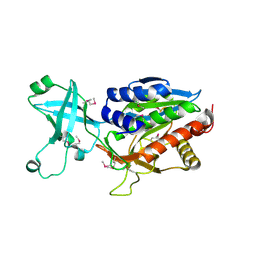
6A8M
DownloadVisualize
BU of 6a8m by Molmil
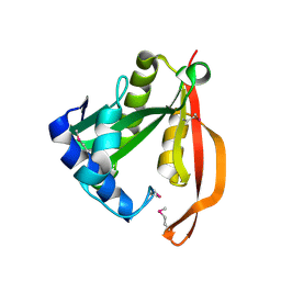
N-terminal domain of FACT complex subunit SPT16 from Eremothecium gossypii (Ashbya gossypii)
Descriptor: FACT complex subunit SPT16
Authors:Gaur, N.K, Are, V.N, Durani, V, Ghosh, B, Kumar, A, Kulkarni, K, Makde, R.D.
Deposit date:2018-07-09
Release date:2018-08-15
Last modified:2023-11-22
Method:X-RAY DIFFRACTION (1.7 Å)
Cite:Evolutionary conservation of protein dynamics: insights from all-atom molecular dynamics simulations of 'peptidase' domain of Spt16.
J.Biomol.Struct.Dyn., 2021
5TMF
DownloadVisualize
BU of 5tmf by Molmil
Re-refinement of thermus thermophilus RNA polymerase
Descriptor: DNA-directed RNA polymerase subunit alpha, DNA-directed RNA polymerase subunit beta, DNA-directed RNA polymerase subunit beta', ...
Authors:Wang, J.
Deposit date:2016-10-12
Release date:2016-11-23
Last modified:2024-03-06
Method:X-RAY DIFFRACTION (3 Å)
Cite:On the validation of crystallographic symmetry and the quality of structures.
Protein Sci., 24, 2015
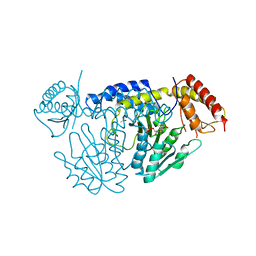
6KG1
DownloadVisualize
BU of 6kg1 by Molmil
NifS from Helicobacter pylori, soaked with L-cysteine for 180 sec
Descriptor: 2-[(3-HYDROXY-2-METHYL-5-PHOSPHONOOXYMETHYL-PYRIDIN-4-YLMETHYL)-AMINO]-PROPIONIC ACID, CHLORIDE ION, Cysteine desulfurase IscS, ...
Authors:Nakamura, R, Takahashi, Y, Fujishiro, T.
Deposit date:2019-07-09
Release date:2019-10-16
Last modified:2023-11-22
Method:X-RAY DIFFRACTION (2.7 Å)
Cite:Snapshots of PLP-substrate and PLP-product external aldimines as intermediates in two types of cysteine desulfurase enzymes.
Febs J., 287, 2020
6KG0
DownloadVisualize
BU of 6kg0 by Molmil
NifS from Helicobacter pylori, soaked with L-cysteine for 118 sec
Descriptor: CHLORIDE ION, Cysteine desulfurase IscS, ISOPROPYL ALCOHOL, ...
Authors:Nakamura, R, Takahashi, Y, Fujishiro, T.
Deposit date:2019-07-09
Release date:2019-10-16
Last modified:2023-11-22
Method:X-RAY DIFFRACTION (2.78 Å)
Cite:Snapshots of PLP-substrate and PLP-product external aldimines as intermediates in two types of cysteine desulfurase enzymes.
Febs J., 287, 2020
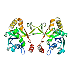
6UE0
DownloadVisualize
BU of 6ue0 by Molmil
Crystal structure of dihydrodipicolinate synthase from Klebsiella pneumoniae bound to pyruvate
Descriptor: 4-hydroxy-tetrahydrodipicolinate synthase, CHLORIDE ION, SULFATE ION
Authors:Impey, R.E, Lee, M, Hawkins, D.A, Sutton, J.M, Panjikar, S, Perugini, M.A, Soares da Costa, T.P.
Deposit date:2019-09-20
Release date:2020-02-05
Last modified:2023-11-29
Method:X-RAY DIFFRACTION (1.892 Å)
Cite:Mis-annotations of a promising antibiotic target in high-priority gram-negative pathogens.
Febs Lett., 594, 2020
7LK3
DownloadVisualize
BU of 7lk3 by Molmil
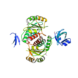
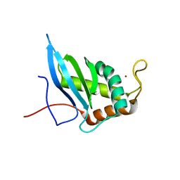
Crystal structure of untwinned human GABARAPL2
Descriptor: 1,2-ETHANEDIOL, Gamma-aminobutyric acid receptor-associated protein-like 2
Authors:Scicluna, K, Dewson, G, Czabotar, P.E, Birkinshaw, R.W.
Deposit date:2021-02-01
Release date:2021-05-12
Last modified:2023-10-18
Method:X-RAY DIFFRACTION (1.9 Å)
Cite:A new crystal form of GABARAPL2.
Acta Crystallogr.,Sect.F, 77, 2021
5ZT0
DownloadVisualize
BU of 5zt0 by Molmil
Crystal Structure of Protein Phosphate 1 Complexed with PP1 binding domain of GL
Descriptor: MANGANESE (II) ION, PHOSPHATE ION, Protein phosphatase 1 regulatory subunit 3B, ...
Authors:Yu, J, Xiang, S.
Deposit date:2018-05-01
Release date:2019-03-13
Last modified:2023-11-22
Method:X-RAY DIFFRACTION (3.3204267 Å)
Cite:Structural basis for protein phosphatase 1 recruitment by glycogen-targeting subunits
FEBS J., 285, 2018
1TEW
DownloadVisualize
BU of 1tew by Molmil
STRUCTURE OF HEXAGONAL TURKEY EGG WHITE LYSOZYME AT 1.65 ANGSTROMS RESOLUTION
Descriptor: THIOCYANATE ION, TURKEY EGG WHITE LYSOZYME
Authors:Howell, P.L.
Deposit date:1994-11-17
Release date:1995-01-26
Last modified:2024-06-05
Method:X-RAY DIFFRACTION (1.65 Å)
Cite:Structure of hexagonal turkey egg-white lysozyme at 1.65A resolution.
Acta Crystallogr.,Sect.D, 51, 1995
1UPN
DownloadVisualize
BU of 1upn by Molmil
COMPLEX OF ECHOVIRUS TYPE 12 WITH DOMAINS 3 AND 4 OF ITS RECEPTOR DECAY ACCELERATING FACTOR (CD55) BY CRYO ELECTRON MICROSCOPY AT 16 A
Descriptor: COMPLEMENT DECAY-ACCELERATING FACTOR, ECHOVIRUS 11 COAT PROTEIN VP1, ECHOVIRUS 11 COAT PROTEIN VP2, ...
Authors:Bhella, D, Goodfellow, I.G, Roversi, P, Pettigrew, D, Chaudry, Y, Evans, D.J, Lea, S.M.
Deposit date:2003-10-08
Release date:2004-01-07
Last modified:2024-10-09
Method:ELECTRON MICROSCOPY (16 Å)
Cite:The Structure of Echovirus Type 12 Bound to a Two-Domain Fragment of its Cellular Attachment Protein Decay-Accelerating Factor (Cd 55)
J.Biol.Chem., 279, 2004
1TE7
DownloadVisualize
BU of 1te7 by Molmil
Solution NMR Structure of Protein yqfB from Escherichia coli. Northeast Structural Genomics Consortium Target ET99
Descriptor: Hypothetical UPF0267 protein yqfB
Authors:Atreya, H.S, Shen, Y, Yee, A, Arrowsmith, C, Szyperski, T, Northeast Structural Genomics Consortium (NESG)
Deposit date:2004-05-24
Release date:2005-01-04
Last modified:2024-05-22
Method:SOLUTION NMR
Cite:G-Matrix Fourier Transform NOESY-Based Protocol for High-Quality Protein Structure Determination
J.Am.Chem.Soc., 127, 2005
4XNJ
DownloadVisualize
BU of 4xnj by Molmil
X-ray structure of PepTst2
Descriptor: (2S)-2,3-DIHYDROXYPROPYL(7Z)-PENTADEC-7-ENOATE, Di-or tripeptide:H+ symporter, PHOSPHATE ION
Authors:Huang, C.Y, Olieric, V, Diederichs, K, Wang, M, Caffrey, M.
Deposit date:2015-01-15
Release date:2015-06-03
Last modified:2024-01-10
Method:X-RAY DIFFRACTION (2.3 Å)
Cite:In meso in situ serial X-ray crystallography of soluble and membrane proteins.
Acta Crystallogr.,Sect.D, 71, 2015
4XNL
DownloadVisualize
BU of 4xnl by Molmil
X-ray structure of AlgE2
Descriptor: (2R)-2,3-DIHYDROXYPROPYL(7Z)-PENTADEC-7-ENOATE, (2S)-2,3-DIHYDROXYPROPYL(7Z)-PENTADEC-7-ENOATE, 3,6,9,12,15,18,21,24-OCTAOXAHEXACOSAN-1-OL, ...
Authors:Ma, P, Huang, C.Y, Olieric, V, Diederichs, K, Wang, M, Caffrey, M.
Deposit date:2015-01-15
Release date:2015-06-03
Last modified:2024-01-10
Method:X-RAY DIFFRACTION (2.9 Å)
Cite:In meso in situ serial X-ray crystallography of soluble and membrane proteins.
Acta Crystallogr.,Sect.D, 71, 2015
4XOS
DownloadVisualize
BU of 4xos by Molmil
ANP32A LRR domain
Descriptor: Acidic leucine-rich nuclear phosphoprotein 32 family member A, CHLORIDE ION, GLYCEROL
Authors:Zamora-Caballero, S, Bravo, J.
Deposit date:2015-01-16
Release date:2015-06-03
Last modified:2024-01-10
Method:X-RAY DIFFRACTION (1.559 Å)
Cite:High-resolution crystal structure of the leucine-rich repeat domain of the human tumour suppressor PP32A (ANP32A).
Acta Crystallogr.,Sect.F, 71, 2015

225946

PDB entries from 2024-10-09

PDB statisticsPDBj update infoContact PDBjnumon