Loading
PDBj
MenuPDBj@FacebookPDBj@TwitterPDBj@YouTubewwPDB FoundationwwPDB
RCSB PDBPDBeBMRBAdv. SearchSearch help

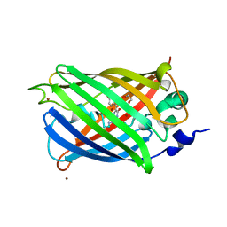
3SSV
DownloadVisualize
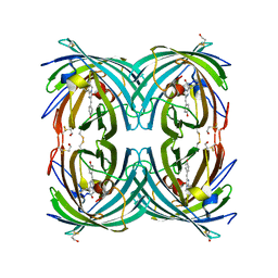
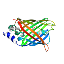
BU of 3ssv by Molmil
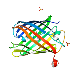
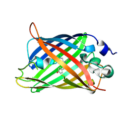
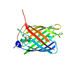
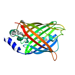
Engineered low-affinity halide-binding protein derived from YFP: fluoride complex
Descriptor: FLUORIDE ION, Green fluorescent protein
Authors:Wang, W, Grimley, J.S, Beese, L.S, Hellinga, H.W.
Deposit date:2011-07-08
Release date:2012-07-11
Last modified:2023-11-15
Method:X-RAY DIFFRACTION (1.861 Å)
Cite:Determination of engineered chloride-binding site structures in fluorescent proteins reveals principles of halide recognition
To be Published
3SVD
DownloadVisualize
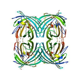
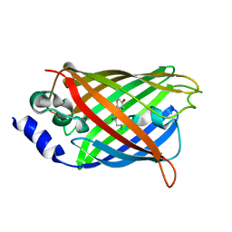
BU of 3svd by Molmil
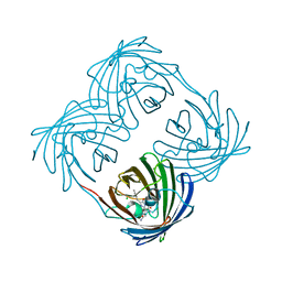
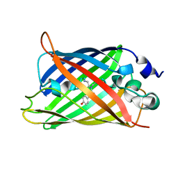
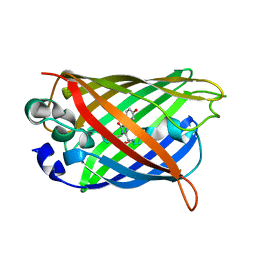
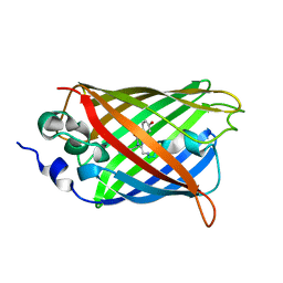
Engineered medium-affinity halide-binding protein derived from YFP: bromide complex
Descriptor: 1,2-ETHANEDIOL, BROMIDE ION, Green fluorescent protein
Authors:Wang, W, Grimley, J.S, Beese, L.S, Hellinga, H.W.
Deposit date:2011-07-12
Release date:2012-07-18
Last modified:2023-11-15
Method:X-RAY DIFFRACTION (1.78 Å)
Cite:Determination of engineered chloride-binding site structures in fluorescent proteins reveals principles of halide recognition
To be Published
3U0N
DownloadVisualize
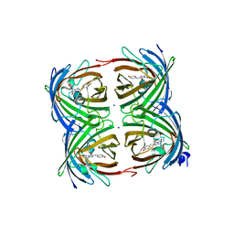
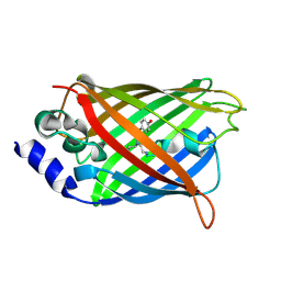
BU of 3u0n by Molmil
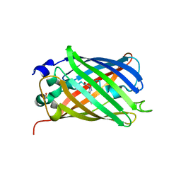
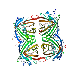
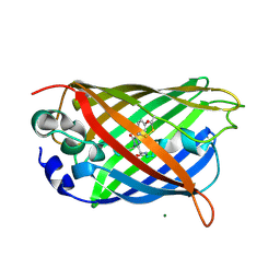
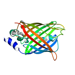
Crystal structure of the engineered fluorescent protein mRuby, crystal form 2
Descriptor: SULFATE ION, mRuby
Authors:Akerboom, J, Looger, L.L, Schreiter, E.R.
Deposit date:2011-09-28
Release date:2012-10-03
Last modified:2023-12-06
Method:X-RAY DIFFRACTION (1.6 Å)
Cite:Genetically encoded calcium indicators for multi-color neural activity imaging and combination with optogenetics.
Front Mol Neurosci, 6, 2013
3VK1
DownloadVisualize
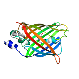
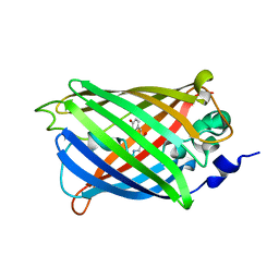
BU of 3vk1 by Molmil
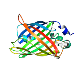
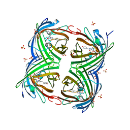
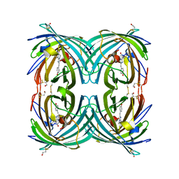
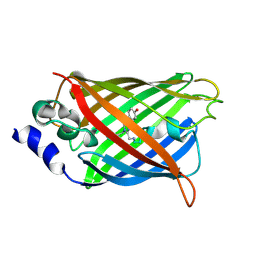
Green-fluorescent variant of the non-fluorescent chromoprotein Rtms5
Descriptor: CHLORIDE ION, GFP-like non-fluorescent chromoprotein, IODIDE ION
Authors:Battad, J.M, Traore, D.A.K, Wilce, M, Byres, M, Rossjohn, J, Devenish, R.J, Prescott, M.
Deposit date:2011-11-07
Release date:2012-06-06
Last modified:2023-11-15
Method:X-RAY DIFFRACTION (2.2 Å)
Cite:A Green Fluorescent Protein Containing a QFG Tri-Peptide Chromophore: Optical Properties and X-Ray Crystal Structure.
Plos One, 7, 2012
3V3D
DownloadVisualize
BU of 3v3d by Molmil
Crystal Structure of an eYFP single mutant
Descriptor: SULFATE ION, Yellow fluorescent protein
Authors:Ngan, N.B, Van Hecke, K, Van Meervelt, L.
Deposit date:2011-12-13
Release date:2012-12-26
Last modified:2023-12-06
Method:X-RAY DIFFRACTION (1.95 Å)
Cite:Improving the second-order nonlinear optical response of fluorescent proteins: the symmetry argument.
J.Am.Chem.Soc., 135, 2013
3UFZ
DownloadVisualize
BU of 3ufz by Molmil
Crystal structure of a Trp-less green fluorescent protein translated by the universal genetic code
Descriptor: Green fluorescent protein
Authors:Kawahara-Kobayashi, A, Araiso, Y, Matsuda, T, Yokoyama, S, Kigawa, T, Nureki, O, Kiga, D.
Deposit date:2011-11-02
Release date:2012-10-17
Last modified:2023-12-06
Method:X-RAY DIFFRACTION (1.85 Å)
Cite:Simplification of the genetic code: restricted diversity of genetically encoded amino acids.
Nucleic Acids Res., 40, 2012
3W1D
DownloadVisualize
BU of 3w1d by Molmil
Structure of a pressure sensitive YFP variant YFP-G3
Descriptor: Green Fluorescent protein
Authors:Imada, K, Yoshizawa, K, Kinoshita, M, Watanabe, T.M.
Deposit date:2012-11-14
Release date:2013-10-02
Last modified:2023-12-06
Method:X-RAY DIFFRACTION (1.5 Å)
Cite:Glycine insertion makes yellow fluorescent protein sensitive to hydrostatic pressure.
Plos One, 8, 2013
3W1C
DownloadVisualize
BU of 3w1c by Molmil
Structure of a pressure sensitive YFP variant YFP-G1
Descriptor: Green Fluorescent protein
Authors:Imada, K, Yoshizawa, K, Kinoshita, M, Watanabe, T.M.
Deposit date:2012-11-14
Release date:2013-10-02
Last modified:2023-12-06
Method:X-RAY DIFFRACTION (1.3 Å)
Cite:Glycine insertion makes yellow fluorescent protein sensitive to hydrostatic pressure.
Plos One, 8, 2013
3TMT
DownloadVisualize
BU of 3tmt by Molmil
IrisFP, distorted chromophore
Descriptor: Green to red photoconvertible GPF-like protein EosFP, SULFATE ION
Authors:Adam, V, Carpentier, P, Roy, A, Field, M, Bourgeois, D.
Deposit date:2011-08-31
Release date:2011-11-16
Last modified:2023-11-15
Method:X-RAY DIFFRACTION (2 Å)
Cite:The nature of transient dark States in a photoactivatable fluorescent protein.
J.Am.Chem.Soc., 133, 2011
3TMR
DownloadVisualize
BU of 3tmr by Molmil
IrisFP, planar chromophore
Descriptor: Green to red photoconvertible GPF-like protein EosFP, SULFATE ION, SULFITE ION
Authors:Adam, V, Carpentier, P, Roy, A, Field, M, Bourgeois, D.
Deposit date:2011-08-31
Release date:2011-11-16
Last modified:2023-11-15
Method:X-RAY DIFFRACTION (2 Å)
Cite:The nature of transient dark States in a photoactivatable fluorescent protein.
J.Am.Chem.Soc., 133, 2011
3WCK
DownloadVisualize
BU of 3wck by Molmil
Crystal structure of monomeric photosensitizing fluorescent protein, Supernova
Descriptor: Monomeric photosenitizing fluorescent protein supernova
Authors:Sakai, N, Matsuda, T, Takemoto, K, Nagai, T.
Deposit date:2013-05-27
Release date:2013-10-02
Last modified:2023-11-15
Method:X-RAY DIFFRACTION (2.3 Å)
Cite:SuperNova, a monomeric photosensitizing fluorescent protein for chromophore-assisted light inactivation
Sci Rep, 3, 2013
3CD9
DownloadVisualize
BU of 3cd9 by Molmil
Development of a family of redox-sensitive green fluorescent protein indicators for use in relatively oxidizing subcellular environments
Descriptor: Green fluorescent protein
Authors:Lohman, J.R.
Deposit date:2008-02-26
Release date:2008-08-19
Last modified:2021-10-20
Method:X-RAY DIFFRACTION (1.502 Å)
Cite:Development of a family of redox-sensitive green fluorescent protein indicators for use in relatively oxidizing subcellular environments
Biochemistry, 47, 2008
3CB9
DownloadVisualize
BU of 3cb9 by Molmil
Development of a family of redox-sensitive green fluorescent protein indicators for use in relatively oxidizing subcellular environments
Descriptor: Green fluorescent protein, MAGNESIUM ION
Authors:Lohman, J.R.
Deposit date:2008-02-21
Release date:2008-08-19
Last modified:2023-11-15
Method:X-RAY DIFFRACTION (1.31 Å)
Cite:Development of a family of redox-sensitive green fluorescent protein indicators for use in relatively oxidizing subcellular environments
Biochemistry, 47, 2008
3CFF
DownloadVisualize
BU of 3cff by Molmil
Photoswitchable red fluorescent protein psRFP, on-state
Descriptor: GFP-like photoswitchable fluorescent protein
Authors:Kachalova, G.S, Gundel, S, Bartunik, H.D, Wiedenmann, J.
Deposit date:2008-03-03
Release date:2009-03-10
Last modified:2023-11-15
Method:X-RAY DIFFRACTION (1.8 Å)
Cite:Structure of Anemonia sulcata red fluorescent protein asRFP
To be Published
3CFA
DownloadVisualize
BU of 3cfa by Molmil
Anemonia sulcata red fluorescent protein asRFP
Descriptor: GFP-like fluorescent protein
Authors:Kachalova, G.S, Gundel, S, Wiedenmann, J, Bartunik, H.D.
Deposit date:2008-03-03
Release date:2009-03-10
Last modified:2023-11-15
Method:X-RAY DIFFRACTION (1.75 Å)
Cite:Structure of Anemonia sulcata red fluorescent protein asRFP
To be Published
3CFH
DownloadVisualize
BU of 3cfh by Molmil
Photoswitchable red fluorescent protein psRFP, off-state
Descriptor: GFP-like photoswitchable fluorescent protein
Authors:Kachalova, G.S, Gundel, S, Bartunik, H.D, Wiedenmann, J.
Deposit date:2008-03-03
Release date:2009-03-10
Last modified:2023-11-15
Method:X-RAY DIFFRACTION (1.75 Å)
Cite:Structure of red fluorescent protein asRFP
To be Published
3CGL
DownloadVisualize
BU of 3cgl by Molmil
Crystal Structure and Raman Studies of dsFP483, a Cyan Fluorescent Protein from Discosoma striata
Descriptor: GFP-like fluorescent chromoprotein dsFP483, SODIUM ION
Authors:Malo, G.D.
Deposit date:2008-03-05
Release date:2008-07-01
Last modified:2023-12-27
Method:X-RAY DIFFRACTION (2.09 Å)
Cite:Crystal structure and Raman studies of dsFP483, a cyan fluorescent protein from Discosoma striata.
J.Mol.Biol., 378, 2008
3DQ7
DownloadVisualize
BU of 3dq7 by Molmil
Structure of the Yellow Fluorescent Protein Citrine Frozen at 1920 Atmospheres Number 1: Structure 17 in a Series of 26 High Pressure Structures
Descriptor: Green fluorescent protein
Authors:Barstow, B, Kim, C.U.
Deposit date:2008-07-09
Release date:2008-09-23
Last modified:2023-11-15
Method:X-RAY DIFFRACTION (1.23 Å)
Cite:Alteration of citrine structure by hydrostatic pressure explains the accompanying spectral shift.
Proc.Natl.Acad.Sci.Usa, 105, 2008
3DQK
DownloadVisualize
BU of 3dqk by Molmil
Structure of the Yellow Fluorescent Protein Citrine Frozen at 1000 Atmospheres Number 2: Structure 6 in a Series of 26 High Pressure Structures
Descriptor: Green fluorescent protein
Authors:Barstow, B, Kim, C.U.
Deposit date:2008-07-09
Release date:2008-09-23
Last modified:2023-11-15
Method:X-RAY DIFFRACTION (1.4 Å)
Cite:Alteration of citrine structure by hydrostatic pressure explains the accompanying spectral shift.
Proc.Natl.Acad.Sci.Usa, 105, 2008
3DPZ
DownloadVisualize
BU of 3dpz by Molmil
Structure of the Yellow Fluorescent Protein Citrine Frozen at 4000 Atmospheres Number 3: Structure 25 in a Series of 26 High Pressure Structures
Descriptor: Green fluorescent protein
Authors:Barstow, B, Kim, C.U.
Deposit date:2008-07-09
Release date:2008-09-23
Last modified:2023-11-15
Method:X-RAY DIFFRACTION (1.7 Å)
Cite:Alteration of citrine structure by hydrostatic pressure explains the accompanying spectral shift.
Proc.Natl.Acad.Sci.Usa, 105, 2008
3DQ8
DownloadVisualize
BU of 3dq8 by Molmil
Structure of the Yellow Fluorescent Protein Citrine Frozen at 1500 Atmospheres Number 2: Structure 16 in a Series of 26 High Pressure Structures
Descriptor: Green fluorescent protein
Authors:Barstow, B, Kim, C.U.
Deposit date:2008-07-09
Release date:2008-09-23
Last modified:2023-11-15
Method:X-RAY DIFFRACTION (1.51 Å)
Cite:Alteration of citrine structure by hydrostatic pressure explains the accompanying spectral shift.
Proc.Natl.Acad.Sci.Usa, 105, 2008
3DPX
DownloadVisualize
BU of 3dpx by Molmil
Structure of the Yellow Fluorescent Protein Citrine Frozen at 5000 Atmospheres: Structure 26 in a Series of 26 High Pressure Structures
Descriptor: Green fluorescent protein
Authors:Barstow, B, Kim, C.U.
Deposit date:2008-07-09
Release date:2008-09-23
Last modified:2023-11-15
Method:X-RAY DIFFRACTION (1.5 Å)
Cite:Alteration of citrine structure by hydrostatic pressure explains the accompanying spectral shift.
Proc.Natl.Acad.Sci.Usa, 105, 2008
3DQ5
DownloadVisualize
BU of 3dq5 by Molmil
Structure of the Yellow Fluorescent Protein Citrine Frozen at 1960 Atmospheres: Structure 19 in a Series of 26 High Pressure Structures
Descriptor: Green fluorescent protein
Authors:Barstow, B, Kim, C.U.
Deposit date:2008-07-09
Release date:2008-09-23
Last modified:2023-11-15
Method:X-RAY DIFFRACTION (1.5 Å)
Cite:Alteration of citrine structure by hydrostatic pressure explains the accompanying spectral shift.
Proc.Natl.Acad.Sci.Usa, 105, 2008
3DQE
DownloadVisualize
BU of 3dqe by Molmil
Structure of the Yellow Fluorescent Protein Citrine Frozen at 1250 Atmospheres Number 1: Structure 11 in a Series of 26 High Pressure Structures
Descriptor: Green fluorescent protein
Authors:Barstow, B, Kim, C.U.
Deposit date:2008-07-09
Release date:2008-09-23
Last modified:2023-11-15
Method:X-RAY DIFFRACTION (1.43 Å)
Cite:Alteration of citrine structure by hydrostatic pressure explains the accompanying spectral shift.
Proc.Natl.Acad.Sci.Usa, 105, 2008
3DPW
DownloadVisualize
BU of 3dpw by Molmil
Structure of the Yellow Fluorescent Protein Citrine Frozen at 1 Atmosphere Number 1: Structure 1 in a Series of 26 High Pressure Structures
Descriptor: Green fluorescent protein
Authors:Barstow, B, Kim, C.U.
Deposit date:2008-07-09
Release date:2008-09-23
Last modified:2023-11-15
Method:X-RAY DIFFRACTION (1.59 Å)
Cite:Alteration of citrine structure by hydrostatic pressure explains the accompanying spectral shift.
Proc.Natl.Acad.Sci.Usa, 105, 2008

223532

PDB entries from 2024-08-07

PDB statisticsPDBj update infoContact PDBjnumon