Loading
PDBj
MenuPDBj@FacebookPDBj@TwitterPDBj@YouTubewwPDB FoundationwwPDB
RCSB PDBPDBeBMRBAdv. SearchSearch help

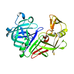
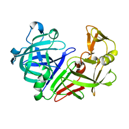
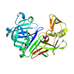
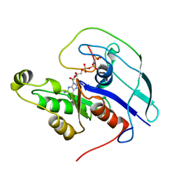
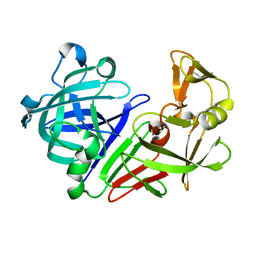
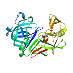
5P0Q
DownloadVisualize
BU of 5p0q by Molmil
Automated refinement of diffraction data obtained from an endothiapepsin crystal treated with fragment 73
Descriptor: endothiapepsin
Authors:Schiebel, J, Heine, A, Klebe, G.
Deposit date:2016-06-28
Release date:2016-08-03
Last modified:2021-11-17
Method:X-RAY DIFFRACTION (1.48 Å)
Cite:High-Throughput Crystallography: Reliable and Efficient Identification of Fragment Hits.
Structure, 24, 2016
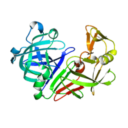
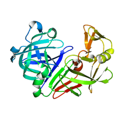
5P4V
DownloadVisualize
BU of 5p4v by Molmil
Automated refinement of diffraction data obtained from an endothiapepsin crystal treated with fragment 220
Descriptor: endothiapepsin
Authors:Schiebel, J, Heine, A, Klebe, G.
Deposit date:2016-06-28
Release date:2016-08-03
Last modified:2021-11-17
Method:X-RAY DIFFRACTION (1.329 Å)
Cite:High-Throughput Crystallography: Reliable and Efficient Identification of Fragment Hits.
Structure, 24, 2016
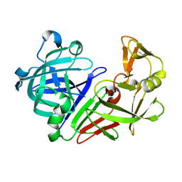
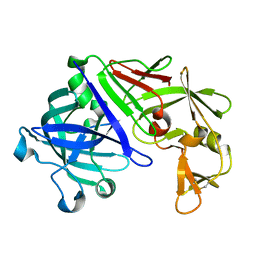
5P59
DownloadVisualize
BU of 5p59 by Molmil
Automated refinement of diffraction data obtained from an endothiapepsin crystal treated with fragment 234
Descriptor: endothiapepsin
Authors:Schiebel, J, Heine, A, Klebe, G.
Deposit date:2016-06-28
Release date:2016-08-03
Last modified:2024-10-16
Method:X-RAY DIFFRACTION (1.18 Å)
Cite:High-Throughput Crystallography: Reliable and Efficient Identification of Fragment Hits.
Structure, 24, 2016
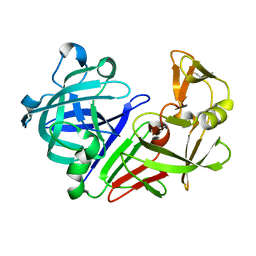
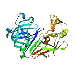
5P5P
DownloadVisualize
BU of 5p5p by Molmil
Automated refinement of diffraction data obtained from an endothiapepsin crystal treated with fragment 250
Descriptor: endothiapepsin
Authors:Schiebel, J, Heine, A, Klebe, G.
Deposit date:2016-06-28
Release date:2016-08-03
Last modified:2021-11-17
Method:X-RAY DIFFRACTION (1.09 Å)
Cite:High-Throughput Crystallography: Reliable and Efficient Identification of Fragment Hits.
Structure, 24, 2016
5P65
DownloadVisualize
BU of 5p65 by Molmil
Automated refinement of diffraction data obtained from an endothiapepsin crystal treated with fragment 266
Descriptor: endothiapepsin
Authors:Schiebel, J, Heine, A, Klebe, G.
Deposit date:2016-06-28
Release date:2016-08-03
Last modified:2021-11-17
Method:X-RAY DIFFRACTION (1.1 Å)
Cite:High-Throughput Crystallography: Reliable and Efficient Identification of Fragment Hits.
Structure, 24, 2016
5P14
DownloadVisualize
BU of 5p14 by Molmil
Automated refinement of diffraction data obtained from an endothiapepsin crystal treated with fragment 87
Descriptor: endothiapepsin
Authors:Schiebel, J, Heine, A, Klebe, G.
Deposit date:2016-06-28
Release date:2016-08-03
Last modified:2024-10-30
Method:X-RAY DIFFRACTION (1.24 Å)
Cite:High-Throughput Crystallography: Reliable and Efficient Identification of Fragment Hits.
Structure, 24, 2016
5P1J
DownloadVisualize
BU of 5p1j by Molmil
Automated refinement of diffraction data obtained from an endothiapepsin crystal treated with fragment 102
Descriptor: endothiapepsin
Authors:Schiebel, J, Heine, A, Klebe, G.
Deposit date:2016-06-28
Release date:2016-08-03
Last modified:2021-11-17
Method:X-RAY DIFFRACTION (1.369 Å)
Cite:High-Throughput Crystallography: Reliable and Efficient Identification of Fragment Hits.
Structure, 24, 2016
5P6J
DownloadVisualize
BU of 5p6j by Molmil
Automated refinement of diffraction data obtained from an endothiapepsin crystal treated with fragment 280
Descriptor: endothiapepsin
Authors:Schiebel, J, Heine, A, Klebe, G.
Deposit date:2016-06-28
Release date:2016-08-03
Last modified:2021-11-17
Method:X-RAY DIFFRACTION (1.259 Å)
Cite:High-Throughput Crystallography: Reliable and Efficient Identification of Fragment Hits.
Structure, 24, 2016
5P6X
DownloadVisualize
BU of 5p6x by Molmil
Automated refinement of diffraction data obtained from an endothiapepsin crystal treated with fragment 294
Descriptor: endothiapepsin
Authors:Schiebel, J, Heine, A, Klebe, G.
Deposit date:2016-06-28
Release date:2016-08-03
Last modified:2024-10-23
Method:X-RAY DIFFRACTION (1.397 Å)
Cite:High-Throughput Crystallography: Reliable and Efficient Identification of Fragment Hits.
Structure, 24, 2016
5P7B
DownloadVisualize
BU of 5p7b by Molmil
Automated refinement of diffraction data obtained from an endothiapepsin crystal treated with fragment 308
Descriptor: endothiapepsin
Authors:Schiebel, J, Heine, A, Klebe, G.
Deposit date:2016-06-28
Release date:2016-08-03
Last modified:2021-11-17
Method:X-RAY DIFFRACTION (1.407 Å)
Cite:High-Throughput Crystallography: Reliable and Efficient Identification of Fragment Hits.
Structure, 24, 2016
5P7P
DownloadVisualize
BU of 5p7p by Molmil
Automated refinement of diffraction data obtained from an endothiapepsin crystal treated with fragment 322
Descriptor: endothiapepsin
Authors:Schiebel, J, Heine, A, Klebe, G.
Deposit date:2016-06-28
Release date:2016-08-03
Last modified:2024-10-16
Method:X-RAY DIFFRACTION (1.439 Å)
Cite:High-Throughput Crystallography: Reliable and Efficient Identification of Fragment Hits.
Structure, 24, 2016
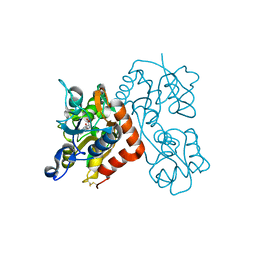
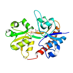
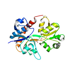
3EN3
DownloadVisualize
BU of 3en3 by Molmil
Crystal Structure of the GluR4 Ligand-Binding domain in complex with kainate
Descriptor: 3-(CARBOXYMETHYL)-4-ISOPROPENYLPROLINE, Glutamate receptor 4,Glutamate receptor
Authors:Gill, A, Madden, D.R.
Deposit date:2008-09-25
Release date:2009-05-19
Last modified:2017-07-26
Method:X-RAY DIFFRACTION (2.43 Å)
Cite:Correlating AMPA receptor activation and cleft closure across subunits: crystal structures of the GluR4 ligand-binding domain in complex with full and partial agonists
Biochemistry, 47, 2008
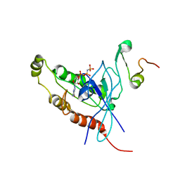
1E0A
DownloadVisualize
BU of 1e0a by Molmil
Cdc42 complexed with the GTPase binding domain of p21 activated kinase
Descriptor: Cell division control protein 42 homolog, MAGNESIUM ION, PHOSPHOAMINOPHOSPHONIC ACID-GUANYLATE ESTER, ...
Authors:Morreale, A, Venkatesan, M, Mott, H.R, Owen, D, Nietlispach, D, Lowe, P.N, Laue, E.D.
Deposit date:2000-03-16
Release date:2000-04-18
Last modified:2024-05-15
Method:SOLUTION NMR
Cite:Solution Structure of Cdc42 Bound to the Gtpase Binding Domian of Pak
Nat.Struct.Biol., 7, 2000
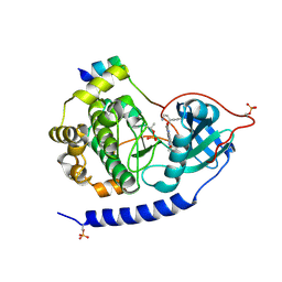
3E8E
DownloadVisualize
BU of 3e8e by Molmil
Crystal structures of the kinase domain of PKA in complex with ATP-competitive inhibitors
Descriptor: 4-[2-(4-amino-2,5-dihydro-1,2,5-oxadiazol-3-yl)-6-{[(1S)-3-amino-1-phenylpropyl]oxy}-1-ethyl-1H-imidazo[4,5-c]pyridin-4-yl]-2-methylbut-3-yn-2-ol, PKI inhibitor peptide, cAMP-dependent protein kinase catalytic subunit alpha
Authors:Concha, N.O, Elkins, P.A, Smallwood, A, Ward, P.
Deposit date:2008-08-19
Release date:2008-11-18
Last modified:2024-10-30
Method:X-RAY DIFFRACTION (2 Å)
Cite:Aminofurazans as potent inhibitors of AKT kinase
Bioorg.Med.Chem.Lett., 19, 2009
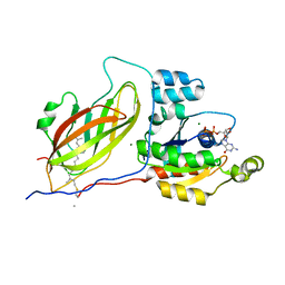
1CF4
DownloadVisualize
BU of 1cf4 by Molmil
CDC42/ACK GTPASE-BINDING DOMAIN COMPLEX
Descriptor: MAGNESIUM ION, PHOSPHOAMINOPHOSPHONIC ACID-GUANYLATE ESTER, PROTEIN (ACTIVATED P21CDC42HS KINASE), ...
Authors:Mott, H.R, Owen, D, Nietlispach, D, Lowe, P.N, Lim, L, Laue, E.D.
Deposit date:1999-03-23
Release date:1999-06-18
Last modified:2023-12-27
Method:SOLUTION NMR
Cite:Structure of the small G protein Cdc42 bound to the GTPase-binding domain of ACK.
Nature, 399, 1999
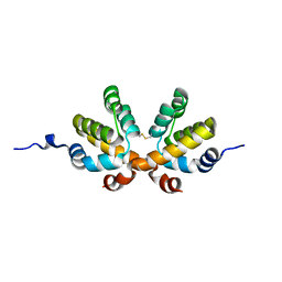
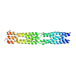
2NZ7
DownloadVisualize
BU of 2nz7 by Molmil
Crystal Structure Analysis of Caspase-recruitment Domain (CARD) of Nod1
Descriptor: Caspase recruitment domain-containing protein 4
Authors:Thiagarajan, S, Robbins, S.L, Dubas, R.L, Park, Y.C.
Deposit date:2006-11-22
Release date:2007-12-04
Last modified:2024-10-16
Method:X-RAY DIFFRACTION (1.9 Å)
Cite:Monomer/dimer transition of the caspase-recruitment domain of human Nod1
Biochemistry, 47, 2008
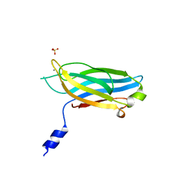
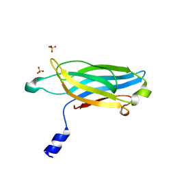
3F05
DownloadVisualize
BU of 3f05 by Molmil
Crystal Structure of Synaptotagmin I C2A domain with Mn(II)
Descriptor: MANGANESE (II) ION, SULFATE ION, Synaptotagmin-1
Authors:Guo, F, Dakshinamurthy, R, Thallapuranam, S.K.K, Sakon, J.
Deposit date:2008-10-24
Release date:2009-10-06
Last modified:2023-12-27
Method:X-RAY DIFFRACTION (1.4 Å)
Cite:Cystal structure of synaptotagmin I C2A
to be published
3DLN
DownloadVisualize
BU of 3dln by Molmil
Crystal structure of the binding domain of the AMPA subunit GluR3 bound to glutamate
Descriptor: GLUTAMIC ACID, Glutamate receptor 3, ZINC ION
Authors:Ahmed, A.H, Wang, Q, Sondermann, H, Oswald, R.E.
Deposit date:2008-06-27
Release date:2008-11-25
Last modified:2024-10-30
Method:X-RAY DIFFRACTION (1.91 Å)
Cite:Structure of the S1S2 glutamate binding domain of GLuR3.
Proteins, 75, 2008
3F04
DownloadVisualize
BU of 3f04 by Molmil
Crystal Structure of Synaptotagmin I C2A domain
Descriptor: SULFATE ION, Synaptotagmin-1
Authors:Guo, F, Dakshinamurthy, R, Thallapuranam, S.K.K, Sakon, J.
Deposit date:2008-10-24
Release date:2009-09-08
Last modified:2023-12-27
Method:X-RAY DIFFRACTION (1.35 Å)
Cite:Cystal structure of synaptotagmin I C2A
to be published
3F6N
DownloadVisualize
BU of 3f6n by Molmil
Crystal structure of the virion-associated protein P3 from Caulimovirus
Descriptor: Virion-associated protein
Authors:Hoh, F, Uzest, M, Blanc, S, Dumas, C.
Deposit date:2008-11-06
Release date:2009-11-24
Last modified:2024-10-30
Method:X-RAY DIFFRACTION (3.1 Å)
Cite:Structural insights into the molecular mechanisms of cauliflower mosaic virus transmission by its insect vector.
J.Virol., 84, 2010
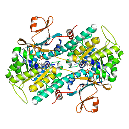
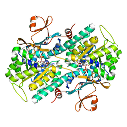
3DHD
DownloadVisualize
BU of 3dhd by Molmil
Crystal structure of human NAMPT complexed with nicotinamide mononucleotide and pyrophosphate
Descriptor: BETA-NICOTINAMIDE RIBOSE MONOPHOSPHATE, MAGNESIUM ION, Nicotinamide phosphoribosyltransferase, ...
Authors:Ho, M, Burgos, E.S, Almo, S.C, Schramm, V.L.
Deposit date:2008-06-17
Release date:2009-08-18
Last modified:2023-08-30
Method:X-RAY DIFFRACTION (2 Å)
Cite:A phosphoenzyme mimic, overlapping catalytic sites and reaction coordinate motion for human NAMPT.
Proc.Natl.Acad.Sci.USA, 106, 2009
1DOA
DownloadVisualize
BU of 1doa by Molmil
Structure of the rho family gtp-binding protein cdc42 in complex with the multifunctional regulator rhogdi
Descriptor: GERAN-8-YL GERAN, GUANOSINE-5'-DIPHOSPHATE, MAGNESIUM ION, ...
Authors:Hoffman, G.R, Nassar, N, Cerione, R.C.
Deposit date:1999-12-20
Release date:2000-02-09
Last modified:2011-07-13
Method:X-RAY DIFFRACTION (2.6 Å)
Cite:Structure of the Rho family GTP-binding protein Cdc42 in complex with the multifunctional regulator RhoGDI.
Cell(Cambridge,Mass.), 100, 2000
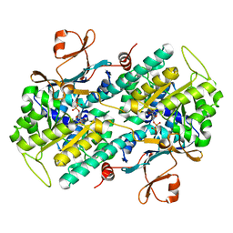
3DKJ
DownloadVisualize
BU of 3dkj by Molmil
Crystal structure of human NAMPT complexed with benzamide and phosphoribosyl pyrophosphate
Descriptor: 1-O-pyrophosphono-5-O-phosphono-alpha-D-ribofuranose, BENZAMIDE, Nicotinamide phosphoribosyltransferase
Authors:Ho, M, Burgos, E.S, Almo, S.C, Schramm, V.L.
Deposit date:2008-06-25
Release date:2009-08-18
Last modified:2023-08-30
Method:X-RAY DIFFRACTION (2 Å)
Cite:A phosphoenzyme mimic, overlapping catalytic sites and reaction coordinate motion for human NAMPT.
Proc.Natl.Acad.Sci.USA, 106, 2009
3DP6
DownloadVisualize
BU of 3dp6 by Molmil
Crystal structure of the binding domain of the AMPA subunit GluR2 bound to glutamate
Descriptor: GLUTAMIC ACID, Glutamate receptor 2, ZINC ION
Authors:Ahmed, A.H, Wang, Q, Sondermann, H, Oswald, R.E.
Deposit date:2008-07-07
Release date:2008-11-25
Last modified:2024-10-30
Method:X-RAY DIFFRACTION (1.55 Å)
Cite:Structure of the S1S2 glutamate binding domain of GLuR3.
Proteins, 75, 2008
3DGR
DownloadVisualize
BU of 3dgr by Molmil
Crystal structure of human NAMPT complexed with ADP analogue
Descriptor: Nicotinamide phosphoribosyltransferase, PHOSPHOMETHYLPHOSPHONIC ACID ADENOSYL ESTER
Authors:Ho, M, Burgos, E.S, Almo, S.C, Schramm, V.L.
Deposit date:2008-06-15
Release date:2009-08-18
Last modified:2023-08-30
Method:X-RAY DIFFRACTION (2.1 Å)
Cite:A phosphoenzyme mimic, overlapping catalytic sites and reaction coordinate motion for human NAMPT.
Proc.Natl.Acad.Sci.USA, 106, 2009

226707

PDB entries from 2024-10-30

PDB statisticsPDBj update infoContact PDBjnumon