Loading
PDBj
MenuPDBj@FacebookPDBj@TwitterPDBj@YouTubewwPDB FoundationwwPDB
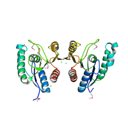
RCSB PDBPDBeBMRBAdv. SearchSearch help

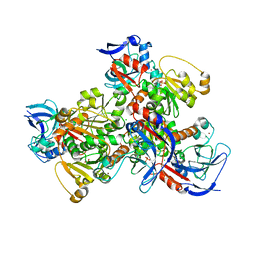
4JBG
DownloadVisualize
BU of 4jbg by Molmil
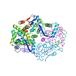
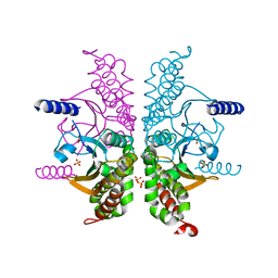
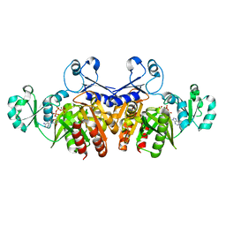
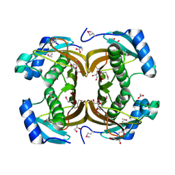
1.75A resolution structure of a thermostable alcohol dehydrogenase from Pyrobaculum aerophilum
Descriptor: Alcohol dehydrogenase (Zinc), CHLORIDE ION, PHOSPHATE ION, ...
Authors:Lovell, S, Battaile, K.P, Vitale, A, Throne, N, Hu, X, Shen, M, D'Auria, S, Auld, D.S.
Deposit date:2013-02-19
Release date:2013-06-19
Last modified:2024-02-28
Method:X-RAY DIFFRACTION (1.75 Å)
Cite:Physicochemical Characterization of a Thermostable Alcohol Dehydrogenase from Pyrobaculum aerophilum.
Plos One, 8, 2013
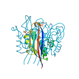
4JRX
DownloadVisualize
BU of 4jrx by Molmil
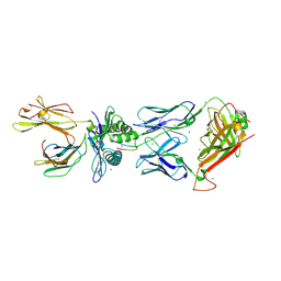
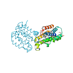
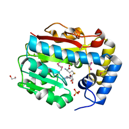
Crystal Structure of CA5 TCR-HLA B*3505-LPEP complex
Descriptor: Beta-2-microglobulin, CA5 TCR alpha chain, CA5 TCR beta chain, ...
Authors:Liu, Y.C, Rossjohn, J, Gras, S.
Deposit date:2013-03-22
Release date:2013-04-10
Last modified:2023-11-08
Method:X-RAY DIFFRACTION (2.3 Å)
Cite:Highly divergent T-cell receptor binding modes underlie specific recognition of a bulged viral peptide bound to a human leukocyte antigen class I molecule.
J.Biol.Chem., 288, 2013
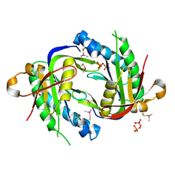
4JTG
DownloadVisualize
BU of 4jtg by Molmil
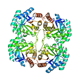
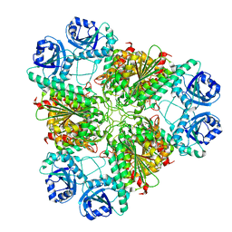
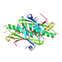
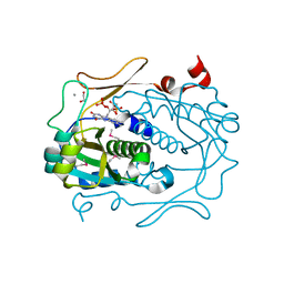
Crystal structure of F114R/R117A mutant of 3-deoxy-D-manno-octulosonate 8-phosphate synthase (KDO8PS) from Neisseria meningitidis
Descriptor: 2-dehydro-3-deoxyphosphooctonate aldolase, CHLORIDE ION, GLYCEROL, ...
Authors:Allison, T.M, Cochrane, F.C, Jameson, G.B, Parker, E.J.
Deposit date:2013-03-23
Release date:2013-06-26
Last modified:2023-09-20
Method:X-RAY DIFFRACTION (1.85 Å)
Cite:Examining the Role of Intersubunit Contacts in Catalysis by 3-Deoxy-d-manno-octulosonate 8-Phosphate Synthase.
Biochemistry, 52, 2013
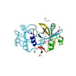
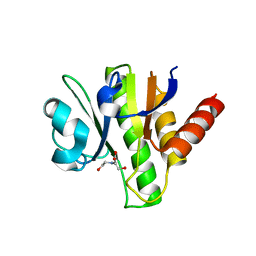
3H4W
DownloadVisualize
BU of 3h4w by Molmil
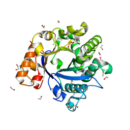
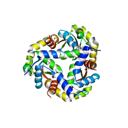
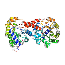
Structure of a Ca+2 dependent Phosphatidylinositol-specific phospholipase C (PI-PLC) Enzyme from Streptomyces antibioticus
Descriptor: ACETYL GROUP, CHLORIDE ION, ETHANOL, ...
Authors:Jackson, M.R, Selby, T.L.
Deposit date:2009-04-21
Release date:2010-04-28
Last modified:2011-07-13
Method:X-RAY DIFFRACTION (1.5 Å)
Cite:Crystal Structure of a Ca2+-dependent PI-PLC
To be Published
3GRE
DownloadVisualize
BU of 3gre by Molmil
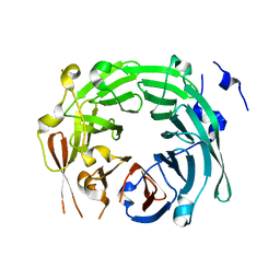
Crystal structure of Saccharomyces cerevisiae Vps15 WD repeat domain
Descriptor: Serine/threonine-protein kinase VPS15
Authors:Vanhooke, J.L, Sondek, J, Betts, L.
Deposit date:2009-03-25
Release date:2009-07-28
Last modified:2024-02-21
Method:X-RAY DIFFRACTION (1.8 Å)
Cite:Structure and function of Vps15 in the endosomal G protein signaling pathway.
Biochemistry, 48, 2009
3H87
DownloadVisualize
BU of 3h87 by Molmil
Rv0301 Rv0300 Toxin Antitoxin Complex from Mycobacterium tuberculosis
Descriptor: BETA-MERCAPTOETHANOL, GLYCEROL, IMIDAZOLE, ...
Authors:Min, A, Sawaya, M.R, Cascio, D, Eisenberg, D, Integrated Center for Structure and Function Innovation (ISFI)
Deposit date:2009-04-28
Release date:2009-05-05
Last modified:2024-02-21
Method:X-RAY DIFFRACTION (1.49 Å)
Cite:The crystal structure of the Rv0301-Rv0300 VapBC-3 toxin-antitoxin complex from M. tuberculosis reveals a Mg(2+) ion in the active site and a putative RNA-binding site.
Protein Sci., 21, 2012
3H8F
DownloadVisualize
BU of 3h8f by Molmil
High pH native structure of leucine aminopeptidase from Pseudomonas putida
Descriptor: BICARBONATE ION, Cytosol aminopeptidase, MANGANESE (II) ION, ...
Authors:Kale, A, Dijkstra, B.W, Sonke, T, Thunnissen, A.M.W.H.
Deposit date:2009-04-29
Release date:2010-04-14
Last modified:2024-02-21
Method:X-RAY DIFFRACTION (2.2 Å)
Cite:Crystal structure of the leucine aminopeptidase from Pseudomonas putida reveals the molecular basis for its enantioselectivity and broad substrate specificity.
J.Mol.Biol., 398, 2010
3EOD
DownloadVisualize
BU of 3eod by Molmil
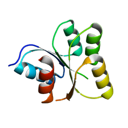
Crystal structure of N-terminal domain of E. coli RssB
Descriptor: Protein hnr
Authors:Levchenko, I, Grant, R.A, Sauer, R.T, Baker, T.A.
Deposit date:2008-09-26
Release date:2009-10-06
Last modified:2024-02-21
Method:X-RAY DIFFRACTION (1.75 Å)
Cite:The structure of RssB, a ClpX adaptor protein that regulates sigma S
To be Published
3E3G
DownloadVisualize
BU of 3e3g by Molmil
H. influenzae beta-carbonic anhydrase, variant G41A
Descriptor: Carbonic anhydrase 2, SULFATE ION, ZINC ION
Authors:Rowlett, R.S, Failing, H.
Deposit date:2008-08-07
Release date:2009-08-18
Last modified:2024-02-21
Method:X-RAY DIFFRACTION (2.3 Å)
Cite:Evidence for a bicarbonate "escort" site in Haemophilus influenzae beta-carbonic anhydrase .
Biochemistry, 49, 2010
2QJW
DownloadVisualize
BU of 2qjw by Molmil
CRYSTAL STRUCTURE OF A PUTATIVE HYDROLASE OF THE ALPHA/BETA SUPERFAMILY (XCC1541) FROM XANTHOMONAS CAMPESTRIS PV. CAMPESTRIS AT 1.35 A RESOLUTION
Descriptor: CHLORIDE ION, DI(HYDROXYETHYL)ETHER, HEXAETHYLENE GLYCOL, ...
Authors:Joint Center for Structural Genomics (JCSG)
Deposit date:2007-07-09
Release date:2007-07-24
Last modified:2023-01-25
Method:X-RAY DIFFRACTION (1.35 Å)
Cite:Crystal structure of uncharacterized protein XCC1541 (NP_636912.1) from Xanthomonas campestris at 1.35 A resolution
To be published
3ESJ
DownloadVisualize
BU of 3esj by Molmil
Crystal structure of 2C-methyl-D-erythritol 2,4-clycodiphosphate synthase complexed with ligand
Descriptor: 2-C-methyl-D-erythritol 2,4-cyclodiphosphate synthase, 4-amino-1-[(2S,4aR,6R,7R,7aS)-2,7-dihydroxy-2-oxidotetrahydro-4H-furo[3,2-d][1,3,2]dioxaphosphinin-6-yl]pyrimidin-2(1H)-one, GERANYL DIPHOSPHATE, ...
Authors:Hunter, W.N, Ramsden, N.L.
Deposit date:2008-10-06
Release date:2009-08-25
Last modified:2023-11-01
Method:X-RAY DIFFRACTION (2.7 Å)
Cite:A structure-based approach to ligand discovery for 2C-methyl-D-erythritol-2,4-cyclodiphosphate synthase: a target for antimicrobial therapy
J.Med.Chem., 52, 2009
3ET6
DownloadVisualize
BU of 3et6 by Molmil
The crystal structure of the catalytic domain of a eukaryotic guanylate cyclase
Descriptor: PHOSPHATE ION, Soluble guanylyl cyclase beta
Authors:Winger, J.A, Derbyshire, E.R, Lamers, M.H, Marletta, M.A, Kuriyan, J.
Deposit date:2008-10-07
Release date:2008-10-14
Last modified:2023-09-06
Method:X-RAY DIFFRACTION (2.55 Å)
Cite:The crystal structure of the catalytic domain of a eukaryotic guanylate cyclase.
Bmc Struct.Biol., 8, 2008
2R0X
DownloadVisualize
BU of 2r0x by Molmil
Crystal structure of a putative flavin reductase (ycdh, hs_1225) from haemophilus somnus 129pt at 1.06 A resolution
Descriptor: 1,2-ETHANEDIOL, ACETATE ION, Possible flavin reductase, ...
Authors:Joint Center for Structural Genomics (JCSG)
Deposit date:2007-08-21
Release date:2007-09-04
Last modified:2023-01-25
Method:X-RAY DIFFRACTION (1.06 Å)
Cite:Crystal structure of putative flavin reductase (YP_719437.1) from Haemophilus somnus 129PT at 1.06 A resolution
To be published
3ETH
DownloadVisualize
BU of 3eth by Molmil
Crystal structure of E. coli Purk in complex with MgATP
Descriptor: ADENOSINE-5'-TRIPHOSPHATE, MAGNESIUM ION, Phosphoribosylaminoimidazole carboxylase ATPase subunit
Authors:Holden, H.M, Thoden, J.B.
Deposit date:2008-10-08
Release date:2008-10-21
Last modified:2023-09-06
Method:X-RAY DIFFRACTION (1.6 Å)
Cite:Structural analysis of the active site geometry of N(5)-Carboxyaminoimidazole ribonucleotide synthetase from Escherichia coli.
Biochemistry, 47, 2008
2Q9R
DownloadVisualize
BU of 2q9r by Molmil
CRYSTAL STRUCTURE OF a DUF416 family protein (SBAL_3149) FROM SHEWANELLA BALTICA OS155 AT 1.91 A RESOLUTION
Descriptor: ACETATE ION, BENZOIC ACID, GLYCEROL, ...
Authors:Joint Center for Structural Genomics (JCSG)
Deposit date:2007-06-13
Release date:2007-06-26
Last modified:2023-01-25
Method:X-RAY DIFFRACTION (1.91 Å)
Cite:Crystal structure of protein of unknown function (YP_001051499.1) from Shewanella baltica OS155 at 1.91 A resolution
To be published
2QL8
DownloadVisualize
BU of 2ql8 by Molmil
Crystal structure of a putative redox protein (lsei_0423) from lactobacillus casei atcc 334 at 1.50 A resolution
Descriptor: 1,2-ETHANEDIOL, BENZOIC ACID, Putative redox protein
Authors:Joint Center for Structural Genomics (JCSG)
Deposit date:2007-07-12
Release date:2007-07-24
Last modified:2023-01-25
Method:X-RAY DIFFRACTION (1.5 Å)
Cite:Crystal structure of putative redox protein (YP_805721.1) from Lactobacillus casei ATCC 334 at 1.50 A resolution
To be published
3H35
DownloadVisualize
BU of 3h35 by Molmil
Structure of the uncharacterized protein ABO_0056 from the hydrocarbon-degrading marine bacterium Alcanivorax borkumensis SK2.
Descriptor: 1,2-ETHANEDIOL, S,R MESO-TARTARIC ACID, uncharacterized protein ABO_0056
Authors:Cuff, M.E, Evdokimova, E, Kagan, O, Edwards, A, Savchenko, A, Joachimiak, A, Midwest Center for Structural Genomics (MCSG)
Deposit date:2009-04-15
Release date:2009-05-12
Last modified:2017-11-01
Method:X-RAY DIFFRACTION (2.15 Å)
Cite:Structure of the uncharacterized protein ABO_0056 from the hydrocarbon-degrading marine bacterium Alcanivorax borkumensis SK2.
TO BE PUBLISHED
3GQE
DownloadVisualize
BU of 3gqe by Molmil
Crystal structure of macro domain of Venezuelan Equine Encephalitis virus
Descriptor: BICINE, Non-structural protein 3
Authors:Jamal, S, Malet, H, Coutard, B, Canard, B.
Deposit date:2009-03-24
Release date:2009-07-21
Last modified:2023-09-06
Method:X-RAY DIFFRACTION (2.3 Å)
Cite:The crystal structures of Chikungunya and Venezuelan equine encephalitis virus nsP3 macro domains define a conserved adenosine binding pocket
J.Virol., 83, 2009
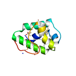
3GSH
DownloadVisualize
BU of 3gsh by Molmil
Three-dimensional structure of a post translational modified barley LTP1
Descriptor: (12E)-10-oxooctadec-12-enoic acid, Non-specific lipid-transfer protein 1, SODIUM ION, ...
Authors:Lascombe, M.B, Prange, T, Bakan, B, Marion, D.
Deposit date:2009-03-27
Release date:2009-12-15
Last modified:2017-11-01
Method:X-RAY DIFFRACTION (1.8 Å)
Cite:The crystal structure of oxylipin-conjugated barley LTP1 highlights the unique plasticity of the hydrophobic cavity of these plant lipid-binding proteins.
Biochem.Biophys.Res.Commun., 390, 2009
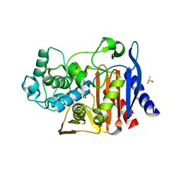
3GQZ
DownloadVisualize
BU of 3gqz by Molmil
AmpC beta-lactamase in complex with Fragment-based Inhibitor
Descriptor: (3S)-1-(4-acetylphenyl)-5-oxopyrrolidine-3-carboxylic acid, Beta-lactamase, DIMETHYL SULFOXIDE
Authors:Teotico, D.T, Shoichet, B.K.
Deposit date:2009-03-24
Release date:2009-04-14
Last modified:2023-09-06
Method:X-RAY DIFFRACTION (1.8 Å)
Cite:Docking for fragment inhibitors of AmpC beta-lactamase
Proc.Natl.Acad.Sci.USA, 106, 2009
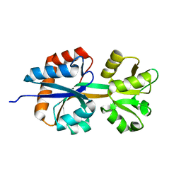
3H7M
DownloadVisualize
BU of 3h7m by Molmil
Crystal Structure of a Histidine Kinase Sensor Domain with Similarity to Periplasmic Binding Proteins
Descriptor: SODIUM ION, Sensor protein
Authors:Cheung, J, Le-Khac, M, Hendrickson, W.A.
Deposit date:2009-04-27
Release date:2009-06-30
Last modified:2024-02-21
Method:X-RAY DIFFRACTION (2.4 Å)
Cite:Crystal structure of a histidine kinase sensor domain with similarity to periplasmic binding proteins.
Proteins, 77, 2009
2QWZ
DownloadVisualize
BU of 2qwz by Molmil
CRYSTAL STRUCTURE OF A PUTATIVE THIOESTERASE (TM1040_1390) FROM SILICIBACTER SP. TM1040 AT 2.15 A RESOLUTION
Descriptor: ACETATE ION, GLYCEROL, Phenylacetic acid degradation-related protein
Authors:Joint Center for Structural Genomics (JCSG)
Deposit date:2007-08-10
Release date:2007-08-21
Last modified:2023-01-25
Method:X-RAY DIFFRACTION (2.15 Å)
Cite:Crystal structure of putative thioesterase (YP_613385.1) from Silicibacter sp. TM1040 at 2.15 A resolution
To be published
3GO4
DownloadVisualize
BU of 3go4 by Molmil
Crystal structure of a duf574 family protein (sav_2177) from streptomyces avermitilis ma-4680 at 1.80 A resolution
Descriptor: 1,2-ETHANEDIOL, Protein of unknown function DUF574, S-ADENOSYL-L-HOMOCYSTEINE, ...
Authors:Joint Center for Structural Genomics (JCSG)
Deposit date:2009-03-18
Release date:2009-04-14
Last modified:2023-11-22
Method:X-RAY DIFFRACTION (1.8 Å)
Cite:Crystal structure of Rossmann-fold protein of unknown function (DUF574) (NP_823353.1) from Streptomyces avermitilis MA-4680 at 1.80 A resolution
To be published
2R01
DownloadVisualize
BU of 2r01 by Molmil
Crystal structure of a putative fmn-dependent nitroreductase (ct0345) from chlorobium tepidum tls at 1.15 A resolution
Descriptor: 1,2-ETHANEDIOL, CALCIUM ION, FLAVIN MONONUCLEOTIDE, ...
Authors:Joint Center for Structural Genomics (JCSG)
Deposit date:2007-08-17
Release date:2007-09-04
Last modified:2023-01-25
Method:X-RAY DIFFRACTION (1.15 Å)
Cite:Crystal structure of putative FMN-dependent nitroreductase (NP_661249.1) from Chlorobium tepidum TLS at 1.15 A resolution
To be published
2QIW
DownloadVisualize
BU of 2qiw by Molmil
Crystal structure of a putative phosphoenolpyruvate phosphonomutase (ncgl1015, cgl1060) from corynebacterium glutamicum atcc 13032 at 1.80 A resolution
Descriptor: CHLORIDE ION, GLYCEROL, HEXAETHYLENE GLYCOL, ...
Authors:Joint Center for Structural Genomics (JCSG)
Deposit date:2007-07-05
Release date:2007-07-17
Last modified:2023-01-25
Method:X-RAY DIFFRACTION (1.8 Å)
Cite:Crystal structure of putative PEP phosphonomutase (NP_600288.1) from Corynebacterium glutamicum ATCC 13032 Kitasato at 1.80 A resolution
To be published

222415

PDB entries from 2024-07-10

PDB statisticsPDBj update infoContact PDBjnumon