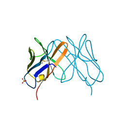
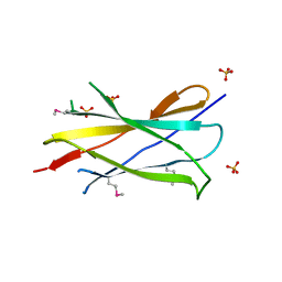
1BAF
 
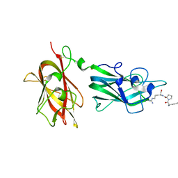
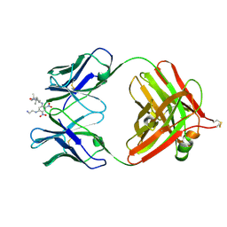 | | 2.9 ANGSTROMS RESOLUTION STRUCTURE OF AN ANTI-DINITROPHENYL-SPIN-LABEL MONOCLONAL ANTIBODY FAB FRAGMENT WITH BOUND HAPTEN | | 分子名称: | IGG1-KAPPA AN02 FAB (HEAVY CHAIN), IGG1-KAPPA AN02 FAB (LIGHT CHAIN), N-(2-AMINO-ETHYL)-4,6-DINITRO-N'-(2,2,6,6-TETRAMETHYL-1-OXY-PIPERIDIN-4-YL)-BENZENE-1,3-DIAMINE | | 著者 | Leahy, D.J, Brunger, A.T, Fox, R.O, Hynes, T.R. | | 登録日 | 1992-01-16 | | 公開日 | 1994-01-31 | | 最終更新日 | 2024-06-05 | | 実験手法 | X-RAY DIFFRACTION (2.9 Å) | | 主引用文献 | 2.9 A resolution structure of an anti-dinitrophenyl-spin-label monoclonal antibody Fab fragment with bound hapten.
J.Mol.Biol., 221, 1991
|
|
1FNF
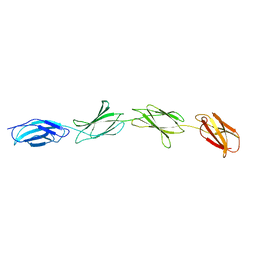 | |
1TEN
 | |
1M6B
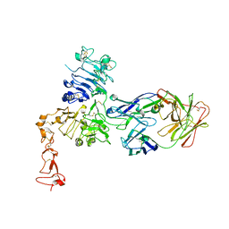
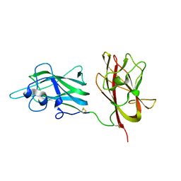
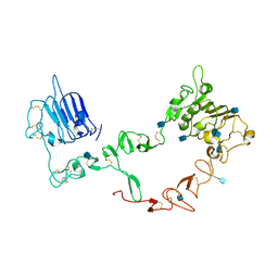 | | Structure of the HER3 (ERBB3) Extracellular Domain | | 分子名称: | 2-acetamido-2-deoxy-beta-D-glucopyranose, 2-acetamido-2-deoxy-beta-D-glucopyranose-(1-4)-2-acetamido-2-deoxy-beta-D-glucopyranose, Receptor protein-tyrosine kinase erbB-3, ... | | 著者 | Leahy, D.J, Cho, H.-S. | | 登録日 | 2002-07-15 | | 公開日 | 2002-08-02 | | 最終更新日 | 2021-10-27 | | 実験手法 | X-RAY DIFFRACTION (2.6 Å) | | 主引用文献 | Structure of the extracellular region of HER3 reveals an interdomain tether.
Science, 297, 2002
|
|
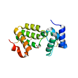
1LFA
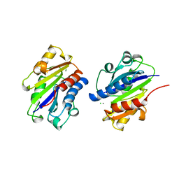 | | CD11A I-DOMAIN WITH BOUND MN++ | | 分子名称: | CD11A, CHLORIDE ION, MANGANESE (II) ION | | 著者 | Leahy, D.J, Qu, A. | | 登録日 | 1995-09-08 | | 公開日 | 1996-01-29 | | 最終更新日 | 2024-02-14 | | 実験手法 | X-RAY DIFFRACTION (1.8 Å) | | 主引用文献 | Crystal structure of the I-domain from the CD11a/CD18 (LFA-1, alpha L beta 2) integrin.
Proc.Natl.Acad.Sci.USA, 92, 1995
|
|
1CD8
 | |
1ZOP
 | | CD11A I-DOMAIN WITH BOUND MAGNESIUM ION | | 分子名称: | CHLORIDE ION, I-DOMAIN FRAGMENT OF LFA-1, MANGANESE (II) ION | | 著者 | Leahy, D.J, Qu, A. | | 登録日 | 1996-06-21 | | 公開日 | 1996-12-07 | | 最終更新日 | 2024-05-22 | | 実験手法 | X-RAY DIFFRACTION (2 Å) | | 主引用文献 | The role of the divalent cation in the structure of the I domain from the CD11a/CD18 integrin.
Structure, 4, 1996
|
|
1ZOO
 | | CD11A I-DOMAIN WITH BOUND MAGNESIUM ION | | 分子名称: | CHLORIDE ION, LEUKOCYTE ADHESION GLYCOPROTEIN, MAGNESIUM ION | | 著者 | Leahy, D.J, Qu, A. | | 登録日 | 1996-06-21 | | 公開日 | 1996-12-07 | | 最終更新日 | 2024-05-22 | | 実験手法 | X-RAY DIFFRACTION (3 Å) | | 主引用文献 | The role of the divalent cation in the structure of the I domain from the CD11a/CD18 integrin.
Structure, 4, 1996
|
|
1ZON
 | | CD11A I-DOMAIN WITHOUT BOUND CATION | | 分子名称: | LEUKOCYTE ADHESION GLYCOPROTEIN | | 著者 | Leahy, D.J, Qu, A. | | 登録日 | 1996-06-20 | | 公開日 | 1996-12-07 | | 最終更新日 | 2024-02-14 | | 実験手法 | X-RAY DIFFRACTION (2 Å) | | 主引用文献 | The role of the divalent cation in the structure of the I domain from the CD11a/CD18 integrin.
Structure, 4, 1996
|
|
6JFV
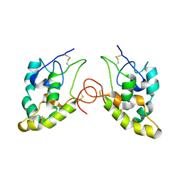
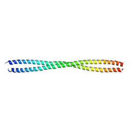 | | The crystal structure of 2B-2B complex from keratins 5 and 14 (C367A mutant of K14) | | 分子名称: | Keratin, type I cytoskeletal 14, type II cytoskeletal 5 | | 著者 | Kim, M.S, Lee, C.H, Coulombe, P.A, Leahy, D.J. | | 登録日 | 2019-02-12 | | 公開日 | 2020-01-22 | | 最終更新日 | 2024-05-29 | | 実験手法 | X-RAY DIFFRACTION (2.6 Å) | | 主引用文献 | Structure-Function Analyses of a Keratin Heterotypic Complex Identify Specific Keratin Regions Involved in Intermediate Filament Assembly.
Structure, 28, 2020
|
|
2IC2
 | |
1SZH
 | | Crystal Structure of C. elegans HER-1 | | 分子名称: | ACETATE ION, Her-1 protein | | 著者 | Hamaoka, B.Y, Dann III, C.E, Geisbrecht, B.V, Leahy, D.J. | | 登録日 | 2004-04-05 | | 公開日 | 2004-08-10 | | 最終更新日 | 2021-10-27 | | 実験手法 | X-RAY DIFFRACTION (1.5 Å) | | 主引用文献 | Crystal structure of Caenorhabditis elegans HER-1 and characterization of the interaction between HER-1 and TRA-2A.
Proc.Natl.Acad.Sci.USA, 101, 2004
|
|
1S78
 | | Insights into ErbB signaling from the structure of the ErbB2-pertuzumab complex | | 分子名称: | 2-acetamido-2-deoxy-beta-D-glucopyranose, 2-acetamido-2-deoxy-beta-D-glucopyranose-(1-4)-2-acetamido-2-deoxy-beta-D-glucopyranose, Pertuzumab Fab heavy chain, ... | | 著者 | Franklin, M.C, Carey, K.D, Vajdos, F.F, Leahy, D.J, de Vos, A.M, Sliwkowski, M.X. | | 登録日 | 2004-01-29 | | 公開日 | 2004-04-27 | | 最終更新日 | 2023-08-23 | | 実験手法 | X-RAY DIFFRACTION (3.25 Å) | | 主引用文献 | Insights into ErbB signaling from the structure of the ErbB2-pertuzumab complex.
Cancer Cell, 5, 2004
|
|
4X3X
 | |
4X3H
 | | CRYSTAL STRUCTURE OF ARC N-LOBE COMPLEXED WITH STARGAZIN PEPTIDE | | 分子名称: | Activity-regulated cytoskeleton-associated protein, VOLTAGE-DEPENDENT CALCIUM CHANNEL GAMMA-2 SUBUNIT | | 著者 | zhang, W, ward, m, leahy, d, worley, p. | | 登録日 | 2014-11-30 | | 公開日 | 2015-06-03 | | 最終更新日 | 2024-02-28 | | 実験手法 | X-RAY DIFFRACTION (2.401 Å) | | 主引用文献 | Structural basis of arc binding to synaptic proteins: implications for cognitive disease.
Neuron, 86, 2015
|
|
4X3I
 | | The crystal structure of Arc N-lobe complexed with CAMK2A fragment | | 分子名称: | Activity-regulated cytoskeleton-associated protein, CALCIUM/CALMODULIN-DEPENDENT PROTEIN KINASE TYPE II SUBUNIT ALPHA | | 著者 | Zhang, W, Ward, M, Leahy, D, Worley, P. | | 登録日 | 2014-11-30 | | 公開日 | 2015-06-03 | | 最終更新日 | 2017-09-27 | | 実験手法 | X-RAY DIFFRACTION (1.8 Å) | | 主引用文献 | Structural basis of arc binding to synaptic proteins: implications for cognitive disease.
Neuron, 86, 2015
|
|
3ODN
 | |
2OGB
 | |
1OT8
 | | Structure of the Ankyrin Domain of the Drosophila Notch Receptor | | 分子名称: | MAGNESIUM ION, Neurogenic locus Notch protein | | 著者 | Zweifel, M.E, Leahy, D.J, Hughson, F.M, Barrick, D. | | 登録日 | 2003-03-21 | | 公開日 | 2003-10-28 | | 最終更新日 | 2024-02-14 | | 実験手法 | X-RAY DIFFRACTION (2 Å) | | 主引用文献 | Structure and stability of the ankyrin domain of the Drosophila Notch receptor
Protein Sci., 12, 2003
|
|
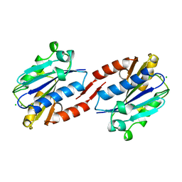
4PFJ
 | | The structure of bi-acetylated SAHH | | 分子名称: | ADENOSINE, Adenosylhomocysteinase, NICOTINAMIDE-ADENINE-DINUCLEOTIDE | | 著者 | Kavran, J.M, Wang, Y, Cole, P.A, Leahy, D.J. | | 登録日 | 2014-04-29 | | 公開日 | 2014-10-01 | | 最終更新日 | 2023-11-15 | | 実験手法 | X-RAY DIFFRACTION (2.3 Å) | | 主引用文献 | Regulation of s-adenosylhomocysteine hydrolase by lysine acetylation.
J.Biol.Chem., 289, 2014
|
|
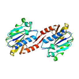
4PGF
 | | The structure of mono-acetylated SAHH | | 分子名称: | ADENOSINE, Adenosylhomocysteinase, NICOTINAMIDE-ADENINE-DINUCLEOTIDE | | 著者 | Kavran, J.M, Wang, Y, Cole, P.A, Leahy, D.J. | | 登録日 | 2014-05-01 | | 公開日 | 2014-10-01 | | 最終更新日 | 2023-11-15 | | 実験手法 | X-RAY DIFFRACTION (2.59 Å) | | 主引用文献 | Regulation of s-adenosylhomocysteine hydrolase by lysine acetylation.
J.Biol.Chem., 289, 2014
|
|
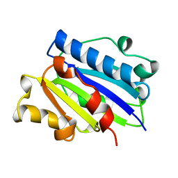
1VHH
 | | A POTENTIAL CATALYTIC SITE WITHIN THE AMINO-TERMINAL SIGNALLING DOMAIN OF SONIC HEDGEHOG | | 分子名称: | SONIC HEDGEHOG, SULFATE ION, ZINC ION | | 著者 | Hall, T.M.T, Porter, J.A, Beachy, P.A, Leahy, D.J. | | 登録日 | 1995-10-03 | | 公開日 | 1996-01-29 | | 最終更新日 | 2024-02-14 | | 実験手法 | X-RAY DIFFRACTION (1.7 Å) | | 主引用文献 | A potential catalytic site revealed by the 1.7-A crystal structure of the amino-terminal signalling domain of Sonic hedgehog.
Nature, 378, 1995
|
|
2ORZ
 | | Structural Basis for Ligand Binding and Heparin Mediated Activation of Neuropilin | | 分子名称: | Neuropilin-1, Tuftsin | | 著者 | Vander Kooi, C.W, Jusino, M.A, Perman, B, Neau, D.B, Bellamy, H.D, Leahy, D.J. | | 登録日 | 2007-02-05 | | 公開日 | 2007-04-03 | | 最終更新日 | 2023-08-30 | | 実験手法 | X-RAY DIFFRACTION (2.15 Å) | | 主引用文献 | Structural basis for ligand and heparin binding to neuropilin B domains.
Proc.Natl.Acad.Sci.Usa, 104, 2007
|
|
2ORX
 | | Structural Basis for Ligand Binding and Heparin Mediated Activation of Neuropilin | | 分子名称: | Neuropilin-1 | | 著者 | Vander Kooi, C.W, Jusino, M.A, Perman, B, Neau, D.B, Bellamy, H.D, Leahy, D.J. | | 登録日 | 2007-02-05 | | 公開日 | 2007-04-03 | | 最終更新日 | 2023-08-30 | | 実験手法 | X-RAY DIFFRACTION (2.4 Å) | | 主引用文献 | Structural basis for ligand and heparin binding to neuropilin B domains
Proc.Natl.Acad.Sci.Usa, 104, 2007
|
|
1IJY
 | | CRYSTAL STRUCTURE OF THE CYSTEINE-RICH DOMAIN OF MOUSE FRIZZLED 8 (MFZ8) | | 分子名称: | FRIZZLED HOMOLOG 8 | | 著者 | Dann III, C.E, Hsieh, J.C, Rattner, A, Sharma, D, Nathans, J, Leahy, D.J. | | 登録日 | 2001-05-01 | | 公開日 | 2001-07-11 | | 最終更新日 | 2023-08-16 | | 実験手法 | X-RAY DIFFRACTION (1.35 Å) | | 主引用文献 | Insights into Wnt binding and signalling from the structures of two Frizzled cysteine-rich domains.
Nature, 412, 2001
|
|


