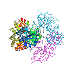
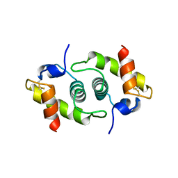
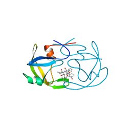
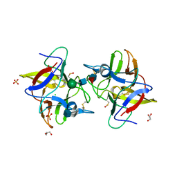
Loading
PDBj
MenuPDBj@FacebookPDBj@TwitterPDBj@YouTubewwPDB FoundationwwPDB
RCSB PDBPDBeBMRBAdv. SearchSearch help
PDB: 358 results

5C9D
DownloadVisualize
BU of 5c9d by Molmil
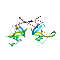
Crystal structure of a retropepsin-like aspartic protease from Rickettsia conorii
Descriptor: ApRick protease, SODIUM ION
Authors:Li, M, Gustchina, A, Cruz, R, Simoes, M, Curto, P, Martinez, J, Faro, C, Simoes, I, Wlodawer, A.
Deposit date:2015-06-26
Release date:2015-10-14
Last modified:2024-03-06
Method:X-RAY DIFFRACTION (2.59 Å)
Cite:Structure of RC1339/APRc from Rickettsia conorii, a retropepsin-like aspartic protease
Acta Crystallogr.,Sect.D, 71, 2015
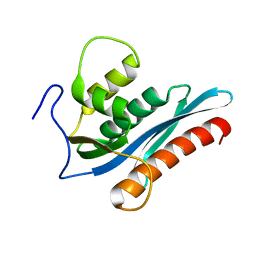
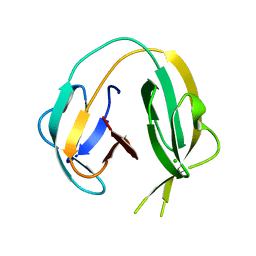
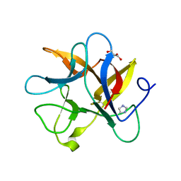
1NAG
DownloadVisualize
BU of 1nag by Molmil
CREVICE-FORMING MUTANTS IN THE RIGID CORE OF BOVINE PANCREATIC TRYPSIN INHIBITOR: CRYSTAL STRUCTURES OF F22A, Y23A, N43G, AND F45A
Descriptor: BOVINE PANCREATIC TRYPSIN INHIBITOR, PHOSPHATE ION
Authors:Danishefsky, A.T, Wlodawer, A, Kim, K.-S, Tao, F, Woodward, C.
Deposit date:1992-08-18
Release date:1993-10-31
Last modified:2019-08-14
Method:X-RAY DIFFRACTION (1.9 Å)
Cite:Crevice-forming mutants in the rigid core of bovine pancreatic trypsin inhibitor: crystal structures of F22A, Y23A, N43G, and F45A.
Protein Sci., 2, 1993
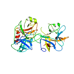
9ARQ
DownloadVisualize
BU of 9arq by Molmil
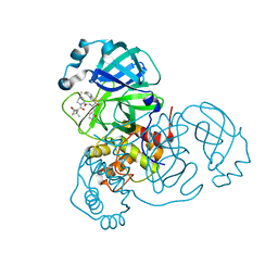
Crystal structure of SARS-CoV-2 main protease (authentic protein) in complex with an inhibitor TKB-245
Descriptor: (1R,2S,5S)-N-{(1S,2S)-1-(4-fluoro-1,3-benzothiazol-2-yl)-1-hydroxy-3-[(3S)-2-oxopyrrolidin-3-yl]propan-2-yl}-6,6-dimethyl-3-[3-methyl-N-(trifluoroacetyl)-L-valyl]-3-azabicyclo[3.1.0]hexane-2-carboxamide, 3C-like proteinase nsp5
Authors:Bulut, H, Hattori, S, Hayashi, H, Hasegawa, K, Li, M, Wlodawer, A, Tamamura, H, Mitsuya, H.
Deposit date:2024-02-23
Release date:2024-04-24
Method:X-RAY DIFFRACTION (2 Å)
Cite:Structural and virologic mechanism of emergence of main protease inhibitor-resistance in SARS-CoV-2 as selected with main protease inhibitors
To Be Published
9AVQ
DownloadVisualize
BU of 9avq by Molmil
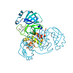
Crystal structure of SARS-CoV-2 main protease A191T mutant in complex with an inhibitor Nirmatrelvir
Descriptor: (1R,2S,5S)-N-{(1E,2S)-1-imino-3-[(3S)-2-oxopyrrolidin-3-yl]propan-2-yl}-6,6-dimethyl-3-[3-methyl-N-(trifluoroacetyl)-L-valyl]-3-azabicyclo[3.1.0]hexane-2-carboxamide, 3C-like proteinase nsp5, DI(HYDROXYETHYL)ETHER
Authors:Bulut, H, Hattori, S, Hayashi, H, Hasegawa, K, Li, M, Wlodawer, A, Tamamura, H, Mitsuya, H.
Deposit date:2024-03-04
Release date:2024-04-24
Method:X-RAY DIFFRACTION (2.58 Å)
Cite:Structural and virologic mechanism of emergence of main protease inhibitor-resistance in SARS-CoV-2 as selected with main protease inhibitors
To Be Published
6PAC
DownloadVisualize
BU of 6pac by Molmil
E. coli L-asparaginase II in complex with L-Asp at pH 5.6
Descriptor: ASPARTIC ACID, IMIDAZOLE, L-asparaginase 2
Authors:Lubkowski, J, Wlodawer, A.
Deposit date:2019-06-11
Release date:2019-09-04
Last modified:2019-10-02
Method:X-RAY DIFFRACTION (1.6 Å)
Cite:Geometric considerations support the double-displacement catalytic mechanism of l-asparaginase.
Protein Sci., 28, 2019
6PAA
DownloadVisualize
BU of 6paa by Molmil
E. coli L-asparaginase II mutant (T12V) in complex with L-Asp at pH 5.5
Descriptor: ASPARTIC ACID, L-asparaginase 2
Authors:Lubkowski, J, Wlodawer, A.
Deposit date:2019-06-11
Release date:2019-09-04
Last modified:2019-10-02
Method:X-RAY DIFFRACTION (1.85 Å)
Cite:Geometric considerations support the double-displacement catalytic mechanism of l-asparaginase.
Protein Sci., 28, 2019
6PA6
DownloadVisualize
BU of 6pa6 by Molmil
ECAII(T89V,K162T) MUTANT IN COMPLEX WITH L-ASN AT PH 8.3 in space group C2
Descriptor: ASPARAGINE, IMIDAZOLE, L-asparaginase 2
Authors:Lubkowski, J, Wlodawer, A.
Deposit date:2019-06-11
Release date:2019-09-04
Last modified:2019-10-02
Method:X-RAY DIFFRACTION (2.12 Å)
Cite:Geometric considerations support the double-displacement catalytic mechanism of l-asparaginase.
Protein Sci., 28, 2019
6PA9
DownloadVisualize
BU of 6pa9 by Molmil
E. coli L-asparaginase II mutant (T12V) in complex with L-Asn at pH 7.0
Descriptor: ASPARAGINE, GLYCEROL, L-asparaginase 2
Authors:Lubkowski, J, Wlodawer, A.
Deposit date:2019-06-11
Release date:2019-09-04
Last modified:2019-10-02
Method:X-RAY DIFFRACTION (1.88 Å)
Cite:Geometric considerations support the double-displacement catalytic mechanism of l-asparaginase.
Protein Sci., 28, 2019
6PA2
DownloadVisualize
BU of 6pa2 by Molmil
E. coli L-asparaginase II mutant (K162M) in complex with L-Asp at pH 5.6
Descriptor: ASPARTIC ACID, GLYCEROL, L-asparaginase 2
Authors:Lubkowski, J, Wlodawer, A.
Deposit date:2019-06-11
Release date:2019-09-04
Last modified:2019-10-02
Method:X-RAY DIFFRACTION (1.9 Å)
Cite:Geometric considerations support the double-displacement catalytic mechanism of l-asparaginase.
Protein Sci., 28, 2019
6PA4
DownloadVisualize
BU of 6pa4 by Molmil
E. coli L-asparaginase II double mutant (T89V,K162T) in complex with L-Asp at pH 7.0
Descriptor: ASPARTIC ACID, GLYCEROL, L-asparaginase 2
Authors:Lubkowski, J, Wlodawer, A.
Deposit date:2019-06-11
Release date:2019-09-04
Last modified:2020-08-19
Method:X-RAY DIFFRACTION (1.85 Å)
Cite:Geometric considerations support the double-displacement catalytic mechanism of l-asparaginase.
Protein Sci., 28, 2019
1BG8
DownloadVisualize
BU of 1bg8 by Molmil
HDEA FROM ESCHERICHIA COLI
Descriptor: HDEA
Authors:Yang, F, Gustafson, K.R, Boyd, M.R, Wlodawer, A.
Deposit date:1998-06-05
Release date:1998-09-16
Last modified:2011-07-13
Method:X-RAY DIFFRACTION (2.2 Å)
Cite:Crystal structure of Escherichia coli HdeA.
Nat.Struct.Biol., 5, 1998
3V1Q
DownloadVisualize
BU of 3v1q by Molmil
Crystal structures of the reverse transcriptase-associated ribonuclease H domain of xenotropic murine leukemia-virus related virus
Descriptor: Reverse transcriptase/ribonuclease H p80
Authors:Zhou, D, Wlodawer, A.
Deposit date:2011-12-09
Release date:2012-03-14
Last modified:2024-02-28
Method:X-RAY DIFFRACTION (2 Å)
Cite:Crystal structures of the reverse transcriptase-associated ribonuclease H domain of xenotropic murine leukemia-virus related virus.
J.Struct.Biol., 177, 2012
6RIX
DownloadVisualize
BU of 6rix by Molmil
Crystal structure of MchDnaB-1 intein
Descriptor: CHLORIDE ION, Replicative DNA helicase
Authors:Beyer, H.M, Lountos, G.T, Mikula, M.K, Wlodawer, A, Iwai, H.
Deposit date:2019-04-25
Release date:2020-05-13
Last modified:2024-01-24
Method:X-RAY DIFFRACTION (1.655 Å)
Cite:The Convergence of the Hedgehog/Intein Fold in Different Protein Splicing Mechanisms.
Int J Mol Sci, 21, 2020
2B7F
DownloadVisualize
BU of 2b7f by Molmil
Crystal structure of human T-cell leukemia virus protease, a novel target for anti-cancer design
Descriptor: (ACE)APQV(STA)VMHP peptide, HTLV protease, PHOSPHATE ION
Authors:Li, M, Laco, G.S, Jaskolski, M, Rozycki, J, Alexandratos, J, Wlodawer, A, Gustchina, A.
Deposit date:2005-10-04
Release date:2005-12-06
Last modified:2023-11-15
Method:X-RAY DIFFRACTION (2.6 Å)
Cite:Crystal structure of human T cell leukemia virus protease, a novel target for anticancer drug design
Proc.Natl.Acad.Sci.Usa, 102, 2005
2FMB
DownloadVisualize
BU of 2fmb by Molmil
EIAV PROTEASE COMPLEXED WITH AN INHIBITOR LP-130
Descriptor: 4-[2-(2-ACETYLAMINO-3-NAPHTALEN-1-YL-PROPIONYLAMINO)-4-METHYL-PENTANOYLAMINO]-3-HYDROXY-6-METHYL-HEPTANOIC ACID [1-(1-CARBAMOYL-2-NAPHTHALEN-1-YL-ETHYLCARBAMOYL)-PROPYL]-AMIDE, EQUINE INFECTIOUS ANEMIA VIRUS PROTEASE
Authors:Kervinen, J, Lubkowski, J, Zdanov, A, Wlodawer, A, Gustchina, A.
Deposit date:1998-07-13
Release date:1999-01-13
Last modified:2024-05-29
Method:X-RAY DIFFRACTION (1.8 Å)
Cite:Toward a universal inhibitor of retroviral proteases: comparative analysis of the interactions of LP-130 complexed with proteases from HIV-1, FIV, and EIAV.
Protein Sci., 7, 1998
6RIY
DownloadVisualize
BU of 6riy by Molmil
Crystal structure of MchDnaB-1 intein (N145AA)
Descriptor: CHLORIDE ION, Replicative DNA helicase
Authors:Beyer, H.M, Lountos, G.T, Mikula, M.K, Wlodawer, A, Iwai, H.
Deposit date:2019-04-25
Release date:2020-05-13
Last modified:2024-01-24
Method:X-RAY DIFFRACTION (1.63 Å)
Cite:The Convergence of the Hedgehog/Intein Fold in Different Protein Splicing Mechanisms.
Int J Mol Sci, 21, 2020
2ANE
DownloadVisualize
BU of 2ane by Molmil
Crystal structure of N-terminal domain of E.Coli Lon Protease
Descriptor: ATP-dependent protease La
Authors:Li, M, Rasulova, F, Melnikov, E.E, Rotanova, T.V, Gustchina, A, Maurizi, M.R, Wlodawer, A.
Deposit date:2005-08-11
Release date:2005-11-01
Last modified:2024-02-14
Method:X-RAY DIFFRACTION (2.03 Å)
Cite:Crystal structure of the N-terminal domain of E. coli Lon protease.
Protein Sci., 14, 2005
2ILK
DownloadVisualize
BU of 2ilk by Molmil
CRYSTAL STRUCTURE OF HUMAN INTERLEUKIN-10 AT 1.6 ANGSTROMS RESOLUTION
Descriptor: INTERLEUKIN-10, SULFATE ION
Authors:Zdanov, A, Schalk-Hihi, C, Wlodawer, A.
Deposit date:1996-07-01
Release date:1996-10-14
Last modified:2011-07-13
Method:X-RAY DIFFRACTION (1.6 Å)
Cite:Crystal structure of human interleukin-10 at 1.6 A resolution and a model of a complex with its soluble receptor.
Protein Sci., 5, 1996
4II0
DownloadVisualize
BU of 4ii0 by Molmil
Crystal structure of CrataBL, a trypsin inhibitor from Crataeva tapia
Descriptor: 2-acetamido-2-deoxy-beta-D-glucopyranose, CrataBL, GLYCEROL, ...
Authors:Zhou, D, Wlodawer, A.
Deposit date:2012-12-19
Release date:2013-07-24
Last modified:2020-07-29
Method:X-RAY DIFFRACTION (1.75 Å)
Cite:Crystal Structure of Crataeva tapia Bark Protein (CrataBL) and Its Effect in Human Prostate Cancer Cell Lines.
Plos One, 8, 2013
4IHZ
DownloadVisualize
BU of 4ihz by Molmil
Crystal structure of CrataBL, a trypsin inhibitor from Crataeva tapia
Descriptor: 2-acetamido-2-deoxy-beta-D-glucopyranose, CHLORIDE ION, CrataBL, ...
Authors:Zhou, D, Wlodawer, A.
Deposit date:2012-12-19
Release date:2013-07-24
Last modified:2020-07-29
Method:X-RAY DIFFRACTION (1.5 Å)
Cite:Crystal Structure of Crataeva tapia Bark Protein (CrataBL) and Its Effect in Human Prostate Cancer Cell Lines.
Plos One, 8, 2013
4J2K
DownloadVisualize
BU of 4j2k by Molmil
Crystal structure of a plant trypsin inhibitor EcTI
Descriptor: GLYCEROL, IMIDAZOLE, Trypsin inhibitor
Authors:Zhou, D, Wlodawer, A.
Deposit date:2013-02-04
Release date:2013-05-08
Last modified:2023-09-20
Method:X-RAY DIFFRACTION (1.75 Å)
Cite:Crystal Structures of a Plant Trypsin Inhibitor from Enterolobium contortisiliquum (EcTI) and of Its Complex with Bovine Trypsin.
Plos One, 8, 2013
4J2Y
DownloadVisualize
BU of 4j2y by Molmil
Crystal structure of a plant trypsin inhibitor EcTI in complex with bovine trypsin.
Descriptor: Cationic trypsin, SULFATE ION, Trypsin inhibitor
Authors:Zhou, D, Wlodawer, A.
Deposit date:2013-02-05
Release date:2013-05-08
Last modified:2023-09-20
Method:X-RAY DIFFRACTION (2 Å)
Cite:Crystal Structures of a Plant Trypsin Inhibitor from Enterolobium contortisiliquum (EcTI) and of Its Complex with Bovine Trypsin.
Plos One, 8, 2013
6V25
DownloadVisualize
BU of 6v25 by Molmil
Complex of mutant (K162M) of E. coli L-asparaginase II with L-Asp
Descriptor: ASPARTIC ACID, L-asparaginase 2
Authors:Lubkowski, J, Wlodawer, A.
Deposit date:2019-11-22
Release date:2020-05-20
Last modified:2023-10-11
Method:X-RAY DIFFRACTION (1.78 Å)
Cite:Mechanism of Catalysis by l-Asparaginase.
Biochemistry, 59, 2020
6V24
DownloadVisualize
BU of 6v24 by Molmil
Complex of mutant (K162M) of E. coli L-asparaginase II with L-Asp. Covalent acyl-enzyme intermediate.
Descriptor: ASPARTIC ACID, L-asparaginase 2
Authors:Lubkowski, J, Wlodawer, A.
Deposit date:2019-11-22
Release date:2020-05-20
Last modified:2023-10-11
Method:X-RAY DIFFRACTION (1.9 Å)
Cite:Mechanism of Catalysis by l-Asparaginase.
Biochemistry, 59, 2020
6V2G
DownloadVisualize
BU of 6v2g by Molmil
Complex of mutant (K162M) of E. coli L-asparaginase II with L-Asp. Covalent acyl-enzyme intermediate and tetrahedral intermediate.
Descriptor: L-asparaginase 2
Authors:Lubkowski, J, Wlodawer, A.
Deposit date:2019-11-22
Release date:2020-05-20
Last modified:2023-10-11
Method:X-RAY DIFFRACTION (1.9 Å)
Cite:Mechanism of Catalysis by l-Asparaginase.
Biochemistry, 59, 2020

220472

PDB entries from 2024-05-29

PDB statisticsPDBj update infoContact PDBjnumon