Loading
PDBj
メニューPDBj@FacebookPDBj@TwitterPDBj@YouTubewwPDB FoundationwwPDB
RCSB PDBPDBeBMRBAdv. SearchSearch help
PDB: 190 件

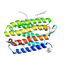
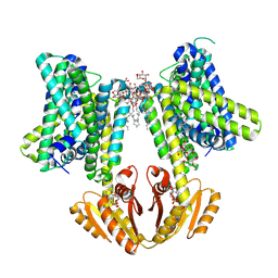
8B70
DownloadVisualize
BU of 8b70 by Molmil
KimA from B. subtilis with nucleotide second-messenger c-di-AMP bound
分子名称: (2R,3R,3aS,5R,7aR,9R,10R,10aS,12R,14aR)-2,9-bis(6-amino-9H-purin-9-yl)octahydro-2H,7H-difuro[3,2-d:3',2'-j][1,3,7,9,2,8 ]tetraoxadiphosphacyclododecine-3,5,10,12-tetrol 5,12-dioxide, DODECYL-BETA-D-MALTOSIDE, POTASSIUM ION, ...
著者Vonck, J, Wieferig, J.P.
登録日2022-09-28
公開日2023-01-18
最終更新日2024-07-24
実験手法ELECTRON MICROSCOPY (3.3 Å)
主引用文献Cyclic di-AMP traps proton-coupled K + transporters of the KUP family in an inward-occluded conformation.
Nat Commun, 14, 2023
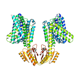
8B71
DownloadVisualize
BU of 8b71 by Molmil
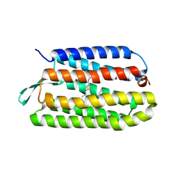
Upright KimA dimer with bound c-di-AMP from B. subtilis
分子名称: (2R,3R,3aS,5R,7aR,9R,10R,10aS,12R,14aR)-2,9-bis(6-amino-9H-purin-9-yl)octahydro-2H,7H-difuro[3,2-d:3',2'-j][1,3,7,9,2,8 ]tetraoxadiphosphacyclododecine-3,5,10,12-tetrol 5,12-dioxide, Potassium transporter KimA
著者Vonck, J, Wieferig, J.P.
登録日2022-09-28
公開日2023-01-18
最終更新日2024-07-24
実験手法ELECTRON MICROSCOPY (3.8 Å)
主引用文献Cyclic di-AMP traps proton-coupled K + transporters of the KUP family in an inward-occluded conformation.
Nat Commun, 14, 2023
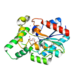
4GXN
DownloadVisualize
BU of 4gxn by Molmil
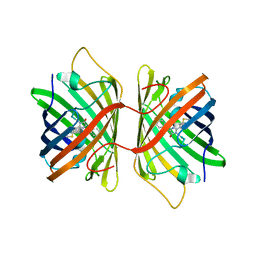
Diethylphosphonate Inhibited Structure of the Proteus mirabilis Lipase
分子名称: CALCIUM ION, DIETHYL PHOSPHONATE, Putative lipase, ...
著者Korman, T.P, Bowie, J.U.
登録日2012-09-04
公開日2013-02-06
最終更新日2024-10-16
実験手法X-RAY DIFFRACTION (2.2 Å)
主引用文献Crystal Structure of Proteus mirabilis Lipase, a Novel Lipase from the Proteus/Psychrophilic Subfamily of Lipase Family I.1.
Plos One, 7, 2012
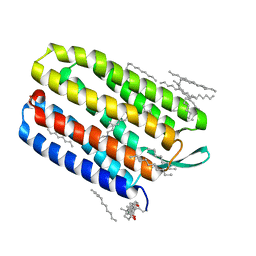
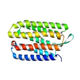
1XJI
DownloadVisualize
BU of 1xji by Molmil
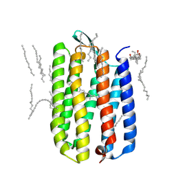
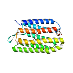
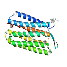
Bacteriorhodopsin crystallized in bicelles at room temperature
分子名称: 3-[(3-CHOLAMIDOPROPYL)DIMETHYLAMMONIO]-1-PROPANESULFONATE, Bacteriorhodopsin, DECANE, ...
著者Faham, S, Boulting, G.L, Massey, E.A, Yohannan, S, Yang, D, Bowie, J.U.
登録日2004-09-23
公開日2005-04-19
最終更新日2024-10-30
実験手法X-RAY DIFFRACTION (2.2 Å)
主引用文献Crystallization of bacteriorhodopsin from bicelle formulations at room temperature
Protein Sci., 14, 2005
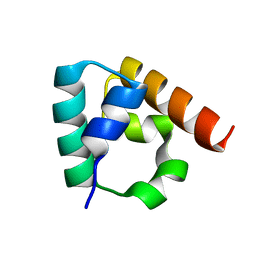
3IDW
DownloadVisualize
BU of 3idw by Molmil
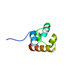
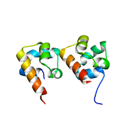
Crystal structure of Sla1 homology domain 2
分子名称: Actin cytoskeleton-regulatory complex protein SLA1
著者Di Pietro, S.M, Cascio, D, Bowie, J.U, Payne, G.S.
登録日2009-07-21
公開日2010-02-23
最終更新日2024-02-21
実験手法X-RAY DIFFRACTION (1.85 Å)
主引用文献Regulation of clathrin adaptor function in endocytosis: novel role for the SAM domain.
Embo J., 29, 2010
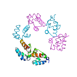
4NJ8
DownloadVisualize
BU of 4nj8 by Molmil
Crystal structure of the human ANKS3 SAM Domain L52A mutant
分子名称: Ankyrin repeat and SAM domain-containing protein 3
著者Leettola, C.N, Cascio, D, Bowie, J.U.
登録日2013-11-08
公開日2014-07-16
最終更新日2024-02-28
実験手法X-RAY DIFFRACTION (1.6 Å)
主引用文献Characterization of the SAM domain of the PKD-related protein ANKS6 and its interaction with ANKS3.
Bmc Struct.Biol., 14, 2014
3COD
DownloadVisualize
BU of 3cod by Molmil
Crystal Structure of T90A/D115A mutant of Bacteriorhodopsin
分子名称: Bacteriorhodopsin, RETINAL
著者Joh, N.H, Min, A, Faham, S, Bowie, J.U.
登録日2008-03-27
公開日2008-04-08
最終更新日2024-10-30
実験手法X-RAY DIFFRACTION (2.7 Å)
主引用文献Modest stabilization by most hydrogen-bonded side-chain interactions in membrane proteins.
Nature, 453, 2008
3HAP
DownloadVisualize
BU of 3hap by Molmil
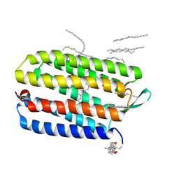
Crystal structure of bacteriorhodopsin mutant L111A crystallized from bicelles
分子名称: 3-[(3-CHOLAMIDOPROPYL)DIMETHYLAMMONIO]-1-PROPANESULFONATE, Bacteriorhodopsin, DECANE, ...
著者Joh, N.H, Yang, D, Bowie, J.U.
登録日2009-05-02
公開日2009-09-22
最終更新日2024-10-30
実験手法X-RAY DIFFRACTION (1.6 Å)
主引用文献Similar energetic contributions of packing in the core of membrane and water-soluble proteins.
J.Am.Chem.Soc., 131, 2009
3HAS
DownloadVisualize
BU of 3has by Molmil
Crystal structure of bacteriorhodopsin mutant L152A crystallized from bicelles
分子名称: 3-[(3-CHOLAMIDOPROPYL)DIMETHYLAMMONIO]-1-PROPANESULFONATE, Bacteriorhodopsin, DECANE, ...
著者Joh, N.H, Yang, D, Bowie, J.U.
登録日2009-05-02
公開日2009-09-22
最終更新日2021-10-13
実験手法X-RAY DIFFRACTION (1.9 Å)
主引用文献Similar energetic contributions of packing in the core of membrane and water-soluble proteins.
J.Am.Chem.Soc., 131, 2009
4NL9
DownloadVisualize
BU of 4nl9 by Molmil
Crystal structure of the human Anks3-SAM/Anks6-SAM heterodimer
分子名称: Ankyrin repeat and SAM domain-containing protein 3, Ankyrin repeat and SAM domain-containing protein 6, MAGNESIUM ION
著者Leettola, C.N, Cascio, D, Bowie, J.U.
登録日2013-11-13
公開日2014-07-16
最終更新日2023-09-20
実験手法X-RAY DIFFRACTION (1.5 Å)
主引用文献Characterization of the SAM domain of the PKD-related protein ANKS6 and its interaction with ANKS3.
Bmc Struct.Biol., 14, 2014
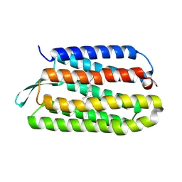
3QH9
DownloadVisualize
BU of 3qh9 by Molmil
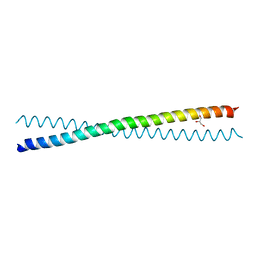
Human Liprin-beta2 Coiled-Coil
分子名称: AMMONIUM ION, GLYCEROL, IODIDE ION, ...
著者Stafford, R.L, Tang, M, Phillips, M.L, Bowie, J.U.
登録日2011-01-25
公開日2011-10-26
最終更新日2024-02-21
実験手法X-RAY DIFFRACTION (2.01 Å)
主引用文献Crystal structure of the central coiled-coil domain from human liprin-beta2
Biochemistry, 50, 2011
3COC
DownloadVisualize
BU of 3coc by Molmil
Crystal Structure of D115A mutant of Bacteriorhodopsin
分子名称: Bacteriorhodopsin, RETINAL
著者Joh, N.H, Faham, S, Bowie, J.U.
登録日2008-03-27
公開日2008-04-08
最終更新日2024-10-30
実験手法X-RAY DIFFRACTION (2.31 Å)
主引用文献Modest stabilization by most hydrogen-bonded side-chain interactions in membrane proteins.
Nature, 453, 2008
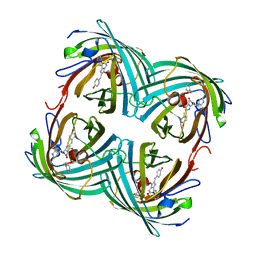
3E5V
DownloadVisualize
BU of 3e5v by Molmil
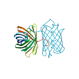
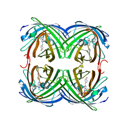
Crystal Structure Analysis of eqFP611 Double Mutant T122R, N143S
分子名称: Red fluorescent protein eqFP611
著者Nar, H, Nienhaus, K, Nienhaus, U, Wiedenmann, J.
登録日2008-08-14
公開日2008-09-23
最終更新日2023-11-15
実験手法X-RAY DIFFRACTION (2.1 Å)
主引用文献Trans-cis isomerization is responsible for the red-shifted fluorescence in variants of the red fluorescent protein eqFP611.
J.Am.Chem.Soc., 130, 2008
3HAQ
DownloadVisualize
BU of 3haq by Molmil
Crystal structure of bacteriorhodopsin mutant I148A crystallized from bicelles
分子名称: 3-[(3-CHOLAMIDOPROPYL)DIMETHYLAMMONIO]-1-PROPANESULFONATE, Bacteriorhodopsin, DECANE, ...
著者Joh, N.H, Yang, D, Bowie, J.U.
登録日2009-05-02
公開日2009-09-22
最終更新日2024-10-30
実験手法X-RAY DIFFRACTION (2.3 Å)
主引用文献Similar energetic contributions of packing in the core of membrane and water-soluble proteins.
J.Am.Chem.Soc., 131, 2009
3E5T
DownloadVisualize
BU of 3e5t by Molmil
Crystal Structure Analysis of FP611
分子名称: Red fluorescent protein eqFP611
著者Nar, H, Nienhaus, K, Nienhaus, U, Wiedenmann, J.
登録日2008-08-14
公開日2008-09-23
最終更新日2023-11-15
実験手法X-RAY DIFFRACTION (1.1 Å)
主引用文献Trans-cis isomerization is responsible for the red-shifted fluorescence in variants of the red fluorescent protein eqFP611.
J.Am.Chem.Soc., 130, 2008
3HAO
DownloadVisualize
BU of 3hao by Molmil
Crystal structure of bacteriorhodopsin mutant L94A crystallized from bicelles
分子名称: Bacteriorhodopsin, RETINAL
著者Joh, N.H, Yang, D, Bowie, J.U.
登録日2009-05-02
公開日2009-09-22
最終更新日2024-10-30
実験手法X-RAY DIFFRACTION (2.49 Å)
主引用文献Similar energetic contributions of packing in the core of membrane and water-soluble proteins.
J.Am.Chem.Soc., 131, 2009
3HAN
DownloadVisualize
BU of 3han by Molmil
Crystal structure of bacteriorhodopsin mutant V49A crystallized from bicelles
分子名称: Bacteriorhodopsin, RETINAL
著者Joh, N.H, Yang, D, Bowie, J.U.
登録日2009-05-02
公開日2009-09-22
最終更新日2024-11-06
実験手法X-RAY DIFFRACTION (2.75 Å)
主引用文献Similar energetic contributions of packing in the core of membrane and water-soluble proteins.
J.Am.Chem.Soc., 131, 2009
3E5W
DownloadVisualize
BU of 3e5w by Molmil
Crystal Structure Analysis of FP611
分子名称: Red fluorescent protein eqFP611
著者Nienhaus, K, Nar, H, Heilker, R, Wiedenmann, J, Nienhaus, G.U.
登録日2008-08-14
公開日2008-09-23
最終更新日2023-11-15
実験手法X-RAY DIFFRACTION (1.71 Å)
主引用文献Trans-cis isomerization is responsible for the red-shifted fluorescence in variants of the red fluorescent protein eqFP611.
J.Am.Chem.Soc., 130, 2008
3HAR
DownloadVisualize
BU of 3har by Molmil
Crystal structure of bacteriorhodopsin mutant I148V crystallized from bicelles
分子名称: 3-[(3-CHOLAMIDOPROPYL)DIMETHYLAMMONIO]-1-PROPANESULFONATE, Bacteriorhodopsin, RETINAL
著者Joh, N.H, Yang, D, Bowie, J.U.
登録日2009-05-02
公開日2009-09-22
最終更新日2021-10-13
実験手法X-RAY DIFFRACTION (1.7 Å)
主引用文献Similar energetic contributions of packing in the core of membrane and water-soluble proteins.
J.Am.Chem.Soc., 131, 2009
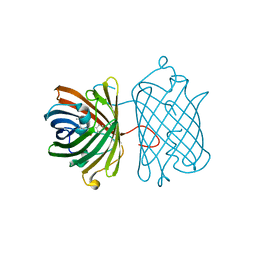
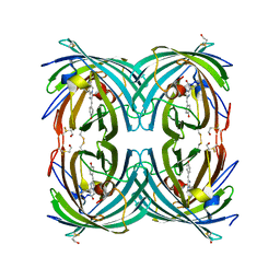
1ZUX
DownloadVisualize
BU of 1zux by Molmil
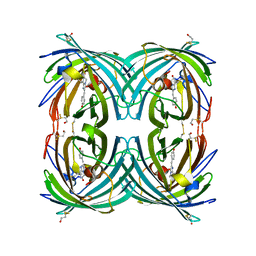
EosFP Fluorescent Protein- Green Form
分子名称: green to red photoconvertible GPF-like protein EosFP
著者Nar, H, Nienhaus, K, Wiedenmann, J, Nienhaus, G.U.
登録日2005-06-01
公開日2005-06-28
最終更新日2024-11-06
実験手法X-RAY DIFFRACTION (1.85 Å)
主引用文献Structural basis for photo-induced protein cleavage and green-to-red conversion of fluorescent protein EosFP.
Proc.Natl.Acad.Sci.Usa, 102, 2005
3CFF
DownloadVisualize
BU of 3cff by Molmil
Photoswitchable red fluorescent protein psRFP, on-state
分子名称: GFP-like photoswitchable fluorescent protein
著者Kachalova, G.S, Gundel, S, Bartunik, H.D, Wiedenmann, J.
登録日2008-03-03
公開日2009-03-10
最終更新日2023-11-15
実験手法X-RAY DIFFRACTION (1.8 Å)
主引用文献Structure of Anemonia sulcata red fluorescent protein asRFP
To be Published
3BQ7
DownloadVisualize
BU of 3bq7 by Molmil
SAM domain of Diacylglycerol Kinase delta1 (E35G)
分子名称: Diacylglycerol kinase delta
著者Knight, M.J, Bowie, J.U, Sawaya, M.R.
登録日2007-12-19
公開日2008-03-25
最終更新日2023-08-30
実験手法X-RAY DIFFRACTION (2.9 Å)
主引用文献Regulation of Enzyme Localization by Polymerization: Polymer Formation by the SAM Domain of Diacylglycerol Kinase delta1
Structure, 16, 2008
2BTJ
DownloadVisualize
BU of 2btj by Molmil
Fluorescent Protein EosFP - red form
分子名称: Green to red photoconvertible GFP-like protein EosFP
著者Nar, H, Nienhaus, K, Wiedenmann, J, Nienhaus, G.U.
登録日2005-06-01
公開日2005-06-23
最終更新日2023-12-13
実験手法X-RAY DIFFRACTION (2 Å)
主引用文献Structural Basis for Photo-Induced Protein Cleavage and Green-to-Red Conversion of Fluorescent Protein Eosfp.
Proc.Natl.Acad.Sci.USA, 102, 2005
3CFA
DownloadVisualize
BU of 3cfa by Molmil
Anemonia sulcata red fluorescent protein asRFP
分子名称: GFP-like fluorescent protein
著者Kachalova, G.S, Gundel, S, Wiedenmann, J, Bartunik, H.D.
登録日2008-03-03
公開日2009-03-10
最終更新日2023-11-15
実験手法X-RAY DIFFRACTION (1.75 Å)
主引用文献Structure of Anemonia sulcata red fluorescent protein asRFP
To be Published
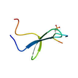
1I8G
DownloadVisualize
BU of 1i8g by Molmil
SOLUTION STRUCTURE OF PIN1 WW DOMAIN COMPLEXED WITH CDC25 PHOSPHOTHREONINE PEPTIDE
分子名称: M-PHASE INDUCER PHOSPHATASE 3, PEPTIDYL-PROLYL CIS-TRANS ISOMERASE NIMA-INTERACTING 1
著者Wintjens, R, Wieruszeski, J.-M, Drobecq, H, Lippens, G, Landrieu, I.
登録日2001-03-14
公開日2001-07-18
最終更新日2022-02-23
実験手法SOLUTION NMR
主引用文献1H NMR study on the binding of Pin1 Trp-Trp domain with phosphothreonine peptides.
J.Biol.Chem., 276, 2001

227111

件を2024-11-06に公開中

PDB statisticsPDBj update infoContact PDBjnumon