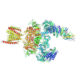
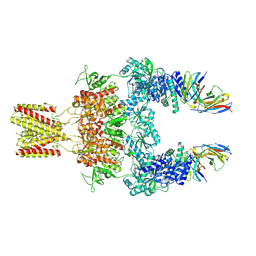
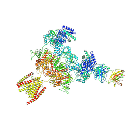
8TYU
 
 | |
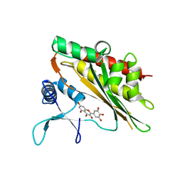
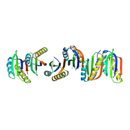
6M89
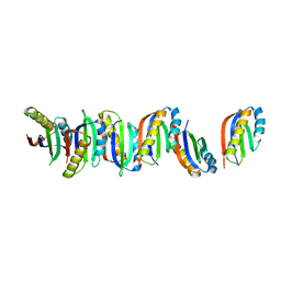
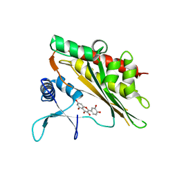 | | Crystal structure of the core catalytic domain of human inositol phosphate multikinase in complex with quercetin | | 分子名称: | 3,5,7,3',4'-PENTAHYDROXYFLAVONE, Inositol polyphosphate multikinase,Inositol polyphosphate multikinase | | 著者 | Wang, H, Shears, S.B. | | 登録日 | 2018-08-21 | | 公開日 | 2019-01-23 | | 最終更新日 | 2023-10-11 | | 実験手法 | X-RAY DIFFRACTION (1.85 Å) | | 主引用文献 | Inhibition of Inositol Polyphosphate Kinases by Quercetin and Related Flavonoids: A Structure-Activity Analysis.
J. Med. Chem., 62, 2019
|
|
6M8D
 | |
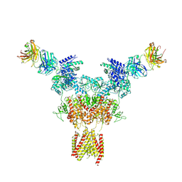
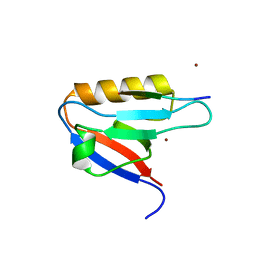
6LMJ
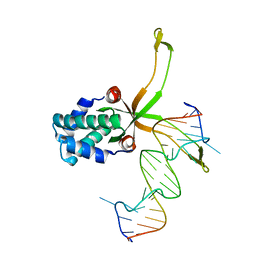 | | ASFV pA104R in complex with double-strand DNA | | 分子名称: | A104R, DNA (5'-D(*TP*GP*CP*TP*TP*AP*TP*CP*AP*AP*TP*TP*TP*GP*TP*TP*GP*CP*A)-3') | | 著者 | Wang, H, Qi, J, Chai, Y, Gao, F, Liu, R. | | 登録日 | 2019-12-25 | | 公開日 | 2020-05-13 | | 最終更新日 | 2023-11-22 | | 実験手法 | X-RAY DIFFRACTION (2.8 Å) | | 主引用文献 | The structural basis of African swine fever virus pA104R binding to DNA and its inhibition by stilbene derivatives.
Proc.Natl.Acad.Sci.USA, 117, 2020
|
|
6LMH
 | |
6M8B
 | |
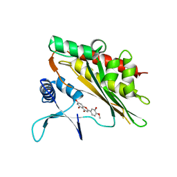
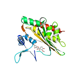
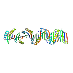
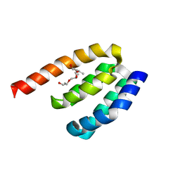
6M88
 | | Crystal structure of the core catalytic domain of human inositol phosphate multikinase in complex with myricetin | | 分子名称: | 3,5,7-TRIHYDROXY-2-(3,4,5-TRIHYDROXYPHENYL)-4H-CHROMEN-4-ONE, Inositol polyphosphate multikinase,Inositol polyphosphate multikinase | | 著者 | Wang, H, Shears, S.B. | | 登録日 | 2018-08-21 | | 公開日 | 2019-01-23 | | 最終更新日 | 2023-10-11 | | 実験手法 | X-RAY DIFFRACTION (1.9 Å) | | 主引用文献 | Inhibition of Inositol Polyphosphate Kinases by Quercetin and Related Flavonoids: A Structure-Activity Analysis.
J. Med. Chem., 62, 2019
|
|
6M8C
 | |
6M8A
 | |
6M8E
 | |
8JG7
 | | Serine decarboxylase | | 分子名称: | GLYCEROL, PYRIDOXAL-5'-PHOSPHATE, Serine decarboxylase, ... | | 著者 | Wang, H, Gong, W. | | 登録日 | 2023-05-19 | | 公開日 | 2024-05-22 | | 実験手法 | X-RAY DIFFRACTION (2.85 Å) | | 主引用文献 | Crystal structure of aserine decarboxylase from Arabidopsis thaliana
To Be Published
|
|
8JIZ
 | |
8JJ2
 | |
8JJ1
 | |
8JJ0
 | |
7WEG
 | | Complex structure of PDZD7 and FCHSD2 | | 分子名称: | FCHSD2, PDZ domain-containing protein 7, ZINC ION | | 著者 | Wang, H, Lin, L, Lu, Q. | | 登録日 | 2021-12-23 | | 公開日 | 2022-11-16 | | 最終更新日 | 2023-11-29 | | 実験手法 | X-RAY DIFFRACTION (2 Å) | | 主引用文献 | Deafness-related protein PDZD7 forms complex with the C-terminal tail of FCHSD2.
Biochem.J., 479, 2022
|
|
7B8V
 | |
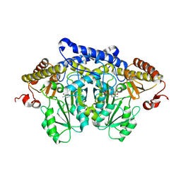
7B90
 | | Circular permutant of ribosomal protein S6, P54-55 truncated, I8A mutant | | 分子名称: | 30S ribosomal protein S6,30S ribosomal protein S6 | | 著者 | Wang, H, Logan, D.T, Oliveberg, M. | | 登録日 | 2020-12-13 | | 公開日 | 2022-06-08 | | 最終更新日 | 2024-01-31 | | 実験手法 | X-RAY DIFFRACTION (2.05 Å) | | 主引用文献 | Circular permutant of ribosomal protein S6, P54-55 truncate, I8A
To Be Published
|
|
7BFG
 | |
7BFC
 | |
7BFF
 | |
7BFE
 | |
7BFD
 | |
7DE7
 | | Crystal structure of PDZD7 HHD domain | | 分子名称: | (2R)-2-{[(2R)-2-{[(2S)-2-{[(2R)-2-hydroxypropyl]oxy}propyl]oxy}propyl]oxy}propan-1-ol, PDZ domain-containing protein 7 | | 著者 | Wang, H, Lin, L, Lu, Q. | | 登録日 | 2020-11-02 | | 公開日 | 2021-08-25 | | 最終更新日 | 2023-11-29 | | 実験手法 | X-RAY DIFFRACTION (1.49 Å) | | 主引用文献 | Structure and Membrane Targeting of the PDZD7 Harmonin Homology Domain (HHD) Associated With Hearing Loss.
Front Cell Dev Biol, 9, 2021
|
|
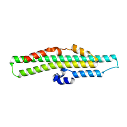
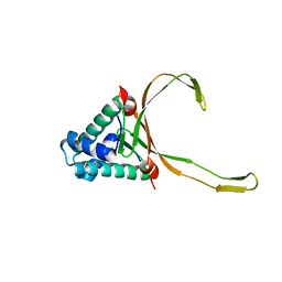
6ISA
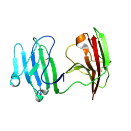 | | mCD226 | | 分子名称: | CD226 antigen | | 著者 | Wang, H, Qi, J, Zhang, S, Li, Y, Tan, S, Gao, G.F. | | 登録日 | 2018-11-16 | | 公開日 | 2018-12-26 | | 最終更新日 | 2023-11-22 | | 実験手法 | X-RAY DIFFRACTION (2 Å) | | 主引用文献 | Binding mode of the side-by-side two-IgV molecule CD226/DNAM-1 to its ligand CD155/Necl-5.
Proc. Natl. Acad. Sci. U.S.A., 116, 2019
|
|


