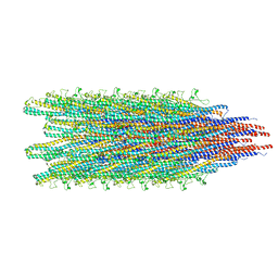
5WJU
 
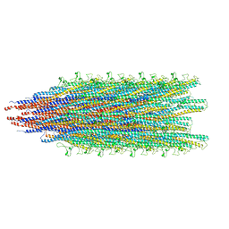
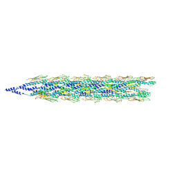
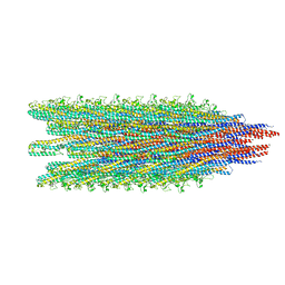 | | Cryo-EM structure of B. subtilis flagellar filaments A39V, N133H | | 分子名称: | Flagellin | | 著者 | Wang, F, Burrage, A.M, Kearns, D.B, Egelman, E.H. | | 登録日 | 2017-07-24 | | 公開日 | 2017-10-25 | | 最終更新日 | 2024-03-13 | | 実験手法 | ELECTRON MICROSCOPY (4.3 Å) | | 主引用文献 | A structural model of flagellar filament switching across multiple bacterial species.
Nat Commun, 8, 2017
|
|
5WJY
 | |
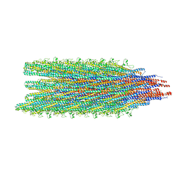
5WJZ
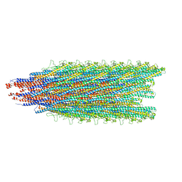
 | | Cryo-EM structure of B. subtilis flagellar filaments E115G | | 分子名称: | Flagellin | | 著者 | Wang, F, Burrage, A.M, Orlova, A, Kearns, D.B, Egelman, E.H. | | 登録日 | 2017-07-24 | | 公開日 | 2017-10-25 | | 最終更新日 | 2024-03-13 | | 実験手法 | ELECTRON MICROSCOPY (5.7 Å) | | 主引用文献 | A structural model of flagellar filament switching across multiple bacterial species.
Nat Commun, 8, 2017
|
|
5WK5
 | |
5WJX
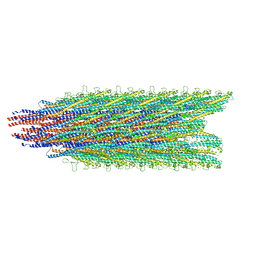
 | | Cryo-EM structure of B. subtilis flagellar filaments S17P | | 分子名称: | Flagellin | | 著者 | Wang, F, Burrage, A.M, Orlova, A, Kearns, D.B, Egelman, E.H. | | 登録日 | 2017-07-24 | | 公開日 | 2017-10-25 | | 最終更新日 | 2024-03-13 | | 実験手法 | ELECTRON MICROSCOPY (6.7 Å) | | 主引用文献 | A structural model of flagellar filament switching across multiple bacterial species.
Nat Commun, 8, 2017
|
|
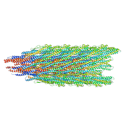
5WJW
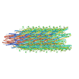
 | | Cryo-EM structure of B. subtilis flagellar filaments H84R | | 分子名称: | Flagellin | | 著者 | Wang, F, Burrage, A.M, Orlova, A, Kearns, D.B, Egelman, E.H. | | 登録日 | 2017-07-24 | | 公開日 | 2017-10-25 | | 最終更新日 | 2024-03-13 | | 実験手法 | ELECTRON MICROSCOPY (4.4 Å) | | 主引用文献 | A structural model of flagellar filament switching across multiple bacterial species.
Nat Commun, 8, 2017
|
|
5WJV
 | |
2NV6
 | |
5WK6
 | |
5WJT
 | |
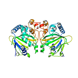
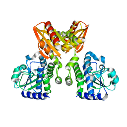
4HPV
 | | Crystal structure of S-Adenosylmethionine synthetase from Sulfolobus solfataricus | | 分子名称: | S-adenosylmethionine synthase | | 著者 | Wang, F, Hurley, K.A, Helmich, K.E, Singh, S, Bingman, C.A, Thorson, J.S, Phillips Jr, G.N, Enzyme Discovery for Natural Product Biosynthesis (NatPro) | | 登録日 | 2012-10-24 | | 公開日 | 2012-11-14 | | 最終更新日 | 2017-11-15 | | 実験手法 | X-RAY DIFFRACTION (2.214 Å) | | 主引用文献 | Understanding molecular recognition of promiscuity of thermophilic methionine adenosyltransferase sMAT from Sulfolobus solfataricus.
Febs J., 281, 2014
|
|
7DE0
 | |
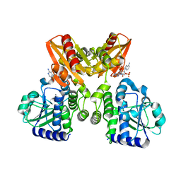
4PIW
 | | Crystal structure of sugar aminotransferase WecE from Escherichia coli K-12 | | 分子名称: | TDP-4-keto-6-deoxy-D-glucose transaminase family protein | | 著者 | Wang, F, Xu, W, Helmich, K.E, Singh, S, Yennamalli, R.M, Miller, M.D, Bingman, C.A, Thorson, J.S, Phillips Jr, G.N, Enzyme Discovery for Natural Product Biosynthesis (NatPro) | | 登録日 | 2014-05-09 | | 公開日 | 2014-07-16 | | 最終更新日 | 2023-11-15 | | 実験手法 | X-RAY DIFFRACTION (2.7 Å) | | 主引用文献 | Crystal structure of sugar aminotransferase WecE from Escherichia coli K-12
To Be Published
|
|
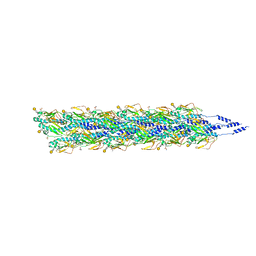
6EF8
 | | Cryo-EM of the OmcS nanowires from Geobacter sulfurreducens | | 分子名称: | C-type cytochrome OmcS, HEME C | | 著者 | Wang, F, Gu, Y, Egelman, E.H, Malvankar, N.S. | | 登録日 | 2018-08-16 | | 公開日 | 2019-04-10 | | 最終更新日 | 2019-11-27 | | 実験手法 | ELECTRON MICROSCOPY (3.7 Å) | | 主引用文献 | Structure of Microbial Nanowires Reveals Stacked Hemes that Transport Electrons over Micrometers.
Cell, 177, 2019
|
|
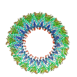
5VXX
 | | Cryo-EM reconstruction of Neisseria gonorrhoeae Type IV pilus | | 分子名称: | Fimbrial protein, PHOSPHORIC ACID MONO-(2-AMINO-ETHYL) ESTER, alpha-D-galactopyranose-(1-3)-2,4-bisacetamido-2,4,6-trideoxy-beta-D-glucopyranose | | 著者 | Wang, F, Orlova, A, Altindal, T, Craig, L, Egelman, E.H. | | 登録日 | 2017-05-24 | | 公開日 | 2017-07-12 | | 最終更新日 | 2020-07-29 | | 実験手法 | ELECTRON MICROSCOPY (5.1 Å) | | 主引用文献 | Cryoelectron Microscopy Reconstructions of the Pseudomonas aeruginosa and Neisseria gonorrhoeae Type IV Pili at Sub-nanometer Resolution.
Structure, 25, 2017
|
|
5VXY
 | | Cryo-EM reconstruction of PAK pilus from Pseudomonas aeruginosa | | 分子名称: | Fimbrial protein | | 著者 | Wang, F, Osinksi, T, Orlova, A, Altindal, T, Craig, L, Egelman, E.H. | | 登録日 | 2017-05-24 | | 公開日 | 2017-07-12 | | 最終更新日 | 2019-12-11 | | 実験手法 | ELECTRON MICROSCOPY (8 Å) | | 主引用文献 | Cryoelectron Microscopy Reconstructions of the Pseudomonas aeruginosa and Neisseria gonorrhoeae Type IV Pili at Sub-nanometer Resolution.
Structure, 25, 2017
|
|
5XBI
 | |
5XBT
 | | The structure of BrlR bound to c-di-GMP | | 分子名称: | 9,9'-[(2R,3R,3aS,5S,7aR,9R,10R,10aS,12S,14aR)-3,5,10,12-tetrahydroxy-5,12-dioxidooctahydro-2H,7H-difuro[3,2-d:3',2'-j][1,3,7,9,2,8]tetraoxadiphosphacyclododecine-2,9-diyl]bis(2-amino-1,9-dihydro-6H-purin-6-one), DI(HYDROXYETHYL)ETHER, GLYCEROL, ... | | 著者 | Wang, F, Qing, H, Gu, L. | | 登録日 | 2017-03-21 | | 公開日 | 2018-05-02 | | 最終更新日 | 2024-03-27 | | 実験手法 | X-RAY DIFFRACTION (2.495 Å) | | 主引用文献 | BrlR from Pseudomonas aeruginosa is a receptor for both cyclic di-GMP and pyocyanin.
Nat Commun, 9, 2018
|
|
5XSN
 | | The catalytic domain of GdpP with c-di-AMP | | 分子名称: | (2R,3R,3aS,5R,7aR,9R,10R,10aS,12R,14aR)-2,9-bis(6-amino-9H-purin-9-yl)octahydro-2H,7H-difuro[3,2-d:3',2'-j][1,3,7,9,2,8 ]tetraoxadiphosphacyclododecine-3,5,10,12-tetrol 5,12-dioxide, MANGANESE (II) ION, Phosphodiesterase acting on cyclic dinucleotides | | 著者 | Wang, F, Gu, L. | | 登録日 | 2017-06-14 | | 公開日 | 2018-01-31 | | 最終更新日 | 2023-11-22 | | 実験手法 | X-RAY DIFFRACTION (2.501 Å) | | 主引用文献 | Structural and biochemical characterization of the catalytic domains of GdpP reveals a unified hydrolysis mechanism for the DHH/DHHA1 phosphodiesterase
Biochem. J., 475, 2018
|
|
5XSI
 | | The catalytic domain of GdpP | | 分子名称: | MANGANESE (II) ION, Phosphodiesterase acting on cyclic dinucleotides | | 著者 | Wang, F, Gu, L. | | 登録日 | 2017-06-14 | | 公開日 | 2018-01-31 | | 最終更新日 | 2024-03-27 | | 実験手法 | X-RAY DIFFRACTION (2.2 Å) | | 主引用文献 | Structural and biochemical characterization of the catalytic domains of GdpP reveals a unified hydrolysis mechanism for the DHH/DHHA1 phosphodiesterase
Biochem. J., 475, 2018
|
|
5XSP
 | | The catalytic domain of GdpP with 5'-pApA | | 分子名称: | ADENOSINE MONOPHOSPHATE, MANGANESE (II) ION, Phosphodiesterase acting on cyclic dinucleotides | | 著者 | Wang, F, Gu, L. | | 登録日 | 2017-06-15 | | 公開日 | 2018-01-31 | | 最終更新日 | 2024-05-29 | | 実験手法 | X-RAY DIFFRACTION (2.146 Å) | | 主引用文献 | Structural and biochemical characterization of the catalytic domains of GdpP reveals a unified hydrolysis mechanism for the DHH/DHHA1 phosphodiesterase
Biochem. J., 475, 2018
|
|
5XT3
 | | The catalytic domain of GdpP with c-di-GMP | | 分子名称: | 9,9'-[(2R,3R,3aS,5S,7aR,9R,10R,10aS,12S,14aR)-3,5,10,12-tetrahydroxy-5,12-dioxidooctahydro-2H,7H-difuro[3,2-d:3',2'-j][1,3,7,9,2,8]tetraoxadiphosphacyclododecine-2,9-diyl]bis(2-amino-1,9-dihydro-6H-purin-6-one), MANGANESE (II) ION, Phosphodiesterase acting on cyclic dinucleotides | | 著者 | Wang, F, Gu, L. | | 登録日 | 2017-06-16 | | 公開日 | 2018-01-31 | | 最終更新日 | 2023-11-22 | | 実験手法 | X-RAY DIFFRACTION (2.591 Å) | | 主引用文献 | Structural and biochemical characterization of the catalytic domains of GdpP reveals a unified hydrolysis mechanism for the DHH/DHHA1 phosphodiesterase
Biochem. J., 475, 2018
|
|
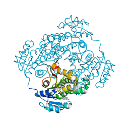
7ESF
 | | The Crystal Structure of human MTH1 from Biortus | | 分子名称: | 7,8-dihydro-8-oxoguanine triphosphatase, DI(HYDROXYETHYL)ETHER, TETRAETHYLENE GLYCOL | | 著者 | Wang, F, Cheng, W, Shang, H, Wang, R, Zhang, B, Tian, F. | | 登録日 | 2021-05-10 | | 公開日 | 2021-05-26 | | 最終更新日 | 2023-11-29 | | 実験手法 | X-RAY DIFFRACTION (1.55 Å) | | 主引用文献 | The Crystal Structure of human MTH1 from Biortus
To Be Published
|
|
6D5F
 | | Cryo-EM reconstruction of membrane-enveloped filamentous virus SFV1 (Sulfolobus filamentous virus 1) | | 分子名称: | DNA (336-MER), Fimbrial protein | | 著者 | Wang, F, Osinski, T, Liu, Y, Krupovic, M, Prangishvili, D, Egelman, E.H. | | 登録日 | 2018-04-19 | | 公開日 | 2018-08-29 | | 最終更新日 | 2020-01-08 | | 実験手法 | ELECTRON MICROSCOPY (3.7 Å) | | 主引用文献 | Structural conservation in a membrane-enveloped filamentous virus infecting a hyperthermophilic acidophile.
Nat Commun, 9, 2018
|
|
3FNH
 | | Crystal structure of InhA bound to triclosan derivative | | 分子名称: | 2-(2,4-DICHLOROPHENOXY)-5-(2-PHENYLETHYL)PHENOL, Enoyl-[acyl-carrier-protein] reductase [NADH], NICOTINAMIDE-ADENINE-DINUCLEOTIDE | | 著者 | Wang, F. | | 登録日 | 2008-12-24 | | 公開日 | 2009-01-20 | | 最終更新日 | 2023-09-06 | | 実験手法 | X-RAY DIFFRACTION (2.8 Å) | | 主引用文献 | Triclosan Derivatives: Towards Potent Inhibitors of Drug-Sensitive and Drug-Resistant Mycobacterium tuberculosis.
Chemmedchem, 4, 2009
|
|


