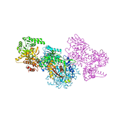
7O1J
 
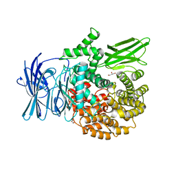
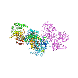 | | Structure of Mycobacterium tuberculosis beta-oxidation trifunctional enzyme beta-C92A mutant | | 分子名称: | 3-hydroxyacyl-CoA dehydrogenase, GLYCEROL, Putative acyltransferase Rv0859, ... | | 著者 | Dalwani, S, Wierenga, R.K, Venkatesan, R. | | 登録日 | 2021-03-29 | | 公開日 | 2021-08-25 | | 最終更新日 | 2024-01-31 | | 実験手法 | X-RAY DIFFRACTION (2.36 Å) | | 主引用文献 | Substrate specificity and conformational flexibility properties of the Mycobacterium tuberculosis beta-oxidation trifunctional enzyme.
J.Struct.Biol., 213, 2021
|
|
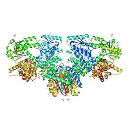
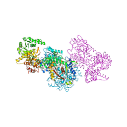
7O1I
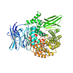
 | | Structure of Mycobacterium tuberculosis beta-oxidation trifunctional enzyme alpha-E141A mutant | | 分子名称: | 3-hydroxyacyl-CoA dehydrogenase, COENZYME A, GLYCEROL, ... | | 著者 | Dalwani, S, Wierenga, R.K, Venkatesan, R. | | 登録日 | 2021-03-29 | | 公開日 | 2021-08-25 | | 最終更新日 | 2024-01-31 | | 実験手法 | X-RAY DIFFRACTION (2.3 Å) | | 主引用文献 | Substrate specificity and conformational flexibility properties of the Mycobacterium tuberculosis beta-oxidation trifunctional enzyme.
J.Struct.Biol., 213, 2021
|
|
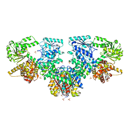
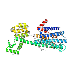
7O1K
 | | Structure of Mycobacterium tuberculosis beta-oxidation trifunctional enzyme alpha-E141A, beta-C92A mutant | | 分子名称: | 3-hydroxyacyl-CoA dehydrogenase, GLYCEROL, Putative acyltransferase Rv0859, ... | | 著者 | Dalwani, S, Wierenga, R.K, Venkatesan, R. | | 登録日 | 2021-03-29 | | 公開日 | 2021-08-25 | | 最終更新日 | 2024-01-31 | | 実験手法 | X-RAY DIFFRACTION (2.86 Å) | | 主引用文献 | Substrate specificity and conformational flexibility properties of the Mycobacterium tuberculosis beta-oxidation trifunctional enzyme.
J.Struct.Biol., 213, 2021
|
|
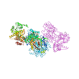
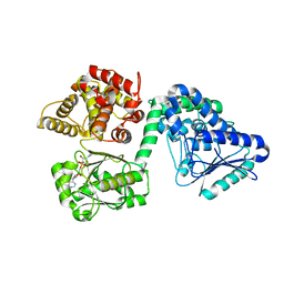
7O4V
 | | Structure of Mycobacterium tuberculosis beta-oxidation trifunctional enzyme in complex with oxidized nicotinamide adenine dinucleotide | | 分子名称: | 3-hydroxyacyl-CoA dehydrogenase, NICOTINAMIDE-ADENINE-DINUCLEOTIDE, Putative acyltransferase Rv0859, ... | | 著者 | Dalwani, S, Wierenga, R.K, Venkatesan, R. | | 登録日 | 2021-04-07 | | 公開日 | 2021-08-25 | | 最終更新日 | 2024-01-31 | | 実験手法 | X-RAY DIFFRACTION (2.42 Å) | | 主引用文献 | Substrate specificity and conformational flexibility properties of the Mycobacterium tuberculosis beta-oxidation trifunctional enzyme.
J.Struct.Biol., 213, 2021
|
|
7O1L
 | | Structure of Mycobacterium tuberculosis beta-oxidation trifunctional enzyme alpha-H462A mutant | | 分子名称: | 3-hydroxyacyl-CoA dehydrogenase, COENZYME A, GLYCEROL, ... | | 著者 | Dalwani, S, Wierenga, R.K, Venkatesan, R. | | 登録日 | 2021-03-29 | | 公開日 | 2021-08-25 | | 最終更新日 | 2024-01-31 | | 実験手法 | X-RAY DIFFRACTION (2.38 Å) | | 主引用文献 | Substrate specificity and conformational flexibility properties of the Mycobacterium tuberculosis beta-oxidation trifunctional enzyme.
J.Struct.Biol., 213, 2021
|
|
7O4Q
 | | Structure of Mycobacterium tuberculosis beta-oxidation trifunctional enzyme in space group C2221 (unliganded) | | 分子名称: | 3-hydroxyacyl-CoA dehydrogenase, GLYCEROL, Putative acyltransferase Rv0859, ... | | 著者 | Dalwani, S, Wierenga, R.K, Venkatesan, R. | | 登録日 | 2021-04-07 | | 公開日 | 2021-08-25 | | 最終更新日 | 2024-01-31 | | 実験手法 | X-RAY DIFFRACTION (2.1 Å) | | 主引用文献 | Substrate specificity and conformational flexibility properties of the Mycobacterium tuberculosis beta-oxidation trifunctional enzyme.
J.Struct.Biol., 213, 2021
|
|
7O4R
 | | Structure of Mycobacterium tuberculosis beta-oxidation trifunctional enzyme with Coenzyme A bound at the thiolase active sites and additional binding site (CoA(HAD/KAT)) | | 分子名称: | 3-hydroxyacyl-CoA dehydrogenase, COENZYME A, GLYCEROL, ... | | 著者 | Dalwani, S, Wierenga, R.K, Venkatesan, R. | | 登録日 | 2021-04-07 | | 公開日 | 2021-08-25 | | 最終更新日 | 2024-01-31 | | 実験手法 | X-RAY DIFFRACTION (2.79 Å) | | 主引用文献 | Substrate specificity and conformational flexibility properties of the Mycobacterium tuberculosis beta-oxidation trifunctional enzyme.
J.Struct.Biol., 213, 2021
|
|
7O4T
 | | Structure of Mycobacterium tuberculosis beta-oxidation trifunctional enzyme with Coenzyme A bound at the hydratase, thiolase active sites and possible additional binding site (CoA(ECH/HAD)) | | 分子名称: | 3'-PHOSPHATE-ADENOSINE-5'-DIPHOSPHATE, 3-hydroxyacyl-CoA dehydrogenase, COENZYME A, ... | | 著者 | Dalwani, S, Wierenga, R.K, Venkatesan, R. | | 登録日 | 2021-04-07 | | 公開日 | 2021-08-25 | | 最終更新日 | 2024-01-31 | | 実験手法 | X-RAY DIFFRACTION (2.1 Å) | | 主引用文献 | Substrate specificity and conformational flexibility properties of the Mycobacterium tuberculosis beta-oxidation trifunctional enzyme.
J.Struct.Biol., 213, 2021
|
|
7O4S
 | | Structure of Mycobacterium tuberculosis beta-oxidation trifunctional enzyme with Coenzyme A bound at the hydratase, thiolase active sites and additional binding site (CoA(ECH2)) | | 分子名称: | 3'-PHOSPHATE-ADENOSINE-5'-DIPHOSPHATE, 3-hydroxyacyl-CoA dehydrogenase, ADENOSINE-5'-DIPHOSPHATE, ... | | 著者 | Dalwani, S, Wierenga, R.K, Venkatesan, R. | | 登録日 | 2021-04-07 | | 公開日 | 2021-08-25 | | 最終更新日 | 2024-01-31 | | 実験手法 | X-RAY DIFFRACTION (2.79 Å) | | 主引用文献 | Substrate specificity and conformational flexibility properties of the Mycobacterium tuberculosis beta-oxidation trifunctional enzyme.
J.Struct.Biol., 213, 2021
|
|
7O1G
 | | Structure of Mycobacterium tuberculosis beta-oxidation trifunctional enzyme alpha-E141A-H462A, beta-C92A mutant | | 分子名称: | 3-hydroxyacyl-CoA dehydrogenase, Putative acyltransferase Rv0859, SULFATE ION | | 著者 | Dalwani, S, Wierenga, R.K, Venkatesan, R. | | 登録日 | 2021-03-29 | | 公開日 | 2021-08-25 | | 最終更新日 | 2024-01-31 | | 実験手法 | X-RAY DIFFRACTION (3.03 Å) | | 主引用文献 | Substrate specificity and conformational flexibility properties of the Mycobacterium tuberculosis beta-oxidation trifunctional enzyme.
J.Struct.Biol., 213, 2021
|
|
7O1M
 | | Structure of Mycobacterium tuberculosis beta-oxidation trifunctional enzyme alpha-H462A, beta-C92A mutant | | 分子名称: | 3-hydroxyacyl-CoA dehydrogenase, GLYCEROL, Putative acyltransferase Rv0859, ... | | 著者 | Dalwani, S, Wierenga, R.K, Venkatesan, R. | | 登録日 | 2021-03-29 | | 公開日 | 2021-08-25 | | 最終更新日 | 2024-01-31 | | 実験手法 | X-RAY DIFFRACTION (2.89 Å) | | 主引用文献 | Substrate specificity and conformational flexibility properties of the Mycobacterium tuberculosis beta-oxidation trifunctional enzyme.
J.Struct.Biol., 213, 2021
|
|
6CM4
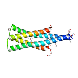
 | | Structure of the D2 Dopamine Receptor Bound to the Atypical Antipsychotic Drug Risperidone | | 分子名称: | 3-[2-[4-(6-fluoranyl-1,2-benzoxazol-3-yl)piperidin-1-yl]ethyl]-2-methyl-6,7,8,9-tetrahydropyrido[1,2-a]pyrimidin-4-one, D(2) dopamine receptor, endolysin chimera, ... | | 著者 | Wang, S, Che, T, Levit, A, Shoichet, B.K, Wacker, D, Roth, B.L. | | 登録日 | 2018-03-02 | | 公開日 | 2018-03-14 | | 最終更新日 | 2023-10-04 | | 実験手法 | X-RAY DIFFRACTION (2.867 Å) | | 主引用文献 | Structure of the D2 dopamine receptor bound to the atypical antipsychotic drug risperidone.
Nature, 555, 2018
|
|
7O4U
 | |
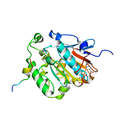
2I8E
 | | Structure of SSO1404, a predicted DNA repair-associated protein from Sulfolobus solfataricus P2 | | 分子名称: | Hypothetical protein, IODIDE ION | | 著者 | Wang, S, Zimmerman, M.D, Kudritska, M, Chruszcz, M, Savchenko, A, Edwards, A, Joachimiak, A, Minor, W, Midwest Center for Structural Genomics (MCSG) | | 登録日 | 2006-09-01 | | 公開日 | 2006-09-26 | | 最終更新日 | 2022-04-13 | | 実験手法 | X-RAY DIFFRACTION (1.59 Å) | | 主引用文献 | A novel family of sequence-specific endoribonucleases associated with the clustered regularly interspaced short palindromic repeats.
J.Biol.Chem., 283, 2008
|
|
3EBG
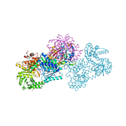
 | | Structure of the M1 Alanylaminopeptidase from malaria | | 分子名称: | GLYCEROL, M1 family aminopeptidase, MAGNESIUM ION, ... | | 著者 | McGowan, S, Porter, C.J, Buckle, A.M, Whisstock, J.C. | | 登録日 | 2008-08-27 | | 公開日 | 2009-01-27 | | 最終更新日 | 2024-02-21 | | 実験手法 | X-RAY DIFFRACTION (2.1 Å) | | 主引用文献 | Structural basis for the inhibition of the essential Plasmodium falciparum M1 neutral aminopeptidase
Proc.Natl.Acad.Sci.USA, 106, 2009
|
|
3EBH
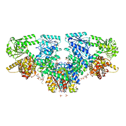
 | | Structure of the M1 Alanylaminopeptidase from malaria complexed with bestatin | | 分子名称: | 2-(3-AMINO-2-HYDROXY-4-PHENYL-BUTYRYLAMINO)-4-METHYL-PENTANOIC ACID, GLYCEROL, M1 family aminopeptidase, ... | | 著者 | McGowan, S, Porter, C.J, Buckle, A.M, Whisstock, J.C. | | 登録日 | 2008-08-27 | | 公開日 | 2009-01-27 | | 最終更新日 | 2024-02-21 | | 実験手法 | X-RAY DIFFRACTION (1.65 Å) | | 主引用文献 | Structural basis for the inhibition of the essential Plasmodium falciparum M1 neutral aminopeptidase
Proc.Natl.Acad.Sci.USA, 106, 2009
|
|
1JRK
 | | Crystal Structure of a Nudix Protein from Pyrobaculum aerophilum Reveals a Dimer with Intertwined Beta Sheets | | 分子名称: | (4S)-2-METHYL-2,4-PENTANEDIOL, Nudix homolog | | 著者 | Wang, S, Mura, C, Sawaya, M.R, Cascio, D, Eisenberg, D. | | 登録日 | 2001-08-13 | | 公開日 | 2002-04-03 | | 最終更新日 | 2024-04-03 | | 実験手法 | X-RAY DIFFRACTION (2.4 Å) | | 主引用文献 | Structure of a Nudix protein from Pyrobaculum aerophilum reveals a dimer with two intersubunit beta-sheets.
Acta Crystallogr.,Sect.D, 58, 2002
|
|
5UKV
 | | DHp domain of PhoR of M. tuberculosis - SeMet | | 分子名称: | 1,2-ETHANEDIOL, 2-(2-METHOXYETHOXY)ETHANOL, ATP-binding protein, ... | | 著者 | Wang, S. | | 登録日 | 2017-01-23 | | 公開日 | 2017-12-06 | | 最終更新日 | 2020-01-01 | | 実験手法 | X-RAY DIFFRACTION (1.9 Å) | | 主引用文献 | Asymmetric Structure of the Dimerization Domain of PhoR, a Sensor Kinase Important for the Virulence of Mycobacterium tuberculosis.
ACS Omega, 2, 2017
|
|
6JBR
 | | Tps1/UDP/T6P complex | | 分子名称: | 6-O-phosphono-alpha-D-glucopyranose-(1-1)-alpha-D-glucopyranose, Trehalose-6-phosphate synthase, URIDINE-5'-DIPHOSPHATE | | 著者 | Wang, S, Zhao, Y, Wang, D, Liu, J. | | 登録日 | 2019-01-26 | | 公開日 | 2019-12-04 | | 最終更新日 | 2023-11-22 | | 実験手法 | X-RAY DIFFRACTION (2.03 Å) | | 主引用文献 | Crystal structures of Magnaporthe oryzae trehalose-6-phosphate synthase (MoTps1) suggest a model for catalytic process of Tps1.
Biochem.J., 476, 2019
|
|
6J19
 | | ATPase | | 分子名称: | ADENOSINE-5'-TRIPHOSPHATE, ESAT-6-like protein EsxB, ESX-1 secretion system protein EccCb1, ... | | 著者 | Wang, S.H, Li, J, Rao, Z.H. | | 登録日 | 2018-12-28 | | 公開日 | 2019-12-04 | | 最終更新日 | 2023-11-22 | | 実験手法 | X-RAY DIFFRACTION (1.978 Å) | | 主引用文献 | Structural insights into substrate recognition by the type VII secretion system.
Protein Cell, 11, 2020
|
|
6J18
 | | ATPase | | 分子名称: | ADENOSINE-5'-TRIPHOSPHATE, ESX-5 secretion system protein EccC5, MAGNESIUM ION | | 著者 | Wang, S.H, Li, J, Rao, Z.H. | | 登録日 | 2018-12-28 | | 公開日 | 2019-12-04 | | 最終更新日 | 2023-11-22 | | 実験手法 | X-RAY DIFFRACTION (2 Å) | | 主引用文献 | Structural insights into substrate recognition by the type VII secretion system.
Protein Cell, 11, 2020
|
|
6JD5
 | | ATPase | | 分子名称: | ADENOSINE-5'-TRIPHOSPHATE, ESX conserved component EccC2. ESX-2 type VII secretion system protein. Possible membrane protein, MAGNESIUM ION | | 著者 | Wang, S.H, Li, J, Rao, Z.H. | | 登録日 | 2019-01-31 | | 公開日 | 2019-12-04 | | 最終更新日 | 2023-11-22 | | 実験手法 | X-RAY DIFFRACTION (2.2 Å) | | 主引用文献 | Structural insights into substrate recognition by the type VII secretion system.
Protein Cell, 11, 2020
|
|
6J17
 | | ATPase | | 分子名称: | ADENOSINE-5'-TRIPHOSPHATE, ESX-3 secretion system protein EccC3, MAGNESIUM ION | | 著者 | Wang, S.H, Li, J, Rao, Z.H. | | 登録日 | 2018-12-28 | | 公開日 | 2019-12-04 | | 最終更新日 | 2023-11-22 | | 実験手法 | X-RAY DIFFRACTION (1.975 Å) | | 主引用文献 | Structural insights into substrate recognition by the type VII secretion system.
Protein Cell, 11, 2020
|
|
6H1E
 | | Crystal structure of C21orf127-TRMT112 in complex with SAH and H4 peptide | | 分子名称: | HemK methyltransferase family member 2, Histone H4 peptide, Multifunctional methyltransferase subunit TRM112-like protein, ... | | 著者 | Wang, S, Hermann, B, Metzger, E, Peng, L, Einsle, O, Schuele, R. | | 登録日 | 2018-07-11 | | 公開日 | 2019-05-22 | | 最終更新日 | 2024-01-17 | | 実験手法 | X-RAY DIFFRACTION (1.9 Å) | | 主引用文献 | KMT9 monomethylates histone H4 lysine 12 and controls proliferation of prostate cancer cells.
Nat.Struct.Mol.Biol., 26, 2019
|
|
6H1D
 | | Crystal structure of C21orf127-TRMT112 in complex with SAH | | 分子名称: | HemK methyltransferase family member 2, Multifunctional methyltransferase subunit TRM112-like protein, S-ADENOSYL-L-HOMOCYSTEINE | | 著者 | Wang, S, Hermann, B, Metzger, E, Peng, L, Einsle, O, Schuele, R. | | 登録日 | 2018-07-11 | | 公開日 | 2019-05-22 | | 最終更新日 | 2024-01-17 | | 実験手法 | X-RAY DIFFRACTION (1.94 Å) | | 主引用文献 | KMT9 monomethylates histone H4 lysine 12 and controls proliferation of prostate cancer cells.
Nat.Struct.Mol.Biol., 26, 2019
|
|


