Loading
PDBj
MenuPDBj@FacebookPDBj@TwitterPDBj@YouTubewwPDB FoundationwwPDB
RCSB PDBPDBeBMRBAdv. SearchSearch help
PDB: 18 results

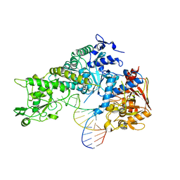
3SNP
DownloadVisualize
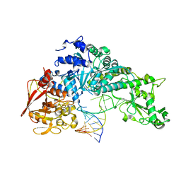
BU of 3snp by Molmil
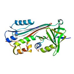
Crystal structure analysis of iron regulatory protein 1 in complex with ferritin H IRE RNA
Descriptor: Cytoplasmic aconitate hydratase, ferritin H IRE RNA
Authors:Volz, K, Selezneva, A.I, Walden, W.E.
Deposit date:2011-06-29
Release date:2011-07-27
Last modified:2023-09-13
Method:X-RAY DIFFRACTION (2.8 Å)
Cite:Structure of dual function iron regulatory protein 1 complexed with ferritin IRE-RNA.
Science, 314, 2006
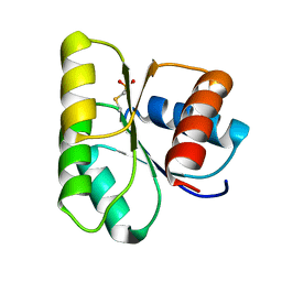
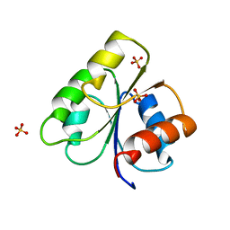
3CHY
DownloadVisualize
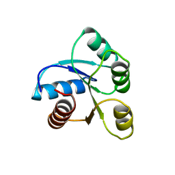
BU of 3chy by Molmil
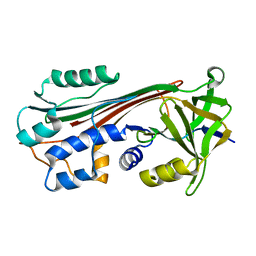
CRYSTAL STRUCTURE OF ESCHERICHIA COLI CHEY REFINED AT 1.7-ANGSTROM RESOLUTION
Descriptor: CHEY, SULFATE ION
Authors:Volz, K, Matsumura, P.
Deposit date:1991-04-22
Release date:1993-01-15
Last modified:2024-02-21
Method:X-RAY DIFFRACTION (1.66 Å)
Cite:Crystal structure of Escherichia coli CheY refined at 1.7-A resolution.
J.Biol.Chem., 266, 1991
3SN2
DownloadVisualize
BU of 3sn2 by Molmil
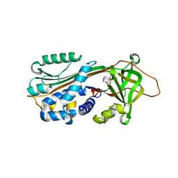
Crystal structure analysis of iron regulatory protein 1 in complex with transferrin receptor IRE B RNA
Descriptor: Cytoplasmic aconitate hydratase, transferrin receptor iron regulatory element B RNA
Authors:Volz, K, Selezneva, A.I, Walden, W.E.
Deposit date:2011-06-28
Release date:2011-11-23
Last modified:2023-09-13
Method:X-RAY DIFFRACTION (2.99 Å)
Cite:Accommodating variety in iron-responsive elements: Crystal structure of transferrin receptor 1 B IRE bound to iron regulatory protein 1.
Febs Lett., 586, 2012
1QU9
DownloadVisualize
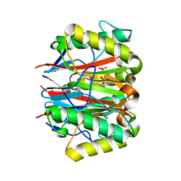
BU of 1qu9 by Molmil
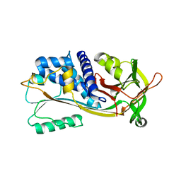
1.2 A CRYSTAL STRUCTURE OF YJGF GENE PRODUCT FROM E. COLI
Descriptor: YJGF PROTEIN
Authors:Volz, K.
Deposit date:1999-07-07
Release date:1999-12-01
Last modified:2018-01-31
Method:X-RAY DIFFRACTION (1.2 Å)
Cite:A test case for structure-based functional assignment: the 1.2 A crystal structure of the yjgF gene product from Escherichia coli
Protein Sci., 8, 1999
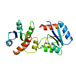
6CHY
DownloadVisualize
BU of 6chy by Molmil
STRUCTURE OF CHEMOTAXIS PROTEIN CHEY
Descriptor: CHEY, SULFATE ION
Authors:Zhu, X, Rebello, J, Matsumura, P, Volz, K.
Deposit date:1996-08-29
Release date:1996-12-07
Last modified:2024-05-22
Method:X-RAY DIFFRACTION (2.33 Å)
Cite:Crystal structures of CheY mutants Y106W and T87I/Y106W. CheY activation correlates with movement of residue 106.
J.Biol.Chem., 272, 1997
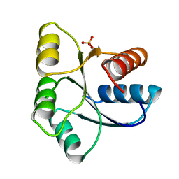
5CHY
DownloadVisualize
BU of 5chy by Molmil
STRUCTURE OF CHEMOTAXIS PROTEIN CHEY
Descriptor: CALCIUM ION, CHEY
Authors:Zhu, X, Rebello, J, Matsumura, P, Volz, K.
Deposit date:1996-08-29
Release date:1996-12-07
Last modified:2024-05-22
Method:X-RAY DIFFRACTION (2 Å)
Cite:Crystal structures of CheY mutants Y106W and T87I/Y106W. CheY activation correlates with movement of residue 106.
J.Biol.Chem., 272, 1997
1C4W
DownloadVisualize
BU of 1c4w by Molmil
1.9 A STRUCTURE OF A-THIOPHOSPHONATE MODIFIED CHEY D57C
Descriptor: CHEMOTAXIS PROTEIN CHEY
Authors:Halkides, C.J, McEvoy, M.M, Matsumura, P, Volz, K, Dahlquist, F.W.
Deposit date:1999-09-28
Release date:2000-05-08
Last modified:2023-12-27
Method:X-RAY DIFFRACTION (1.84 Å)
Cite:The 1.9 A resolution crystal structure of phosphono-CheY, an analogue of the active form of the response regulator, CheY.
Biochemistry, 39, 2000
1VLZ
DownloadVisualize
BU of 1vlz by Molmil
UNCOUPLED PHOSPHORYLATION AND ACTIVATION IN BACTERIAL CHEMOTAXIS: THE 2.1 ANGSTROM STRUCTURE OF A THREONINE TO ISOLEUCINE MUTANT AT POSITION 87 OF CHEY
Descriptor: CHEY
Authors:Ganguli, S, Wang, H, Matsumura, P, Volz, K.
Deposit date:1995-04-21
Release date:1995-07-10
Last modified:2024-02-14
Method:X-RAY DIFFRACTION (2.05 Å)
Cite:Uncoupled phosphorylation and activation in bacterial chemotaxis. The 2.1-A structure of a threonine to isoleucine mutant at position 87 of CheY.
J.Biol.Chem., 270, 1995
1EHC
DownloadVisualize
BU of 1ehc by Molmil
STRUCTURE OF SIGNAL TRANSDUCTION PROTEIN CHEY
Descriptor: CHEY, SULFATE ION
Authors:Jiang, M, Bourret, R, Simon, M, Volz, K.
Deposit date:1996-03-05
Release date:1997-05-15
Last modified:2024-02-07
Method:X-RAY DIFFRACTION (2.26 Å)
Cite:Uncoupled phosphorylation and activation in bacterial chemotaxis. The 2.3 A structure of an aspartate to lysine mutant at position 13 of CheY.
J.Biol.Chem., 272, 1997
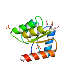
1JBE
DownloadVisualize
BU of 1jbe by Molmil
1.08 A Structure of apo-Chey reveals meta-active conformation
Descriptor: Chemotaxis protein CheY, GLYCEROL, SULFATE ION
Authors:Simonovic, M, Volz, K.
Deposit date:2001-06-04
Release date:2001-08-08
Last modified:2024-02-07
Method:X-RAY DIFFRACTION (1.08 Å)
Cite:A distinct meta-active conformation in the 1.1-A resolution structure of wild-type ApoCheY.
J.Biol.Chem., 276, 2001
1M93
DownloadVisualize
BU of 1m93 by Molmil
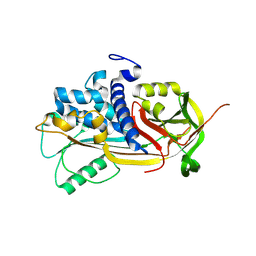
1.65 A Structure of Cleaved Viral Serpin CRMA
Descriptor: PHOSPHATE ION, Serine proteinase inhibitor 2
Authors:Simonovic, M, Gettins, P.G.W, Volz, K.
Deposit date:2002-07-26
Release date:2003-08-05
Last modified:2024-02-14
Method:X-RAY DIFFRACTION (1.65 Å)
Cite:Crystal structure of viral serpin crmA provides insights into its mechanism of cysteine proteinase inhibition
Protein Sci., 9, 2000
1C8O
DownloadVisualize
BU of 1c8o by Molmil
2.9 A STRUCTURE OF CLEAVED VIRAL SERPIN CRMA
Descriptor: ICE INHIBITOR
Authors:Simonovic, M, Gettins, P.G.W, Volz, K.
Deposit date:2000-06-01
Release date:2000-09-06
Last modified:2023-12-27
Method:X-RAY DIFFRACTION (2.9 Å)
Cite:Crystal structure of viral serpin crmA provides insights into its mechanism of cysteine proteinase inhibition.
Protein Sci., 9, 2000
1XQG
DownloadVisualize
BU of 1xqg by Molmil
3.10 A crystal structure of maspin, Space group P 4 21 2
Descriptor: Maspin
Authors:Al-Ayyoubi, M, Gettins, P.G, Volz, K.
Deposit date:2004-10-12
Release date:2004-10-26
Last modified:2023-08-23
Method:X-RAY DIFFRACTION (3.1 Å)
Cite:Crystal Structure of Human Maspin, a Serpin with Antitumor Properties: Reactive Center Loop of Mapsin is Exposed but Constrained
J.Biol.Chem., 279, 2004
1XQJ
DownloadVisualize
BU of 1xqj by Molmil
3.10 A Crystal structure of maspin, space group I 4 2 2
Descriptor: Maspin
Authors:Al-Ayyoubi, M, Gettins, P.G, Volz, K.
Deposit date:2004-10-12
Release date:2004-10-26
Last modified:2023-08-23
Method:X-RAY DIFFRACTION (3.1 Å)
Cite:Crystal Structure of Human Maspin, a Serpin with Antitumor Properties: Reactive Center Loop of Mapsin is Exposed but Constrained
J.Biol.Chem., 279, 2004
1J8E
DownloadVisualize
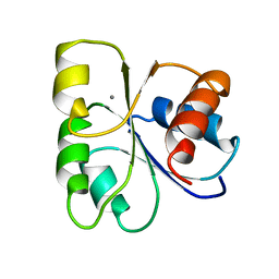
BU of 1j8e by Molmil
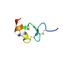
Crystal structure of ligand-binding repeat CR7 from LRP
Descriptor: CALCIUM ION, LOW-DENSITY LIPOPROTEIN RECEPTOR-RELATED PROTEIN 1
Authors:Simonovic, M, Dolmer, K, Huang, W, Strickland, D.K, Volz, K, Gettins, P.G.W.
Deposit date:2001-05-21
Release date:2001-12-19
Last modified:2024-10-30
Method:X-RAY DIFFRACTION (1.85 Å)
Cite:Calcium coordination and pH dependence of the calcium affinity of ligand-binding repeat CR7 from the LRP. Comparison with related domains from the LRP and the LDL receptor.
Biochemistry, 40, 2001
1OO8
DownloadVisualize
BU of 1oo8 by Molmil
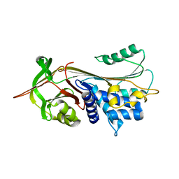
CRYSTAL STRUCTURE OF A1PI-PITTSBURGH IN THE NATIVE CONFORMATION
Descriptor: Alpha-1-antitrypsin precursor
Authors:Dementiev, A, Simonovic, M, Volz, K, Gettins, P.G.
Deposit date:2003-03-03
Release date:2003-08-05
Last modified:2023-08-16
Method:X-RAY DIFFRACTION (2.65 Å)
Cite:Canonical inhibitor-like interactions explain reactivity of alpha1-proteinase inhibitor Pittsburgh and antithrombin with proteinases
J.Biol.Chem., 278, 2003
1OPH
DownloadVisualize
BU of 1oph by Molmil
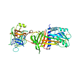
NON-COVALENT COMPLEX BETWEEN ALPHA-1-PI-PITTSBURGH AND S195A TRYPSIN
Descriptor: Alpha-1-antitrypsin precursor, Trypsinogen, cationic precursor
Authors:Dementiev, A, Simonovic, M, Volz, K, Gettins, P.G.
Deposit date:2003-03-05
Release date:2003-08-05
Last modified:2023-08-16
Method:X-RAY DIFFRACTION (2.3 Å)
Cite:Canonical inhibitor-like interactions explain reactivity of alpha1-proteinase inhibitor Pittsburgh and antithrombin with proteinases
J.Biol.Chem., 278, 2003
1IMV
DownloadVisualize
BU of 1imv by Molmil
2.85 A crystal structure of PEDF
Descriptor: 2-acetamido-2-deoxy-beta-D-glucopyranose, PIGMENT EPITHELIUM-DERIVED FACTOR
Authors:Simonovic, M, Gettins, P.G.W, Volz, K.
Deposit date:2001-05-11
Release date:2001-09-26
Last modified:2024-10-30
Method:X-RAY DIFFRACTION (2.85 Å)
Cite:Crystal structure of human PEDF, a potent anti-angiogenic and neurite growth-promoting factor.
Proc.Natl.Acad.Sci.USA, 98, 2001

226707

건을2024-10-30부터공개중

PDB statisticsPDBj update infoContact PDBjnumon