Loading
PDBj
MenuPDBj@FacebookPDBj@TwitterPDBj@YouTubewwPDB FoundationwwPDB
RCSB PDBPDBeBMRBAdv. SearchSearch help
PDB: 11 results

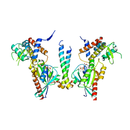
4UJ5
DownloadVisualize
BU of 4uj5 by Molmil
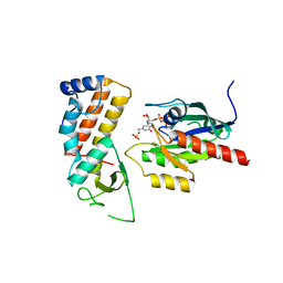
Crystal structure of human Rab11-Rabin8-FIP3
Descriptor: MAGNESIUM ION, PHOSPHOAMINOPHOSPHONIC ACID-GUANYLATE ESTER, RAB-3A-INTERACTING PROTEIN, ...
Authors:Vetter, M, Lorentzen, E.
Deposit date:2015-04-08
Release date:2015-08-12
Last modified:2024-01-10
Method:X-RAY DIFFRACTION (2.604 Å)
Cite:Structure of Rab11-Fip3-Rabin8 Reveals Simultaneous Binding of Fip3 and Rabin8 Effectors to Rab11.
Nat.Struct.Mol.Biol., 22, 2015
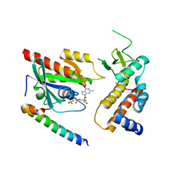
4UJ4
DownloadVisualize
BU of 4uj4 by Molmil
Crystal structure of human Rab11-Rabin8-FIP3
Descriptor: MAGNESIUM ION, PHOSPHOAMINOPHOSPHONIC ACID-GUANYLATE ESTER, Rab-3A-interacting protein, ...
Authors:Vetter, M, Lorentzen, E.
Deposit date:2015-04-08
Release date:2015-08-12
Last modified:2024-01-10
Method:X-RAY DIFFRACTION (4.2 Å)
Cite:Structure of Rab11-FIP3-Rabin8 reveals simultaneous binding of FIP3 and Rabin8 effectors to Rab11.
Nat. Struct. Mol. Biol., 22, 2015
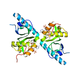
4UJ3
DownloadVisualize
BU of 4uj3 by Molmil
Crystal structure of human Rab11-Rabin8-FIP3
Descriptor: MAGNESIUM ION, PHOSPHOAMINOPHOSPHONIC ACID-GUANYLATE ESTER, RAB-3A-INTERACTING PROTEIN, ...
Authors:Vetter, M, Lorentzen, E.
Deposit date:2015-04-08
Release date:2015-08-12
Last modified:2024-01-10
Method:X-RAY DIFFRACTION (3 Å)
Cite:Structure of Rab11-Fip3-Rabin8 Reveals Simultaneous Binding of Fip3 and Rabin8 Effectors to Rab11.
Nat.Struct.Mol.Biol., 22, 2015
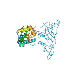
4B5P
DownloadVisualize
BU of 4b5p by Molmil
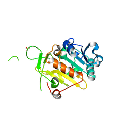
Crystal structure of human alpha tubulin acetyltransferase catalytic domain Q58A variant
Descriptor: ACETYL COENZYME *A, ALPHA-TUBULIN N-ACETYLTRANSFERASE, SODIUM ION
Authors:Taschner, M, Vetter, M, Lorentzen, E.
Deposit date:2012-08-07
Release date:2012-10-24
Last modified:2023-12-20
Method:X-RAY DIFFRACTION (1.6 Å)
Cite:Atomic Resolution Structure of Human Alpha-Tubulin Acetyltransferase Bound to Acetyl-Coa.
Proc.Natl.Acad.Sci.USA, 109, 2012
4B5O
DownloadVisualize
BU of 4b5o by Molmil
Crystal structure of human alpha tubulin acetyltransferase catalytic domain
Descriptor: ACETYL COENZYME *A, ALPHA-TUBULIN N-ACETYLTRANSFERASE, SODIUM ION
Authors:Taschner, M, Vetter, M, Lorentzen, E.
Deposit date:2012-08-07
Release date:2012-10-24
Last modified:2012-12-12
Method:X-RAY DIFFRACTION (1.05 Å)
Cite:Atomic Resolution Structure of Human Alpha-Tubulin Acetyltransferase Bound to Acetyl-Coa.
Proc.Natl.Acad.Sci.USA, 109, 2012
5FMR
DownloadVisualize
BU of 5fmr by Molmil
crIFT52 N-terminal domain
Descriptor: INTRAFLAGELLAR TRANSPORT PROTEIN COMPONENT IFT52, SULFATE ION
Authors:Mourao, A, Vetter, M, Lorentzen, E.
Deposit date:2015-11-09
Release date:2016-03-09
Last modified:2017-08-23
Method:X-RAY DIFFRACTION (2 Å)
Cite:Intraflagellar Transport Proteins 172, 80, 57, 54, 38, and 20 Form a Stable Tubulin-Binding Ift-B2 Complex.
Embo J., 35, 2016
6S1R
DownloadVisualize
BU of 6s1r by Molmil
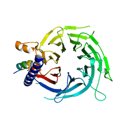
Structure of fission yeast Mis16 bound to histone H4
Descriptor: Histone H4, Histone acetyltransferase type B subunit 2
Authors:Lefevre, S, Korntner-Vetter, M, Singleton, M.R.
Deposit date:2019-06-19
Release date:2019-08-07
Last modified:2024-01-24
Method:X-RAY DIFFRACTION (1.8 Å)
Cite:Subunit interactions and arrangements in the fission yeast Mis16-Mis18-Mis19 complex.
Life Sci Alliance, 2, 2019
6S1L
DownloadVisualize
BU of 6s1l by Molmil
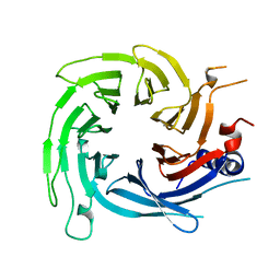
Structure of fission yeast Mis16
Descriptor: Histone acetyltransferase type B subunit 2
Authors:Lefevre, S, Korntner-Vetter, M, Singleton, M.R.
Deposit date:2019-06-19
Release date:2019-08-07
Last modified:2024-01-24
Method:X-RAY DIFFRACTION (1.94 Å)
Cite:Subunit interactions and arrangements in the fission yeast Mis16-Mis18-Mis19 complex.
Life Sci Alliance, 2, 2019
6S29
DownloadVisualize
BU of 6s29 by Molmil
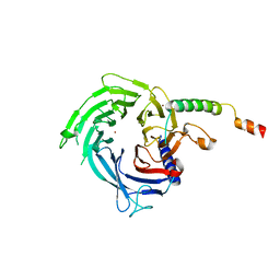
Structure of fission yeast Mis16-Mis19 complex
Descriptor: BROMIDE ION, CENP-A recruiting complex protein mis19, Histone acetyltransferase type B subunit 2
Authors:Lefevre, S, Korntner-Vetter, M, Singleton, M.R.
Deposit date:2019-06-20
Release date:2019-08-07
Last modified:2024-01-24
Method:X-RAY DIFFRACTION (1.988 Å)
Cite:Subunit interactions and arrangements in the fission yeast Mis16-Mis18-Mis19 complex.
Life Sci Alliance, 2, 2019
6F6P
DownloadVisualize
BU of 6f6p by Molmil
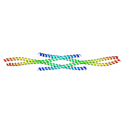
Crystal structure of tetrameric human Rabin8 GEF domain
Descriptor: Rab-3A-interacting protein
Authors:Lorentzen, E, Vetter, M.
Deposit date:2017-12-06
Release date:2017-12-13
Last modified:2018-03-14
Method:X-RAY DIFFRACTION (2.45 Å)
Cite:Crystal structure of tetrameric human Rabin8 GEF domain.
Proteins, 86, 2018
5MY6
DownloadVisualize
BU of 5my6 by Molmil
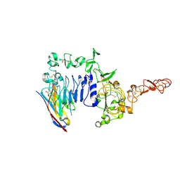
Crystal structure of a HER2-Nb complex
Descriptor: 2-acetamido-2-deoxy-beta-D-glucopyranose, 2-acetamido-2-deoxy-beta-D-glucopyranose-(1-4)-2-acetamido-2-deoxy-beta-D-glucopyranose, GLYCEROL, ...
Authors:Sterckx, Y.G.-J, D'Huyvetter, M, Devoogdt, N.
Deposit date:2017-01-25
Release date:2017-08-09
Last modified:2024-01-17
Method:X-RAY DIFFRACTION (2.246 Å)
Cite:(131)I-labeled Anti-HER2 Camelid sdAb as a Theranostic Tool in Cancer Treatment.
Clin. Cancer Res., 23, 2017

219140

PDB entries from 2024-05-01

PDB statisticsPDBj update infoContact PDBjnumon