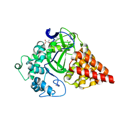
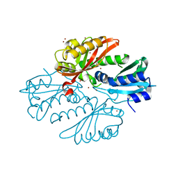
1ADU
 
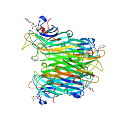
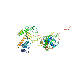 | | EARLY E2A DNA-BINDING PROTEIN | | 分子名称: | ADENOVIRUS SINGLE-STRANDED DNA-BINDING PROTEIN, ZINC ION | | 著者 | Tucker, P.A, Kanellopoulos, P.N, Tsernoglou, D, Van Der Vliet, P.C. | | 登録日 | 1995-05-11 | | 公開日 | 1996-06-20 | | 最終更新日 | 2024-02-07 | | 実験手法 | X-RAY DIFFRACTION (3 Å) | | 主引用文献 | Alternative arrangements of the protein chain are possible for the adenovirus single-stranded DNA binding protein.
J.Mol.Biol., 257, 1996
|
|
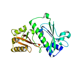
1L0Z
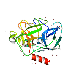 | | THE STRUCTURE OF PORCINE PANCREATIC ELASTASE COMPLEXED WITH XENON AND BROMIDE, CRYOPROTECTED WITH DRY PARAFFIN OIL | | 分子名称: | BROMIDE ION, ELASTASE 1, SODIUM ION, ... | | 著者 | Tucker, P.A, Panjikar, S. | | 登録日 | 2002-02-14 | | 公開日 | 2002-08-28 | | 最終更新日 | 2023-08-16 | | 実験手法 | X-RAY DIFFRACTION (1.5 Å) | | 主引用文献 | Xenon derivatization of halide-soaked protein crystals.
Acta Crystallogr.,Sect.D, 58, 2002
|
|
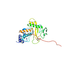
1NI0
 | |
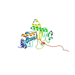
3ETI
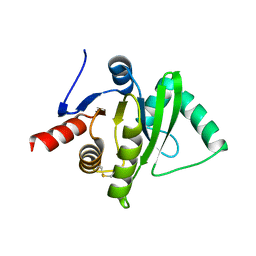 | |
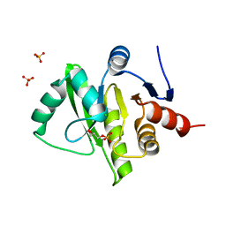
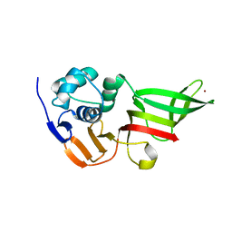
2W9Y
 | | The structure of the lipid binding protein Ce-FAR-7 from Caenorhabditis elegans | | 分子名称: | FATTY ACID/RETINOL BINDING PROTEIN PROTEIN 7, ISOFORM A, CONFIRMED BY TRANSCRIPT EVIDENCE, ... | | 著者 | Jordanova, R, Groves, M.R, Tucker, P.A. | | 登録日 | 2009-01-30 | | 公開日 | 2009-10-20 | | 最終更新日 | 2015-04-29 | | 実験手法 | X-RAY DIFFRACTION (1.8 Å) | | 主引用文献 | Fatty Acid and Retinoid Binding Proteins Have Distinct Binding Pockets for the Two Types of Cargo
J.Biol.Chem., 284, 2009
|
|
3RU0
 | | Cocrystal structure of human SMYD3 with inhibitor Sinefungin bound | | 分子名称: | SET and MYND domain-containing protein 3, SINEFUNGIN, ZINC ION | | 著者 | Foreman, K.W, Brown, M, Park, F, Emtage, S, Harriss, J, Das, C, Zhu, L, Crew, A, Arnold, L, Shaaban, S, Tucker, P. | | 登録日 | 2011-05-04 | | 公開日 | 2011-05-18 | | 最終更新日 | 2024-02-28 | | 実験手法 | X-RAY DIFFRACTION (1.849 Å) | | 主引用文献 | Structural and Functional Profiling of the Human Histone Methyltransferase SMYD3.
Plos One, 6, 2011
|
|
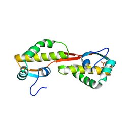
3EW5
 | | Structure of the tetragonal crystal form of X (ADRP) domain from FCoV | | 分子名称: | CHLORIDE ION, SN-GLYCEROL-1-PHOSPHATE, SULFATE ION, ... | | 著者 | Wojdyla, J.A, Manolaridis, I, Tucker, P.A. | | 登録日 | 2008-10-14 | | 公開日 | 2009-10-27 | | 最終更新日 | 2023-12-27 | | 実験手法 | X-RAY DIFFRACTION (3.1 Å) | | 主引用文献 | Structure of the X (ADRP) domain of nsp3 from feline coronavirus
Acta Crystallogr.,Sect.D, 65, 2009
|
|
3GZF
 | | Structure of the C-terminal domain of nsp4 from Feline Coronavirus | | 分子名称: | Replicase polyprotein 1ab, SULFATE ION | | 著者 | Manolaridis, I, Wojdyla, J.A, Panjikar, S, Snijder, E.J, Gorbalenya, A.E, Coutard, B, Tucker, P.A. | | 登録日 | 2009-04-07 | | 公開日 | 2009-08-18 | | 最終更新日 | 2024-03-20 | | 実験手法 | X-RAY DIFFRACTION (2.756 Å) | | 主引用文献 | Structure of the C-terminal domain of nsp4 from feline coronavirus
Acta Crystallogr.,Sect.D, 65, 2009
|
|
2L8K
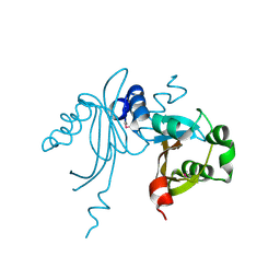
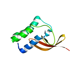 | | NMR Structure of the Arterivirus nonstructural protein 7 alpha (nsp7 alpha) | | 分子名称: | Non-structural protein 7 | | 著者 | Conte, M.R, Gaudin, C, Manolaridis, I, Tucker, P.W, Kelly, G. | | 登録日 | 2011-01-19 | | 公開日 | 2011-07-06 | | 最終更新日 | 2024-05-15 | | 実験手法 | SOLUTION NMR | | 主引用文献 | Structure and Genetic Analysis of the Arterivirus Nonstructural Protein 7{alpha}.
J.Virol., 85, 2011
|
|
1ANV
 | | ADENOVIRUS 5 DBP/URANYL FLUORIDE SOAK | | 分子名称: | ADENOVIRUS SINGLE-STRANDED DNA-BINDING PROTEIN, URANYL (VI) ION, ZINC ION | | 著者 | Kanellopoulos, P.N, Tsernoglou, D, Van Der Vliet, P.C, Tucker, P.A. | | 登録日 | 1996-03-14 | | 公開日 | 1997-02-12 | | 最終更新日 | 2024-05-22 | | 実験手法 | X-RAY DIFFRACTION (2.7 Å) | | 主引用文献 | Conformational change of the adenovirus DNA-binding protein induced by soaking crystals with K3UO2F5 solutions.
Acta Crystallogr.,Sect.D, 52, 1996
|
|
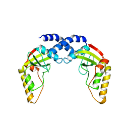
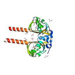
2YKF
 | | Sensor region of a sensor histidine kinase | | 分子名称: | BROMIDE ION, PROBABLE SENSOR HISTIDINE KINASE PDTAS, SULFATE ION | | 著者 | Preu, J, Panjikar, S, Morth, P, Jaiswal, R, Karunakar, P, Tucker, P.A. | | 登録日 | 2011-05-26 | | 公開日 | 2012-06-06 | | 最終更新日 | 2024-05-08 | | 実験手法 | X-RAY DIFFRACTION (2 Å) | | 主引用文献 | The Sensor Region of the Ubiquitous Cytosolic Sensor Kinase, Pdtas, Contains Pas and Gaf Domain Sensing Modules.
J.Struct.Biol., 177, 2012
|
|
1ADV
 | | EARLY E2A DNA-BINDING PROTEIN | | 分子名称: | ADENOVIRUS SINGLE-STRANDED DNA-BINDING PROTEIN, ZINC ION | | 著者 | Kanellopoulos, P.N, Tsernoglou, D, Van Der Vliet, P.C, Tucker, P.A. | | 登録日 | 1995-05-12 | | 公開日 | 1996-06-20 | | 最終更新日 | 2024-02-07 | | 実験手法 | X-RAY DIFFRACTION (3.2 Å) | | 主引用文献 | Alternative arrangements of the protein chain are possible for the adenovirus single-stranded DNA binding protein.
J.Mol.Biol., 257, 1996
|
|
2YKH
 | | Sensor region of a sensor histidine kinase | | 分子名称: | PROBABLE SENSOR HISTIDINE KINASE PDTAS | | 著者 | Preu, J, Panjikar, S, Morth, P, Jaiswal, R, Karunakar, P, Tucker, P.A. | | 登録日 | 2011-05-27 | | 公開日 | 2012-06-06 | | 最終更新日 | 2023-12-20 | | 実験手法 | X-RAY DIFFRACTION (2.78 Å) | | 主引用文献 | The Sensor Region of the Ubiquitous Cytosolic Sensor Kinase, Pdtas, Contains Pas and Gaf Domain Sensing Modules.
J.Struct.Biol., 177, 2012
|
|
2WAZ
 | |
2WB0
 | |
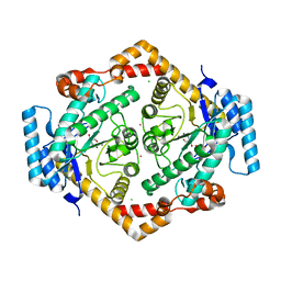
1CJP
 | | CONCANAVALIN A COMPLEX WITH 4'-METHYLUMBELLIFERYL-ALPHA-D-GLUCOPYRANOSIDE | | 分子名称: | 4-METHYLUMBELLIFERYL-ALPHA-D-GLUCOSE, CALCIUM ION, CONCANAVALIN A, ... | | 著者 | Hamodrakas, S.J, Kanellopoulos, P.N, Tucker, P.A. | | 登録日 | 1996-10-03 | | 公開日 | 1997-10-15 | | 最終更新日 | 2024-05-22 | | 実験手法 | X-RAY DIFFRACTION (2.78 Å) | | 主引用文献 | The crystal structure of the complex of concanavalin A with 4'-methylumbelliferyl-alpha-D-glucopyranoside.
J.Struct.Biol., 118, 1997
|
|
3K3C
 | |
3MP2
 | |
1O1H
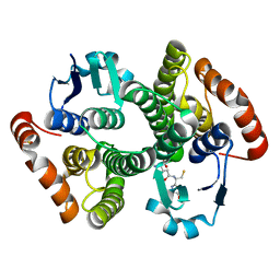
 | | STRUCTURE OF GLUCOSE ISOMERASE DERIVATIZED WITH KR. | | 分子名称: | (4S)-2-METHYL-2,4-PENTANEDIOL, 2-AMINO-2-HYDROXYMETHYL-PROPANE-1,3-DIOL, CALCIUM ION, ... | | 著者 | Nowak, E, Panjikar, S, Tucker, P.A. | | 登録日 | 2002-11-07 | | 公開日 | 2002-11-27 | | 最終更新日 | 2023-08-16 | | 実験手法 | X-RAY DIFFRACTION (1.4 Å) | | 主引用文献 | Structure of Glucose Isomerase Derivatized with Kr.
To be Published
|
|
1K9T
 | | Chitinase a complexed with tetra-N-acetylchitotriose | | 分子名称: | 2-acetamido-2-deoxy-beta-D-glucopyranose-(1-4)-2-acetamido-2-deoxy-beta-D-glucopyranose-(1-4)-2-acetamido-2-deoxy-beta-D-glucopyranose-(1-4)-2-acetamido-2-deoxy-beta-D-glucopyranose, CHITINASE A | | 著者 | Prag, G, Tucker, P.A, Oppenheim, A.B. | | 登録日 | 2001-10-30 | | 公開日 | 2002-11-06 | | 最終更新日 | 2023-10-25 | | 実験手法 | X-RAY DIFFRACTION (1.8 Å) | | 主引用文献 | Complex Structures of Chitinase A Mutant with Oligonag Provide Insight Into the Enzymatic Mechanism
To be Published
|
|
1M0U
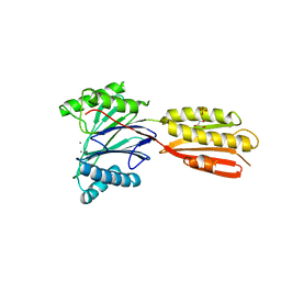
 | | Crystal Structure of the Drosophila Glutathione S-transferase-2 in Complex with Glutathione | | 分子名称: | GLUTATHIONE, GST2 gene product, SULFATE ION | | 著者 | Agianian, B, Tucker, P.A, Schouten, A, Leonard, K, Bullard, B, Gros, P. | | 登録日 | 2002-06-14 | | 公開日 | 2003-02-11 | | 最終更新日 | 2024-02-14 | | 実験手法 | X-RAY DIFFRACTION (1.75 Å) | | 主引用文献 | Structure of a Drosophila Sigma Class Glutathione S-transferase Reveals a Novel
Active Site Topography Suited for Lipid Peroxidation Products
J.Mol.Biol., 326, 2003
|
|
3KE6
 | |
3JZT
 | | Structure of a cubic crystal form of X (ADRP) domain from FCoV with ADP-ribose | | 分子名称: | ADENOSINE-5-DIPHOSPHORIBOSE, CHLORIDE ION, SODIUM ION, ... | | 著者 | Wojdyla, J.A, Manolaridis, I, Tucker, P.A. | | 登録日 | 2009-09-24 | | 公開日 | 2010-01-12 | | 最終更新日 | 2011-07-13 | | 実験手法 | X-RAY DIFFRACTION (3.91 Å) | | 主引用文献 | Structure of the X (ADRP) domain of nsp3 from feline coronavirus
Acta Crystallogr.,Sect.D, 65, 2009
|
|
2WK4
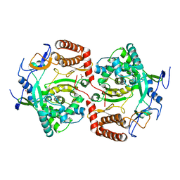 | | Dimeric structure of D347G D348G mutant of the sapporovirus RNA dependent RNA polymerase | | 分子名称: | GLYCEROL, PROTEASE-POLYMERASE P70 | | 著者 | Fullerton, S.W.B, Robel, I, Schuldt, L, Gebhardt, J, Tucker, P.A, Rohayem, J. | | 登録日 | 2009-06-05 | | 公開日 | 2010-09-01 | | 最終更新日 | 2023-12-13 | | 実験手法 | X-RAY DIFFRACTION (2.98 Å) | | 主引用文献 | Dimeric Structure of D347G D348G Mutant of the Sapporovirus Sapporovirus RNA Dependent RNA Polymerase
To be Published
|
|
3K3D
 | |


