Loading
PDBj
MenuPDBj@FacebookPDBj@TwitterPDBj@YouTubewwPDB FoundationwwPDB
RCSB PDBPDBeBMRBAdv. SearchSearch help
PDB: 491 results

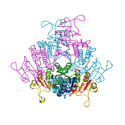
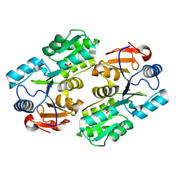
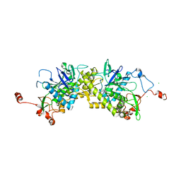
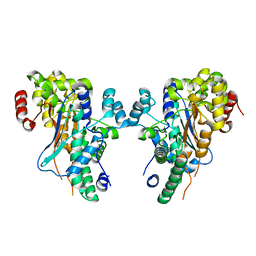
1YBF
DownloadVisualize
BU of 1ybf by Molmil
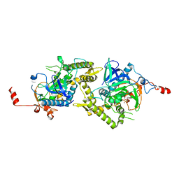
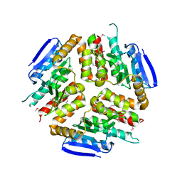
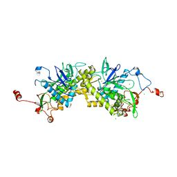
Crystal structure of AMP nucleosidase from Bacteroides thetaiotaomicron VPI-5482
Descriptor: AMP nucleosidase
Authors:Krishnamurthy, N.R, Swaminathan, S, Burley, S.K, New York SGX Research Center for Structural Genomics (NYSGXRC)
Deposit date:2004-12-20
Release date:2005-01-04
Last modified:2021-02-03
Method:X-RAY DIFFRACTION (2.9 Å)
Cite:Crystal structure of AMP nucleosidase from Bacteroides thetaiotaomicron
To be published
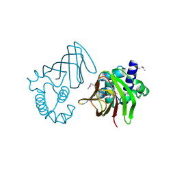
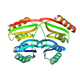
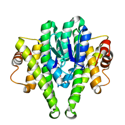
1YBE
DownloadVisualize
BU of 1ybe by Molmil
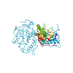
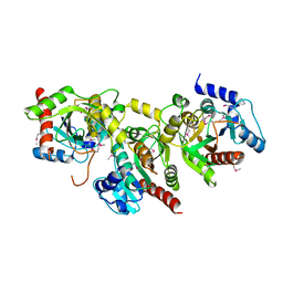
Crystal Structure of a Nicotinate phosphoribosyltransferase
Descriptor: Nicotinate phosphoribosyltransferase
Authors:Seetharaman, J, Swaminathan, S, Burley, S.K, New York SGX Research Center for Structural Genomics (NYSGXRC)
Deposit date:2004-12-20
Release date:2005-01-11
Last modified:2024-02-14
Method:X-RAY DIFFRACTION (2.5 Å)
Cite:Crystal Structure of a Nicotinate phosphoribosyltransferase
To be Published
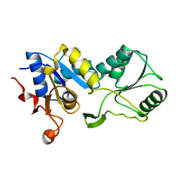
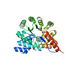
1YBD
DownloadVisualize
BU of 1ybd by Molmil
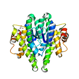
Crystal structure analysis of uridylate kinase from Neisseria meningitidis
Descriptor: FORMIC ACID, GLYCEROL, Uridylate kinase
Authors:Kumaran, D, Swaminathan, S, Burley, S.K, New York SGX Research Center for Structural Genomics (NYSGXRC)
Deposit date:2004-12-20
Release date:2005-02-15
Last modified:2021-02-03
Method:X-RAY DIFFRACTION (2.6 Å)
Cite:Crystal structure analysis of uridylate kinase from Neisseria meningitidis
To be Published
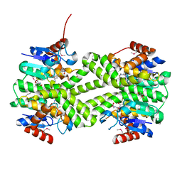
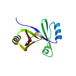
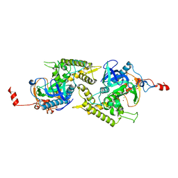
1Y9Q
DownloadVisualize
BU of 1y9q by Molmil
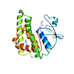
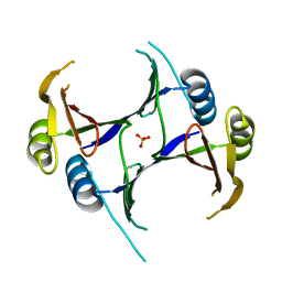
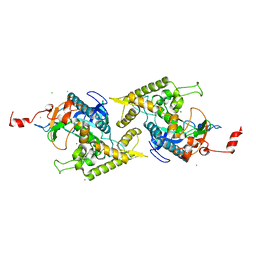
Crystal Structure of HTH_3 family Transcriptional Regulator from Vibrio cholerae
Descriptor: D-METHIONINE, ZINC ION, transcriptional regulator, ...
Authors:Kumaran, D, Swaminathan, S, Burley, S.K, New York SGX Research Center for Structural Genomics (NYSGXRC)
Deposit date:2004-12-16
Release date:2005-01-04
Last modified:2021-02-03
Method:X-RAY DIFFRACTION (1.9 Å)
Cite:Crystal Structure of HTH_3 family Transcriptional Regulator from Vibrio cholerae
To be Published
1YDF
DownloadVisualize
BU of 1ydf by Molmil
Crystal structure of a HAD-like phosphatase from Streptococcus pneumoniae
Descriptor: MAGNESIUM ION, hydrolase, haloacid dehalogenase-like family
Authors:Seetharaman, J, Swaminathan, S, Burley, S.K, New York SGX Research Center for Structural Genomics (NYSGXRC)
Deposit date:2004-12-23
Release date:2005-01-18
Last modified:2024-02-14
Method:X-RAY DIFFRACTION (2.6 Å)
Cite:Crystal Structure of a Hypothetical protein, hydrolase haloacid dehalogenase-like family
To be Published
3LSZ
DownloadVisualize
BU of 3lsz by Molmil
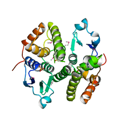
Crystal structure of glutathione s-transferase from Rhodobacter sphaeroides
Descriptor: 2-AMINO-2-HYDROXYMETHYL-PROPANE-1,3-DIOL, GLUTATHIONE, GLYCEROL, ...
Authors:Eswaramoorthy, S, Burley, S.K, Swaminathan, S, New York SGX Research Center for Structural Genomics (NYSGXRC)
Deposit date:2010-02-14
Release date:2010-03-23
Last modified:2021-02-10
Method:X-RAY DIFFRACTION (1.7 Å)
Cite:Crystal structure of glutathione s-transferase from Rhodobacter sphaeroides
To be Published
3M3M
DownloadVisualize
BU of 3m3m by Molmil
Crystal structure of glutathione S-transferase from Pseudomonas fluorescens [Pf-5]
Descriptor: 1,2-ETHANEDIOL, GLUTATHIONE, Glutathione S-transferase, ...
Authors:Bagaria, A, Burley, S.K, Swaminathan, S, New York SGX Research Center for Structural Genomics (NYSGXRC)
Deposit date:2010-03-09
Release date:2010-03-16
Last modified:2021-02-10
Method:X-RAY DIFFRACTION (1.75 Å)
Cite:Crystal structure of glutathione S-transferase from Pseudomonas fluorescens [Pf-5]
To be Published
3M8N
DownloadVisualize
BU of 3m8n by Molmil
Crystal structure of a possible gutathione S-tranferase from Rhodopseudomonas palustris
Descriptor: Possible glutathione S-transferase, SULFATE ION
Authors:Damodharan, L, Burley, S.K, Swaminathan, S, New York SGX Research Center for Structural Genomics (NYSGXRC)
Deposit date:2010-03-18
Release date:2010-04-07
Last modified:2021-02-10
Method:X-RAY DIFFRACTION (2.04 Å)
Cite:Crystal structure of a possible gutathione S-tranferase from Rhodopseudomonas palustris
To be Published
1ZL6
DownloadVisualize
BU of 1zl6 by Molmil
Crystal structure of Tyr350Ala mutant of Clostridium botulinum neurotoxin E catalytic domain
Descriptor: SULFATE ION, ZINC ION, botulinum neurotoxin type E
Authors:Agarwal, R, Binz, T, Swaminathan, S.
Deposit date:2005-05-05
Release date:2005-06-28
Last modified:2023-08-23
Method:X-RAY DIFFRACTION (2.4 Å)
Cite:Analysis of Active Site Residues of Botulinum Neurotoxin E by Mutational, Functional, and Structural Studies: Glu335Gln Is an Apoenzyme.
Biochemistry, 44, 2005
1YVG
DownloadVisualize
BU of 1yvg by Molmil
Structural analysis of the catalytic domain of tetanus neurotoxin
Descriptor: Tetanus toxin, light chain, ZINC ION
Authors:Rao, K.N, Kumaran, D, Binz, T, Swaminathan, S.
Deposit date:2005-02-15
Release date:2005-03-22
Last modified:2023-08-23
Method:X-RAY DIFFRACTION (2.6 Å)
Cite:Structural analysis of the catalytic domain of tetanus neurotoxin.
Toxicon, 45, 2005
1YV9
DownloadVisualize
BU of 1yv9 by Molmil
Crystal structure of a HAD-like phosphatase from Enterococcus faecalis V583
Descriptor: PHOSPHATE ION, hydrolase, haloacid dehalogenase family
Authors:Seetharaman, J, Swaminathan, S, Burley, S.K, New York SGX Research Center for Structural Genomics (NYSGXRC)
Deposit date:2005-02-15
Release date:2005-03-15
Last modified:2023-08-23
Method:X-RAY DIFFRACTION (2.8 Å)
Cite:Crystal Structure of a Hypothetical protein, hydrolase haloacid dehalogenase-like family
To be Published
1U8S
DownloadVisualize
BU of 1u8s by Molmil
Crystal structure of putative glycine cleavage system transcriptional repressor
Descriptor: glycine cleavage system transcriptional repressor, putative
Authors:Kumaran, D, Swaminathan, S.
Deposit date:2004-08-06
Release date:2004-08-17
Last modified:2024-02-14
Method:X-RAY DIFFRACTION (2.45 Å)
Cite:Crystal structure of putative glycine cleavage system transcriptional repressor
To be Published
3HDP
DownloadVisualize
BU of 3hdp by Molmil
Crystal structure of the NI(II)-bound Glyoxalase-I from Clostridium acetobutylicum
Descriptor: Glyoxalase-I, NICKEL (II) ION
Authors:Satyanarayana, L, Eswaramoorthy, S, Burley, S.K, Swaminathan, S, New York SGX Research Center for Structural Genomics (NYSGXRC)
Deposit date:2009-05-07
Release date:2009-05-26
Last modified:2023-09-06
Method:X-RAY DIFFRACTION (2.06 Å)
Cite:Crystal structure of the NI(II)-bound Glyoxalase-I from Clostridium acetobutylicum.
To be Published
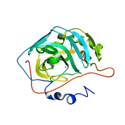
1XEV
DownloadVisualize
BU of 1xev by Molmil
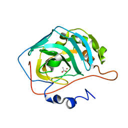
Crystal structure of human carbonic anhydrase II in a new crystal form
Descriptor: Carbonic anhydrase II, ZINC ION
Authors:Mazumdar, P.A, Kumaran, D, Das, A.K, Swaminathan, S.
Deposit date:2004-09-13
Release date:2005-09-20
Last modified:2023-08-23
Method:X-RAY DIFFRACTION (2.2 Å)
Cite:Crystal structure of human carbonic anhydrase II in a new crystal form
To be Published
1XEG
DownloadVisualize
BU of 1xeg by Molmil
Crystal structure of human carbonic anhydrase II complexed with an acetate ion
Descriptor: ACETATE ION, Carbonic anhydrase II, ZINC ION
Authors:Mazumdar, P.A, Kumaran, D, Das, A.K, Swaminathan, S.
Deposit date:2004-09-10
Release date:2005-09-27
Last modified:2024-02-14
Method:X-RAY DIFFRACTION (1.81 Å)
Cite:A novel acetate-bound complex of human carbonic anhydrase II.
Acta Crystallogr.,Sect.F, 64, 2008
3L3S
DownloadVisualize
BU of 3l3s by Molmil
Crystal structure of an enoyl-CoA hydrotase/isomerase family protein from Silicibacter pomeroyi
Descriptor: Enoyl-CoA hydratase/isomerase family protein
Authors:Eswaramoorthy, S, Silberstein, M, Burley, S.K, Swaminathan, S, New York SGX Research Center for Structural Genomics (NYSGXRC)
Deposit date:2009-12-17
Release date:2010-01-05
Last modified:2023-11-22
Method:X-RAY DIFFRACTION (2.32 Å)
Cite:Crystal structure of an enoyl-CoA hydrotase/isomerase family protein from Silicibacter pomeroyi
To be Published
3LXT
DownloadVisualize
BU of 3lxt by Molmil
Crystal structure of Glutathione S Transferase from Pseudomonas fluorescens
Descriptor: CHLORIDE ION, GLYCEROL, Glutathione S Transferase
Authors:Agarwal, R, Burley, S.K, Swaminathan, S, New York SGX Research Center for Structural Genomics (NYSGXRC)
Deposit date:2010-02-25
Release date:2010-03-09
Last modified:2024-02-21
Method:X-RAY DIFFRACTION (1.76 Å)
Cite:Crystal structure of Glutathione S Transferase from Pseudomonas fluorescens
To be Published
3G12
DownloadVisualize
BU of 3g12 by Molmil
Crystal structure of a putative lactoylglutathione lyase from Bdellovibrio bacteriovorus
Descriptor: Putative lactoylglutathione lyase, SULFATE ION
Authors:Patskovsky, Y, Madegowda, M, Gilmore, M, Chang, S, Maletic, M, Smith, D, Sauder, J.M, Burley, S.K, Swaminathan, S, Almo, S.C, New York SGX Research Center for Structural Genomics (NYSGXRC)
Deposit date:2009-01-29
Release date:2009-02-10
Last modified:2024-02-21
Method:X-RAY DIFFRACTION (2.58 Å)
Cite:Crystal structure of a putative lactoylglutathione lyase from Bdellovibrio bacteriovorus
To be Published
1ZKX
DownloadVisualize
BU of 1zkx by Molmil
Crystal structure of Glu158Ala/Thr159Ala/Asn160Ala- a triple mutant of Clostridium botulinum neurotoxin E catalytic domain
Descriptor: CHLORIDE ION, ZINC ION, botulinum neurotoxin type E
Authors:Agarwal, R, Binz, T, Swaminathan, S.
Deposit date:2005-05-04
Release date:2005-07-05
Last modified:2023-08-23
Method:X-RAY DIFFRACTION (2.52 Å)
Cite:Analysis of Active Site Residues of Botulinum Neurotoxin E by Mutational, Functional, and Structural Studies: Glu335Gln Is an Apoenzyme.
Biochemistry, 44, 2005
3M0F
DownloadVisualize
BU of 3m0f by Molmil
Crystal structure of Glutathione S Transferase in complex with glutathione from Pseudomonas fluorescens
Descriptor: GLUTATHIONE, uncharacterized protein GST_N
Authors:Agarwal, R, Burley, S.K, Swaminathan, S, New York SGX Research Center for Structural Genomics (NYSGXRC)
Deposit date:2010-03-03
Release date:2010-03-16
Last modified:2024-02-21
Method:X-RAY DIFFRACTION (1.6 Å)
Cite:Crystal structure of Glutathione S Transferase in complex with glutathione from Pseudomonas fluorescens
To be Published
3RYS
DownloadVisualize
BU of 3rys by Molmil
The crystal structure of adenine deaminase (AAur1117) from Arthrobacter aurescens
Descriptor: ADENINE, Adenosine deaminase 1, ZINC ION
Authors:Zhang, Z, Goble, A.M, Raushel, F.M, Swaminathan, S.
Deposit date:2011-05-11
Release date:2011-05-25
Last modified:2023-09-13
Method:X-RAY DIFFRACTION (2.601 Å)
Cite:The crystal structure of adenine deaminase (AAur1117) from Arthrobacter aurescens
To be Published
1ZN3
DownloadVisualize
BU of 1zn3 by Molmil
Crystal structure of Glu335Ala mutant of Clostridium botulinum neurotoxin type E
Descriptor: CHLORIDE ION, ZINC ION, botulinum neurotoxin type E
Authors:Agarwal, R, Binz, T, Swaminathan, S.
Deposit date:2005-05-11
Release date:2005-07-05
Last modified:2023-08-23
Method:X-RAY DIFFRACTION (2.6 Å)
Cite:Analysis of Active Site Residues of Botulinum Neurotoxin E by Mutational, Functional, and Structural Studies: Glu335Gln Is an Apoenzyme.
Biochemistry, 44, 2005
1ZKW
DownloadVisualize
BU of 1zkw by Molmil
Crystal structure of Arg347Ala mutant of botulinum neurotoxin E catalytic domain
Descriptor: CHLORIDE ION, ZINC ION, botulinum neurotoxin type E
Authors:Agarwal, R, Binz, T, Swaminathan, S.
Deposit date:2005-05-04
Release date:2005-06-28
Last modified:2023-08-23
Method:X-RAY DIFFRACTION (2.17 Å)
Cite:Analysis of Active Site Residues of Botulinum Neurotoxin E by Mutational, Functional, and Structural Studies: Glu335Gln Is an Apoenzyme.
Biochemistry, 44, 2005
1ZL5
DownloadVisualize
BU of 1zl5 by Molmil
Crystal structure of Glu335Gln mutant of Clostridium botulinum neurotoxin E catalytic domain
Descriptor: CHLORIDE ION, botulinum neurotoxin type E
Authors:Agarwal, R, Binz, T, Swaminathan, S.
Deposit date:2005-05-05
Release date:2005-07-05
Last modified:2023-08-23
Method:X-RAY DIFFRACTION (2.6 Å)
Cite:Analysis of Active Site Residues of Botulinum Neurotoxin E by Mutational, Functional, and Structural Studies: Glu335Gln Is an Apoenzyme.
Biochemistry, 44, 2005
3BE3
DownloadVisualize
BU of 3be3 by Molmil
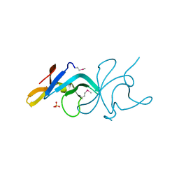
Crystal structure of a protein belonging to pfam DUF1653 from Bordetella bronchiseptica
Descriptor: SULFATE ION, Uncharacterized protein
Authors:Tyagi, R, Eswaramoorthy, S, Burley, S.K, Swaminathan, S, New York SGX Research Center for Structural Genomics (NYSGXRC)
Deposit date:2007-11-16
Release date:2007-11-27
Last modified:2021-02-03
Method:X-RAY DIFFRACTION (2.04 Å)
Cite:Crystal structure of a protein belonging to pfam DUF1653 from Bordetella bronchiseptica.
To be Published

220472

건을2024-05-29부터공개중

PDB statisticsPDBj update infoContact PDBjnumon