4OZY
 
 | |
4B6J
 | |
1XMM
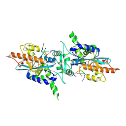
 | | Structure of human Dcps bound to m7GDP | | 分子名称: | 7N-METHYL-8-HYDROGUANOSINE-5'-DIPHOSPHATE, N7-METHYL-GUANOSINE-5'-MONOPHOSPHATE, PHOSPHATE ION, ... | | 著者 | Chen, N, Song, H. | | 登録日 | 2004-10-04 | | 公開日 | 2005-03-22 | | 最終更新日 | 2024-03-06 | | 実験手法 | X-RAY DIFFRACTION (2.5 Å) | | 主引用文献 | Crystal structures of human DcpS in ligand-free and m7GDP-bound forms suggest a dynamic mechanism for scavenger mRNA decapping.
J.Mol.Biol., 347, 2005
|
|
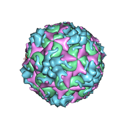
7C4T
 | | Cryo-EM structure of A particle Coxsackievirus A10 at pH 7.4 | | 分子名称: | Capsid protein VP1, Capsid protein VP2, Capsid protein VP3 | | 著者 | Cui, Y, Peng, R, Song, H, Tong, Z, Gao, G.F, Qi, J. | | 登録日 | 2020-05-18 | | 公開日 | 2020-07-22 | | 最終更新日 | 2024-03-27 | | 実験手法 | ELECTRON MICROSCOPY (3.6 Å) | | 主引用文献 | Molecular basis of Coxsackievirus A10 entry using the two-in-one attachment and uncoating receptor KRM1.
Proc.Natl.Acad.Sci.USA, 117, 2020
|
|
7C4W
 | | Cryo-EM structure of A particle Coxsackievirus A10 at pH 5.5 | | 分子名称: | Capsid protein VP1, Capsid protein VP2, Capsid protein VP3 | | 著者 | Cui, Y, Peng, R, Song, H, Tong, Z, Gao, G.F, Qi, J. | | 登録日 | 2020-05-18 | | 公開日 | 2020-07-22 | | 最終更新日 | 2024-03-27 | | 実験手法 | ELECTRON MICROSCOPY (3.4 Å) | | 主引用文献 | Molecular basis of Coxsackievirus A10 entry using the two-in-one attachment and uncoating receptor KRM1.
Proc.Natl.Acad.Sci.USA, 117, 2020
|
|
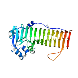
1JXV
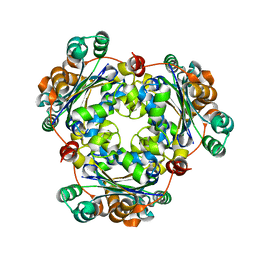 | | Crystal Structure of Human Nucleoside Diphosphate Kinase A | | 分子名称: | Nucleoside Diphosphate Kinase A | | 著者 | Min, K, Song, H.K, Chang, C, Kim, S.Y, Lee, K.J, Suh, S.W. | | 登録日 | 2001-09-10 | | 公開日 | 2002-04-10 | | 最終更新日 | 2023-10-25 | | 実験手法 | X-RAY DIFFRACTION (2.2 Å) | | 主引用文献 | Crystal structure of human nucleoside diphosphate kinase A, a metastasis suppressor.
Proteins, 46, 2002
|
|
4P7L
 | | Structure of Escherichia coli PgaB C-terminal domain, P212121 crystal form | | 分子名称: | 1,2-ETHANEDIOL, CHLORIDE ION, Poly-beta-1,6-N-acetyl-D-glucosamine N-deacetylase | | 著者 | Little, D.J, Li, G, Ing, C, DiFrancesco, B, Bamford, N.C, Robinson, H, Nitz, M, Pomes, R, Howell, P.L. | | 登録日 | 2014-03-27 | | 公開日 | 2014-07-02 | | 最終更新日 | 2023-09-27 | | 実験手法 | X-RAY DIFFRACTION (1.802 Å) | | 主引用文献 | Modification and periplasmic translocation of the biofilm exopolysaccharide poly-beta-1,6-N-acetyl-D-glucosamine.
Proc.Natl.Acad.Sci.USA, 111, 2014
|
|
2AVX
 | | solution structure of E coli SdiA1-171 | | 分子名称: | N-(2-OXOTETRAHYDROFURAN-3-YL)OCTANAMIDE, Regulatory protein sdiA | | 著者 | Yao, Y, Martinez-Yamout, M.A, Dickerson, T.J, Brogan, A.P, Wright, P.E, Dyson, H.J. | | 登録日 | 2005-08-30 | | 公開日 | 2006-06-20 | | 最終更新日 | 2024-05-22 | | 実験手法 | SOLUTION NMR | | 主引用文献 | Structure of the Escherichia coli quorum sensing protein SdiA: activation of the folding switch by acyl homoserine lactones.
J.Mol.Biol., 355, 2006
|
|
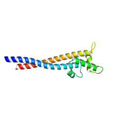
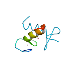
1KBH
 | | Mutual Synergistic Folding in the Interaction Between Nuclear Receptor Coactivators CBP and ACTR | | 分子名称: | CREB-BINDING PROTEIN, nuclear receptor coactivator | | 著者 | Demarest, S.J, Martinez-Yamout, M, Chung, J, Chen, H, Xu, W, Dyson, H.J, Evans, R.M, Wright, P.E. | | 登録日 | 2001-11-06 | | 公開日 | 2002-02-06 | | 最終更新日 | 2024-05-22 | | 実験手法 | SOLUTION NMR | | 主引用文献 | Mutual synergistic folding in recruitment of CBP/p300 by p160 nuclear receptor coactivators.
Nature, 415, 2002
|
|
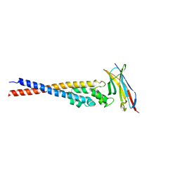
7F9N
 | | Crystal structure of the variable region of Plasmodium RIFIN #4 (PF3D7_1000500) in complex with LAIR1 | | 分子名称: | Leukocyte-associated immunoglobulin-like receptor 1, Rifin | | 著者 | Xie, Y, Song, H, Li, X, Qi, J, Gao, G.F. | | 登録日 | 2021-07-04 | | 公開日 | 2021-08-18 | | 最終更新日 | 2023-11-29 | | 実験手法 | X-RAY DIFFRACTION (3 Å) | | 主引用文献 | Structural basis of malarial parasite RIFIN-mediated immune escape against LAIR1.
Cell Rep, 36, 2021
|
|
7F9K
 | | Crystal structure of the variable region of Plasmodium RIFIN #6(PF3D7_1400600) | | 分子名称: | Rifin | | 著者 | Xie, Y, Song, H, Li, X, Qi, J, Gao, G.F. | | 登録日 | 2021-07-04 | | 公開日 | 2021-08-18 | | 最終更新日 | 2023-11-29 | | 実験手法 | X-RAY DIFFRACTION (2.18 Å) | | 主引用文献 | Structural basis of malarial parasite RIFIN-mediated immune escape against LAIR1.
Cell Rep, 36, 2021
|
|
7F9M
 | | Crystal structure of the variable region of Plasmodium RIFIN #4 (PF3D7_1000500) in complex with LAIR1 (with T67L, N69S and A77T mutations) | | 分子名称: | Leukocyte-associated immunoglobulin-like receptor 1, Rifin | | 著者 | Xie, Y, Song, H, Li, X, Qi, J, Gao, G.F. | | 登録日 | 2021-07-04 | | 公開日 | 2021-08-18 | | 最終更新日 | 2023-11-29 | | 実験手法 | X-RAY DIFFRACTION (2.9 Å) | | 主引用文献 | Structural basis of malarial parasite RIFIN-mediated immune escape against LAIR1.
Cell Rep, 36, 2021
|
|
7C4Y
 | | Cryo-EM structure of empty Coxsackievirus A10 at pH 7.4 | | 分子名称: | Capsid protein VP1, Capsid protein VP2, Capsid protein VP3 | | 著者 | Cui, Y, Peng, R, Song, H, Tong, Z, Gao, G.F, Qi, J. | | 登録日 | 2020-05-18 | | 公開日 | 2020-07-22 | | 最終更新日 | 2024-03-27 | | 実験手法 | ELECTRON MICROSCOPY (3.5 Å) | | 主引用文献 | Molecular basis of Coxsackievirus A10 entry using the two-in-one attachment and uncoating receptor KRM1.
Proc.Natl.Acad.Sci.USA, 117, 2020
|
|
1SSU
 | | Structural and biochemical evidence for disulfide bond heterogeneity in active forms of the somatomedin B domain of human vitronectin | | 分子名称: | Vitronectin | | 著者 | Kamikubo, Y, De Guzman, R, Kroon, G, Curriden, S, Neels, J.G, Churchill, M.J, Dawson, P, Oldziej, S, Jagielska, A, Scheraga, H.A, Loskutoff, D.J, Dyson, H.J. | | 登録日 | 2004-03-24 | | 公開日 | 2004-07-27 | | 最終更新日 | 2022-03-02 | | 実験手法 | SOLUTION NMR | | 主引用文献 | Disulfide bonding arrangements in active forms of the somatomedin B domain of human vitronectin.
Biochemistry, 43, 2004
|
|
2AFF
 | | The solution structure of the Ki67FHA/hNIFK(226-269)3P complex | | 分子名称: | Antigen KI-67, MKI67 FHA domain interacting nucleolar phosphoprotein | | 著者 | Byeon, I.-J.L, Li, H, Song, H, Gronenborn, A.M, Tsai, M.D. | | 登録日 | 2005-07-25 | | 公開日 | 2005-10-25 | | 最終更新日 | 2022-03-09 | | 実験手法 | SOLUTION NMR | | 主引用文献 | Sequential phosphorylation and multisite interactions characterize specific target recognition by the FHA domain of Ki67.
Nat.Struct.Mol.Biol., 12, 2005
|
|
2A1R
 | | Crystal structure of PARN nuclease domain | | 分子名称: | 5'-R(*AP*AP*A)-3', Poly(A)-specific ribonuclease PARN | | 著者 | Wu, M, Song, H. | | 登録日 | 2005-06-21 | | 公開日 | 2005-12-20 | | 最終更新日 | 2017-10-11 | | 実験手法 | X-RAY DIFFRACTION (2.6 Å) | | 主引用文献 | Structural insight into poly(A) binding and catalytic mechanism of human PARN
Embo J., 24, 2005
|
|
2A1S
 | |
1F77
 | | STAPHYLOCOCCAL ENTEROTOXIN H DETERMINED TO 2.4 A RESOLUTION | | 分子名称: | ENTEROTOXIN H, SULFATE ION | | 著者 | Hakansson, M, Petersson, K, Nilsson, H, Forsberg, G, Bjork, P. | | 登録日 | 2000-06-26 | | 公開日 | 2000-07-19 | | 最終更新日 | 2017-10-04 | | 実験手法 | X-RAY DIFFRACTION (2.4 Å) | | 主引用文献 | The crystal structure of staphylococcal enterotoxin H: implications for binding properties to MHC class II and TcR molecules.
J.Mol.Biol., 302, 2000
|
|
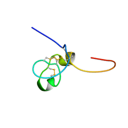
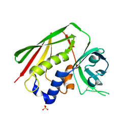
2DS6
 | | Structure of the ZBD in the tetragonal crystal form | | 分子名称: | ATP-dependent Clp protease ATP-binding subunit clpX, ZINC ION | | 著者 | Park, E.Y, Lee, B.G, Hong, S.B, Song, H.K. | | 登録日 | 2006-06-22 | | 公開日 | 2007-02-13 | | 最終更新日 | 2023-10-25 | | 実験手法 | X-RAY DIFFRACTION (2 Å) | | 主引用文献 | Structural Basis of SspB-tail Recognition by the Zinc Binding Domain of ClpX.
J.Mol.Biol., 367, 2007
|
|
2B9C
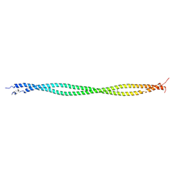 | | Structure of tropomyosin's mid-region: bending and binding sites for actin | | 分子名称: | striated-muscle alpha tropomyosin | | 著者 | Brown, J.H, Zhou, Z, Reshetnikova, L, Robinson, H, Yammani, R.D, Tobacman, L.S, Cohen, C. | | 登録日 | 2005-10-11 | | 公開日 | 2006-01-03 | | 最終更新日 | 2022-12-21 | | 実験手法 | X-RAY DIFFRACTION (2.3 Å) | | 主引用文献 | Structure of the mid-region of tropomyosin: Bending and binding sites for actin.
Proc.Natl.Acad.Sci.Usa, 102, 2005
|
|
2DS7
 | | Structure of the ZBD in the hexagonal crystal form | | 分子名称: | ATP-dependent Clp protease ATP-binding subunit clpX, ZINC ION | | 著者 | Park, E.Y, Lee, B.G, Hong, S.B, Song, H.K. | | 登録日 | 2006-06-22 | | 公開日 | 2007-02-13 | | 最終更新日 | 2021-11-10 | | 実験手法 | X-RAY DIFFRACTION (2.5 Å) | | 主引用文献 | Structural Basis of SspB-tail Recognition by the Zinc Binding Domain of ClpX.
J.Mol.Biol., 367, 2007
|
|
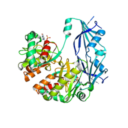
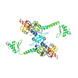
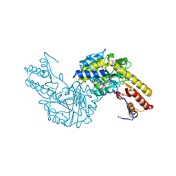
4B3F
 | | crystal structure of Ighmbp2 helicase | | 分子名称: | DNA-BINDING PROTEIN SMUBP-2, PHOSPHATE ION | | 著者 | Lim, S.C, Song, H. | | 登録日 | 2012-07-24 | | 公開日 | 2012-09-26 | | 最終更新日 | 2024-05-08 | | 実験手法 | X-RAY DIFFRACTION (2.5 Å) | | 主引用文献 | The Ighmbp2 Helicase Structure Reveals the Molecular Basis for Disease-Causing Mutations in Dmsa1.
Nucleic Acids Res., 40, 2012
|
|
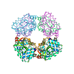
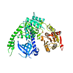
3PDX
 | | Crystal structural of mouse tyrosine aminotransferase | | 分子名称: | Tyrosine aminotransferase | | 著者 | Mehere, P.V, Han, Q, Lemkul, J.A, Robinson, H, Bevan, D.R, Li, J. | | 登録日 | 2010-10-25 | | 公開日 | 2010-11-03 | | 最終更新日 | 2023-12-06 | | 実験手法 | X-RAY DIFFRACTION (2.91 Å) | | 主引用文献 | Tyrosine aminotransferase: biochemical and structural properties and molecular dynamics simulations.
Protein Cell, 1, 2010
|
|
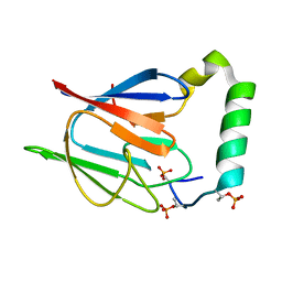
4H6J
 | | Identification of Cys 255 in HIF-1 as a novel site for development of covalent inhibitors of HIF-1 /ARNT PasB domain protein-protein interaction. | | 分子名称: | ARYL HYDROCARBON NUCLEAR TRANSLOCATOR, HYPOXIA INDUCIBLE FACTOR 1-ALPHA | | 著者 | Cardoso, R, Love, R.A, Nilsson, C, Bergqvist, S, Nowlin, D, Yan, J, Liu, K, Zhu, J, Chen, P, Deng, Y.-L, Dyson, H.J, Greig, M.J, Brooun, A. | | 登録日 | 2012-09-19 | | 公開日 | 2012-12-05 | | 最終更新日 | 2024-02-28 | | 実験手法 | X-RAY DIFFRACTION (1.52 Å) | | 主引用文献 | Identification of Cys255 in HIF-1 alpha as a novel site for development of covalent inhibitors of HIF-1 alpha /ARNT PasB domain protein-protein interaction.
Protein Sci., 21, 2012
|
|
3PME
 | | Crystal structure of the receptor binding domain of botulinum neurotoxin C/D mosaic serotype | | 分子名称: | GLYCEROL, SULFATE ION, Type C neurotoxin | | 著者 | Zhang, Y, Buchko, G.W, Qin, L, Robinson, H, Varnum, S.M, Seattle Structural Genomics Center for Infectious Disease (SSGCID) | | 登録日 | 2010-11-16 | | 公開日 | 2010-12-15 | | 最終更新日 | 2011-07-13 | | 実験手法 | X-RAY DIFFRACTION (1.56 Å) | | 主引用文献 | Crystal structure of the receptor binding domain of the botulinum C-D mosaic neurotoxin reveals potential roles of lysines 1118 and 1136 in membrane interactions.
Biochem.Biophys.Res.Commun., 404, 2011
|
|


